Age-associated gadolinium leakage into ocular structures in patients with acute traumatic brain injury
- PMID: 39088894
- PMCID: PMC11348874
- DOI: 10.1016/j.jns.2024.123149
Age-associated gadolinium leakage into ocular structures in patients with acute traumatic brain injury
Abstract
Background: Gadolinium Leakage into Ocular Structures (GLOS) is common following acute cerebrovascular events. The objective of this study was to investigate the occurrence of GLOS in an acute traumatic brain injury (TBI) cohort without acute cerebrovascular injury and to explore associated factors.
Methods: Enrolled acute TBI patients had a baseline MRI ≤48 h of injury (TP1) and follow-up MRI ≤72 h after baseline (TP2). Vitreous chamber enhancement and signal intensity ratios (SIRs) were calculated using pre- and post-contrast Fluid Attenuated Inversion Recovery (FLAIR). White matter hyperintensities (WMHs) were assessed using the Fazekas scale.
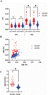
Results: Of the 128 TBI patients included, median age was 47 years, 70% male, and 66% presented with Glasgow Coma Scale of 15. No GLOS was detected at TP1 but was present in 23% of patients at TP2. GLOS+ patients were older (68 years [56-76] vs 39 years [27-53], p < 0.001), more likely to report falls as injury mechanism (62% vs 36%, p = 0.006), report history of hypertension (41% vs 19%, p = 0.025), and had a higher burden of WMHs (59% vs 14% with a total Fazekas ≥2, p < 0.001). Quantitative SIRs confirmed qualitative assessments: GLOS+ patients had higher SIRs at TP2 (0.43 vs 0.22, p < 0.001). Age (OR 3.28, 95%CI [1.88-5.71], p < 0.001) and prior TBI history (OR 4.99, 95%CI [1.46-17.06], p = 0.010) were independent predictors of GLOS. When age was removed, total Fazekas score (OR 2.53, 95%CI [1.60-4.00], p < 0.001) was an independent predictor of GLOS.
Conclusions: GLOS is primarily associated with age and may serve as another imaging marker of chronic vascular disease.
Keywords: Blood-ocular barrier disruption; Cerebral small vessel disease; Gadolinium leakage into ocular structures; MRI; Traumatic brain injury.
Copyright © 2024. Published by Elsevier B.V.
Conflict of interest statement
Declaration of competing interest None.
Figures


References
-
- London A, Benhar I, Schwartz M. The retina as a window to the brain-from eye research to CNS disorders. Nat Rev Neurol 2013;9:44–53. - PubMed
-
- Streilein JW. Ocular immune privilege: therapeutic opportunities from an experiment of nature. Nat Rev Immunol 2003;3:879–889. - PubMed
-
- Cunha-Vaz J The blood-ocular barriers. Surv Ophthalmol 1979;23:279–296. - PubMed
MeSH terms
Substances
Grants and funding
LinkOut - more resources
Full Text Sources
Medical

