Review
. 2024 Sep:141:103731.
doi: 10.1016/j.dnarep.2024.103731. Epub 2024 Jul 22.

Mechanisms and regulation of replication fork reversal

Affiliations
Review

Mechanisms and regulation of replication fork reversal

Madison B Adolph et al. DNA Repair (Amst). 2024 Sep.

Abstract

DNA replication is remarkably accurate with estimates of only a handful of mutations per human genome per cell division cycle. Replication stress caused by DNA lesions, transcription-replication conflicts, and other obstacles to the replication machinery must be efficiently overcome in ways that minimize errors and maximize completion of DNA synthesis. Replication fork reversal is one mechanism that helps cells tolerate replication stress. This process involves reannealing of parental template DNA strands and generation of a nascent-nascent DNA duplex. While fork reversal may be beneficial by facilitating DNA repair or template switching, it must be confined to the appropriate contexts to preserve genome stability. Many enzymes have been implicated in this process including ATP-dependent DNA translocases like SMARCAL1, ZRANB3, HLTF, and the helicase FBH1. In addition, the RAD51 recombinase is required. Many additional factors and regulatory activities also act to ensure reversal is beneficial instead of yielding undesirable outcomes. Finally, reversed forks must also be stabilized and often need to be restarted to complete DNA synthesis. Disruption or deregulation of fork reversal causes a variety of human diseases. In this review we will describe the latest models for reversal and key mechanisms of regulation.

Keywords: DNA damage; DNA replication; Fork protection; Fork reversal; Genome stability; Replication stress.


Conflict of interest statement

Declaration of Competing Interest The authors declare that they have no known competing financial interests or personal relationships that could have appeared to influence the work reported in this paper.

Figures

Figure 1.
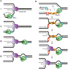
Figure 1.. Replication fork reversal.
(A) Replication fork reversal places a template DNA lesion back into duplex DNA. This process requires RAD51 and is mediated by ATP-dependent translocases. (B) Potential outcomes of fork reversal are illustrated and described further in the text. During template switching, the dashed line indicates DNA synthesis where the nascent DNA strand is used as the template strand. Pink and purple shapes represent endo and exonucleases that degrade nascent DNA or can induce breaks.
Figure 2.
Figure 2.. Assays used to measure replication fork reversal in vitro and in cells.
(A) In vitro branch migration assays utilize model replication fork substrates to observe the conversion of the fork substrate to products. DNA products are visualized through resolution by native polyacrylamide gel electrophoresis. (B) DNA combing and fiber assays allow for analysis of replication fork dynamics. Fork slowing due to reversal in the presence of replication stress would yield shorter labeled fibers. (C) In fork protection assays cells are treated for equal times with CldU and IdU then exposed to replication stress. After processing, DNA track lengths are measured and the IdU over CldU ratio is calculated to monitor nascent strand degradation which is dependent on reversal. (D) Electron microscopy allows for visualization of DNA replication intermediates. After processing, DNA replication forks can be visualized where the daughter strands (D) are of equal length due to restriction digest. Reversed replication forks show the reversed arm (R), and the parental arm (P). (E) Replication intermediates from Xenopus egg extracts can be visualized by 2D-gel electrophoresis and southern blot analysis. Replication fork reversal does not affect the mass but does affect the shape of the DNA. Replication forks migrate in a characteristic Y-arc. Fork stalling results in accumulation of a specific size and shape fragment and reversed forks result in a vertical spike as illustrated.
Figure 3.
Figure 3.. Important enzymes and mechanisms for fork reversal.
(A) Schematic of domain organization of SNF2 translocases SMARCAL1, ZRANB3, HLTF, PICH and RAD54L. (B) Schematic of domain organization of FBH1, RAD51 and FANCM. (C) A speculative model illustrating how the SMARCAL1, HLTF, and ZRANB3 proteins may cooperate to promote reversal. SMARCAL1 recruitment is regulated by RPA, HLTF preferentially binds 3’OH DNA ends, and ZRANB3 is recruited by poly-ubiquitylated PCNA. RAD51 is required for both the initial steps of fork reversal and to protect the regressed nascent DNA arm from degradation by nucleases. RAD51 filaments are stabilized by BRCA2. (D) FBH1 is a 3’−5’ UvrD helicase that can unwind DNA and disassemble RAD51 filaments. It also cooperates with a Cullin complex to act as an E3 ligase to ubiquitylate free RAD51 and prevent its reassociation with the DNA.
Figure 4.
Figure 4.. Model for the bypass of CMG during replication fork reversal.
(A) Potential placements of the CMG helicase during fork reversal. (B) A model for fork reversal that retains CMG at the fork. In this model, RAD51 assembles on the ssDNA and catalyzes a strand exchange reaction into the duplex DNA of the sister chromatid to initiate fork reversal. This is followed by branch migration by the fork reversal translocases to generate the nascent-nascent duplex and reversed fork. This process traps the CMG in the parental DNA duplex.
Figure 5.
Figure 5.. Mechanisms of Fork Restart.
In the presence of replication stress, PARP-dependent poly-ADP ribosylation inhibits RECQ1 to allow reversed forks to accumulate. Subsequently, restart can happen through multiple pathways. (I) RECQ1 can perform branch migration to restore the 3-way fork structure. (II) SMARCAL1 or other reversal enzymes can switch directions by binding to a daughter strand duplex leading to branch migration and fork restoration. (III) RAD51 can promote a strand invasion reaction to promote fork restart. Restart in these latter two pathways would be promoted by end-resection dependent on DNA2 and WRN.

References

    1. Zeman MK and Cimprich KA, Causes and consequences of replication stress. Nat Cell Biol, 2014. 16(1): p. 2–9. - PMC - PubMed
    1. Cortez D., Replication-Coupled DNA Repair. Mol Cell, 2019. 74(5): p. 866–876. - PMC - PubMed
    1. Joseph SA, et al. , Time for remodeling: SNF2-family DNA translocases in replication fork metabolism and human disease. DNA Repair (Amst), 2020. 95: p. 102943. - PMC - PubMed
    1. Conti BA and Smogorzewska A, Mechanisms of direct replication restart at stressed replisomes. DNA Repair (Amst), 2020. 95: p. 102947. - PMC - PubMed
    1. Neelsen KJ and Lopes M, Replication fork reversal in eukaryotes: from dead end to dynamic response. Nature Reviews Molecular Cell Biology, 2015. 16(4): p. 207–220. - PubMed

Publication types

LinkOut - more resources