. 2024 Aug 8;111(8):1643-1655.
doi: 10.1016/j.ajhg.2024.07.005. Epub 2024 Jul 31.

Identification of a DNA methylation episignature for recurrent constellations of embryonic malformations

Affiliations

Identification of a DNA methylation episignature for recurrent constellations of embryonic malformations

Sadegheh Haghshenas et al. Am J Hum Genet. .

Abstract

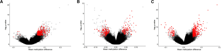
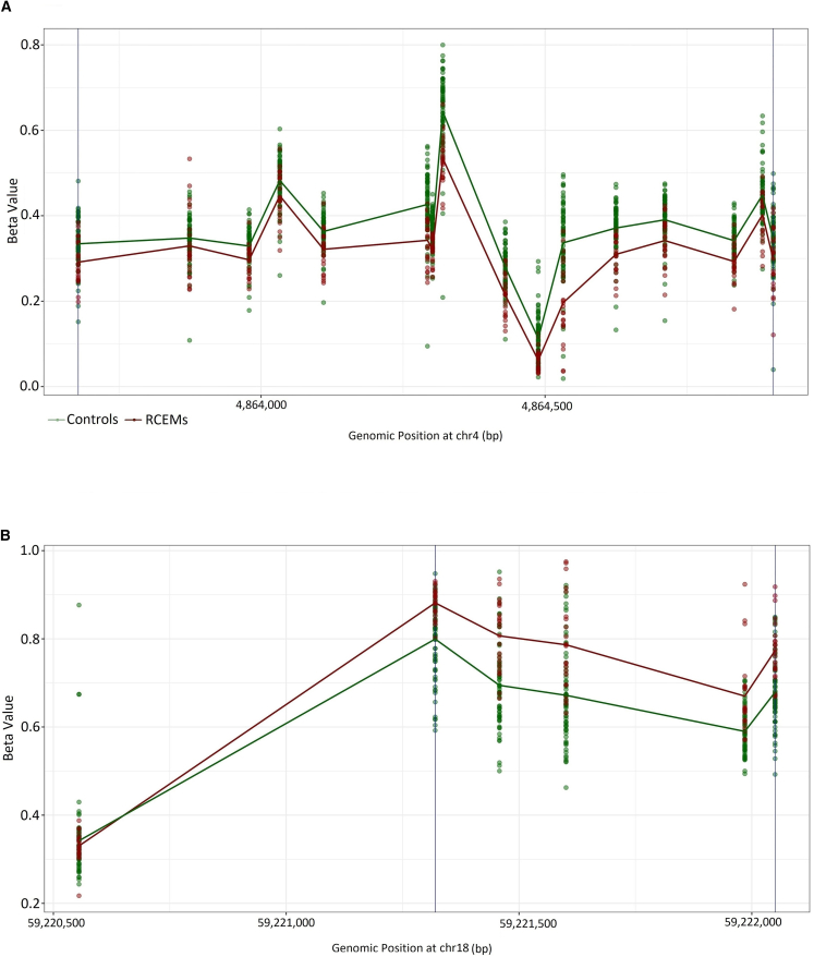
The term "recurrent constellations of embryonic malformations" (RCEM) is used to describe a number of multiple malformation associations that affect three or more body structures. The causes of these disorders are currently unknown, and no diagnostic marker has been identified. Consequently, providing a definitive diagnosis in suspected individuals is challenging. In this study, genome-wide DNA methylation analysis was conducted on DNA samples obtained from the peripheral blood of 53 individuals with RCEM characterized by clinical features recognized as VACTERL and/or oculoauriculovertebral spectrum association. We identified a common DNA methylation episignature in 40 out of the 53 individuals. Subsequently, a sensitive and specific binary classifier was developed based on the DNA methylation episignature. This classifier can facilitate the use of RCEM episignature as a diagnostic biomarker in a clinical setting. The study also investigated the functional correlation of RCEM DNA methylation relative to other genetic disorders with known episignatures, highlighting the common genomic regulatory pathways involved in the pathophysiology of RCEM.

Keywords: DNA methylation; OAV; VACTERL; epigenetics; episignature; recurrent constellations of embryonic malformations.


Conflict of interest statement

Declaration of interests B.S. is a shareholder in EpiSign Inc. involved in commercial uses of EpiSign technology.

Figures

Figure 1
Figure 1
Verification of the probes selected for the combined episignature (A) Hierarchical clustering was performed, with red representing individuals with a VACTERL phenotype, orange representing individuals with OAV, purple representing individuals with the overlapping phenotype, and blue representing control individuals. (B) MDS analysis was conducted using the same color scheme as (A). (C) The summary of MVP scores generated during LOOCV was visualized. Case testing samples are represented by red circles and other database samples by black circles. (D) MVP scores calculated by the SVM classifier are plotted, with blue circles representing training samples and gray circles representing testing samples. Abbreviations of all cohorts ae listed in Table S3.
Figure 2
Figure 2
Visualization of mean methylation differences and the negative logarithm of p values for the three episignatures (A) The robust episignature, (B) the intermediate episignature, and (C) the combined episignature. The selected probes are represented by red circles while other probes are marked with black circles.
Figure 3
Figure 3
Overlap of DMPs among episignature disorders Heatmap presenting the percentage of probes shared between each pair of cohorts. Colors range from white (indicating zero overlap) to red (indicated full overlap), representing the percentage of probes from the x axis cohort that are also present in the y axis cohort’s probes. Abbreviations for all cohorts are listed in Table S3.
Figure 4
Figure 4
Functional correlation analysis of the robust group (A) Methylation differences of all DMPs for each cohort. Red lines indicate mean methylation difference. Each circle represents one probe. (B) Tree and leaf plot of hierarchical clustering of all 56 cohorts using the top DMPs for each cohort. A leaf node represents a cohort, with node sizes illustrating relative scales of the number of selected DMPs for the corresponding cohort, and node colors are indicative of the overall mean methylation difference. (C) DMPs annotated in relation to CGIs (upper side) and genes (lower side). Abbreviations of all cohorts are listed in Table S3.
Figure 5
Figure 5
DMRs in the intermediate and robust groups (A) A general hypomethylation pattern was observed in the single DMR identified for the intermediate cluster located on chromosome 4. (B) A predominant hypermethylation was observed in 65 out of 66 DMRs detected in the robust cluster. The DMR located on chromosome 18 was presented as an example. The details of other DMRs are provided in Table S4.

References

    1. Adam A.P., Curry C.J., Hall J.G., Keppler-Noreuil K.M., Adam M.P., Dobyns W.B. Recurrent constellations of embryonic malformations re-conceptualized as an overlapping group of disorders with shared pathogenesis. Am. J. Med. Genet. 2020;182:2646–2661. doi: 10.1002/ajmg.a.61847. - DOI - PubMed
    1. Stevenson R.E. Common pathogenesis for sirenomelia, OEIS complex, limb-body wall defect, and other malformations of caudal structures. Am. J. Med. Genet. 2021;185:1379–1387. doi: 10.1002/ajmg.a.62103. - DOI - PubMed
    1. Mark P.R. NAD+ deficiency in human congenital malformations and miscarriage: A new model of pleiotropy. Am. J. Med. Genet. 2022;188:2834–2849. doi: 10.1002/ajmg.a.62764. - DOI - PMC - PubMed
    1. Thomas M., Bedard T., Crawford S., Grevers X., Lowry R. Craniofacial Microsomia, Associated Congenital Anomalies, and Risk Factors in 63 Cases from the Alberta Congenital Anomalies Surveillance System. J. Pediatr. 2023;261 - PubMed
    1. Stevens S.J.C., Stumpel C.T.R.M., Diderich K.E.M., van Slegtenhorst M.A., Abbott M.A., Manning C., Balciuniene J., Pyle L.C., Leonard J., Murrell J.R., et al. The broader phenotypic spectrum of congenital caudal abnormalities associated with mutations in the caudal type homeobox 2 gene. Clin. Genet. 2022;101:183–189. doi: 10.1111/cge.14076. - DOI - PMC - PubMed

LinkOut - more resources