. 2024 Sep;300(9):107617.
doi: 10.1016/j.jbc.2024.107617. Epub 2024 Jul 30.

Lack of prion transmission barrier in human PrP transgenic Drosophila

Affiliations

Lack of prion transmission barrier in human PrP transgenic Drosophila

Alana M Thackray et al. J Biol Chem. 2024 Sep.

Abstract

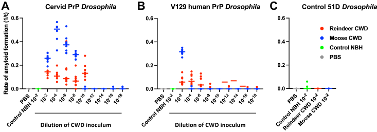
While animal prion diseases are a threat to human health, their zoonotic potential is generally inefficient because of interspecies prion transmission barriers. New animal models are required to provide an understanding of these prion transmission barriers and to assess the zoonotic potential of animal prion diseases. To address this goal, we generated Drosophila transgenic for human or nonhuman primate prion protein (PrP) and determined their susceptibility to known pathogenic prion diseases, namely varient Creutzfeldt-Jakob disease (vCJD) and classical bovine spongiform encephalopathy (BSE), and that with unknown pathogenic potential, namely chronic wasting disease (CWD). Adult Drosophila transgenic for M129 or V129 human PrP or nonhuman primate PrP developed a neurotoxic phenotype and showed an accelerated loss of survival after exposure to vCJD, classical BSE, or CWD prions at the larval stage. vCJD prion strain identity was retained after passage in both M129 and V129 human PrP Drosophila. All of the primate PrP fly lines accumulated prion seeding activity and concomitantly developed a neurotoxic phenotype, generally including accelerated loss of survival, after exposure to CWD prions derived from different cervid species, including North American white-tailed deer and muntjac, and European reindeer and moose. These novel studies show that primate PrP transgenic Drosophila lack known prion transmission barriers since, in mammalian hosts, V129 human PrP is associated with severe resistance to classical BSE prions, while both human and cynomolgus macaque PrP are associated with resistance to CWD prions. Significantly, our data suggest that interspecies differences in the amino acid sequence of PrP may not be a principal determinant of the prion transmission barrier.

Keywords: BSE; Drosophila; chronic wasting disease; human; neurodegenerative disease; nonhuman primate; prion; transgenic; vCJD; zoonotic.


Conflict of interest statement

Conflict of interest The authors declare that they have no conflicts of interest with the contents of this article.

Figures

Figure 1
Figure 1
Amino acid sequence comparison of primate PrP. Amino acid sequence alignment of human (M129) and nonhuman primate PrP sequences. The nonhuman primate species used in the comparison were chimpanzee, cynomolgus macaque (C. macaque), squirrel monkey (S. monkey), and lemur. The PrP sequence for each species corresponds to the amino acid sequences for the mature PrP and the subsequent GPI anchor signal sequence. The nonhuman primate PrP amino acid sequences were compared to that of human PrP. Consensus residues are represented by a dot (.), while nonconsensus residues are indicated by the corresponding amino acid colored in red. The numbers above the sequence lines correspond to the amino acid numbering for the full-length human PrP, while the numbers to the right of each sequence line correspond to the amino acid numbering for the full-length PrP of the corresponding species. GPI, glycosylphosphatidylinositol; PrP, prion protein.
Figure 2
Figure 2
Western blot detection of prion protein expression in primate PrP Drosophila.Elav x primate PrP or Elav x control 51D Drosophila were harvested at 5 days of age. Fly head homogenates were prepared and analyzed by SDS-PAGE and Western blot with anti-PrP mAb Sha31 as described in the Experimental procedures. Molecular mass marker values (kDa) are shown on the left-hand side.
Figure 3
Figure 3
Accumulation of prion seeding activity in prion-exposed primate PrP Drosophila.Elav x primate PrP Drosophila were exposed to human vCJD (A), bovine classical BSE (B), or cervid CWD (white-tailed deer CWD prion-infected) (C), or prion-free human normal brain homogenate (NBH) (D), bovine NBH (E), or cervid NBH (F) at the larval stage. Adult Drosophila were collected at the indicated time points after hatching (see colored key), and head homogenate was prepared and used as seed in RT-QuIC reactions. Known RT-QuIC positive and negative fly head homogenates were included as controls for the assay (data not shown). The data shown are rate of amyloid formation 1/time (1/t) for each treatment group. Statistical analysis of Figure 3 is shown in Table S1. BSE, bovine spongiform encephalopathy; CWD, chronic wasting disease; vCJD, variant Creutzfeldt-Jakob disease; PrP, prion protein; RT-QuIC, real-time quaking-induced conversion.
Figure 4
Figure 4
Accelerated decline of locomotor activity in prion-exposed primate PrP Drosophila.Elav x primate PrP Drosophila were exposed to human vCJD, bovine classical BSE or white-tailed deer CWD prion-infected, or prion-free human, bovine or cervid brain material at the larval stage. After hatching, flies were assessed for locomotor activity by a negative geotaxis climbing assay. The mean performance index is shown for three groups of n = 15 flies of each genotype per time point. Statistical analysis of the linear regression plots was performed using an unpaired (two-tailed) Student t test. All prion-exposed primate PrP Drosophila treatment group plots were significantly different (p < 0.05) from their respective prion-free control plots over the whole of the climbing assay time course. M129 human PrP Drosophila (A), or V129 human PrP Drosophila (B), chimpanzee PrP Drosophila (C), cynomolgus macaque PrP Drosophila (D), squirrel monkey PrP Drosophila (E) and lemur PrP Drosophila (F). The mean performance index for control 51D Drosophila is shown in Figure S1. Statistical analysis of Figure 4 is shown in Table S2. BSE, bovine spongiform encephalopathy; CWD, chronic wasting disease; PrP, prion protein; vCJD, variant Creutzfeldt-Jakob disease.
Figure 5
Figure 5
Accelerated decline of loss of survival of prion-exposed primate PrP Drosophila.Elav x primate PrP Drosophila or Elav x control 51D Drosophila were exposed to human vCJD, bovine classical BSE or white-tailed deer CWD prion-infected, or prion-free human, bovine or cervid brain material, or PBS at the larval stage, respectively. After hatching, the number of surviving flies was recorded three times a week and the data shown as Kaplan–Meier plots. (A) M129 human PrP Drosophila, or (B) V129 human PrP Drosophila, (C) chimpanzee PrP Drosophila, (D) cynomolgus macaque PrP Drosophila, (E) squirrel monkey PrP Drosophila, and (F) lemur PrP Drosophila. The survival curves for control 51D Drosophila are shown in Figure S2. Statistical analysis of Figure 5 and Figure S2 is shown in Table S3. BSE, bovine spongiform encephalopathy; CWD, chronic wasting disease; PrP, prion protein; vCJD, variant Creutzfeldt-Jakob disease.
Figure 6
Figure 6
Authentic vCJD prion strain propagation in human PrP Drosophila.Elav x M129 and Elav x V129 human PrP Drosophila, and Elav x control 51D Drosophila, were exposed to vCJD prion inoculum at the larval stage and harvested at 40 days post hatching when head homogenate was prepared from harvested flies and intracerebrally inoculated into bovine PrP transgenic (tgBov) mice. As a control, the original vCJD prion inoculum was also inoculated into tgBov mice. Inoculated mice were euthanized when they showed clinical signs of prion infection or after 700 days for those that did not develop clinical disease. Western blot analysis of inoculated tgBov mouse brains is shown in Figure 6A. Mice were considered positive for prion disease when proteinase K-resistant PrP 27–30 was detected in brain tissue by Western blot. Lane 1: sheep scrapie DAW isolate (positive control); lane 2: vCJD isolate (positive control); lane 3: direct transmission of vCJD isolate in tgBov; lanes 4 and 5: vCJD passaged in Met129 human PrP Drosophila transmitted to tgBov (representing two different mouse samples); lanes 6 and 7: vCJD passaged in Val129 human PrP Drosophila transmitted to tgBov (representing two different mouse samples); and lanes 8 and 9: vCJD passaged in control 51D Drosophila transmitted to tgBov (representing two different mouse samples). The attack rate (number of prion-positive mice/total number of mice inoculated) and the incubation time for inoculated mice, which represents the average period from inoculation to euthanasia for each inoculated group of animals, shown in days ± SD, is reported for each treatment group and are shown in Figure 6B. Lesion profile analysis of prion-diseased mice was established by scoring the vacuolar changes observed in predefined brain areas and is shown in Figure 6C: filled circle, vCJD isolate; open square, vCJD passaged in Met129 human PrP Drosophila; and open triangle, vCJD passaged in Val129 human PrP Drosophila. The data shown are mean lesion profile scores (three to six mouse brains examined for each isolate) for the following areas of the brain: for gray (G) matter, G1, dorsal medulla nuclei; G2, cerebellar cortex of the folia, including the granular layer, adjacent to the fourth ventricle; G3, cortex of the superior colliculus; G4, hypothalamus; G5, thalamus; G6, hippocampus; G7, septal nuclei of the paraterminal body; G8, cerebral cortex (at the level of G4 and G5); G9, cerebral cortex (at the level of G7); for white (W) matter, W1, cerebellar peduncles; W2, white matter in lateral tegmentum; W3, cerebellar peduncle/internal capsule. PrP, prion protein; vCJD, variant Creutzfeldt-Jakob disease.
Figure 7
Figure 7
Prion seeding activity in CWD prion-exposed human PrP Drosophila.Elav x M129 (A), Elav x V129 (B), human PrP Drosophila, or Elav x control 51D Drosophila (C) were exposed to CWD-infected North American (white-tailed deer or muntjac) or European (Norwegian reindeer or moose) brain material, or Norwegian reindeer lymph node (LN) material or prion-free control cervid normal brain homogenate (control NBH) at the larval stage. Adult Drosophila were collected at the indicated time points after hatching (see colored key), and head homogenate was prepared and used as seed in RT-QuIC reactions. Known RT-QuIC positive and negative fly head homogenates were included as controls for the assay (data not shown). The data shown are rate of amyloid formation 1/time (1/t) for each treatment group. Statistical analysis of Figure 7 is shown in Table S4. CWD, chronic wasting disease; PrP, prion protein; RT-QuIC, real-time quaking-induced conversion.
Figure 8
Figure 8
Accelerated loss of locomotor activity in CWD prion-exposed human PrP Drosophila.Elav x M129 (A) or Elav x V129 (B) human PrP or Elav x control 51D (C) Drosophila were exposed to CWD-infected North American (white-tailed deer or muntjac) or European (Norwegian reindeer or moose) brain material, or Norwegian reindeer lymph node (LN) material or prion-free control cervid normal brain homogenate (control NBH), or PBS, at the larval stage. After hatching, flies were assessed for locomotor activity by a negative geotaxis climbing assay. The mean performance index is shown for three groups of n = 15 flies of each genotype per time point. Statistical analysis of the linear regression plots was performed using an unpaired (two-tailed) Student t test. All prion-exposed Elav x M129 or Elav x V129 human PrP Drosophila treatment group plots were significantly different (p < 0.05) from their respective prion-free control plots over the whole of the climbing assay time course. Statistical analysis of Figure 8 is shown in Table S5. CWD, chronic wasting disease; PrP, prion protein.
Figure 9
Figure 9
Accelerated loss of survival of CWD prion-exposed human PrP Drosophila.Elav x M129 (A) or Elav x V129 (B) human PrP or Elav x control 51D (C) Drosophila were exposed to CWD-infected North American (white-tailed deer or muntjac) or European (Norwegian reindeer or moose) brain material, or Norwegian reindeer lymph node (LN) material or prion-free control cervid normal brain homogenate (control NBH), or PBS, at the larval stage. After hatching, the number of surviving flies was recorded three times a week and the data shown as Kaplan–Meier plots. Median survival and statistical analysis of Figure 9 are shown in Table S6. CWD, chronic wasting disease; PrP, prion protein.
Figure 10
Figure 10
Accelerated loss of locomotor activity induced by reindeer or moose CWD prions.Elav x cervid PrP (A) and (D), Elav x V129 human PrP (B) and (E), or Elav x control 51D (C) and (F) Drosophila were exposed to a 10−2 dilution series of European (Norwegian reindeer or moose) CWD-infected brain material, or a 10−2 dilution of prion-free control cervid normal brain homogenate (control NBH), or PBS, at the larval stage. After hatching, flies were assessed for locomotor activity by a negative geotaxis climbing assay. The mean performance index is shown for three groups of n = 15 flies of each genotype per time point. Statistical analysis of the linear regression plots was performed using an unpaired (two-tailed) Student t test. All prion-exposed Elav x cervid PrP and Elav x V129 human PrP Drosophila treatment group plots were significantly different (p < 0.05) from their respective prion-free control plots over the whole of the climbing assay time course. Reindeer CWD prions (A-C) and moose CWD prions (D-F). Statistical analysis of Figure 10 is shown in Table S7. Day 50 mean performance index data from Figure 10 are shown plotted against the 10−2 dilution series of the relevant CWD prion inoculum in Fig. S3. CWD, chronic wasting disease; PrP, prion protein.
Figure 11
Figure 11
Differences in prion seeding activity accumulation induced by Norwegian reindeer or moose CWD inoculum.Elav x cervid PrP (A), Elav x V129 human PrP (B), or Elav x control 51D (C) Drosophila were exposed to a 10−2 dilution series of European (Norwegian reindeer or moose) CWD-infected brain material, or 10−2 prion-free control cervid normal brain homogenate (control NBH), or PBS, at the larval stage. Adult Drosophila were collected at 40 days post hatching and head homogenate was prepared and used as seed in RT-QuIC reactions. The data shown are rate of amyloid formation 1/time (1/t) for each treatment group. Statistical analysis of Figure 11 is shown in Table S8. CWD, chronic wasting disease; PrP, prion protein; RT-QuIC, real-time quaking-induced conversion.
Figure 12
Figure 12
Moose CWD prions do not affect survival of V129 human PrP Drosophila.Elav x cervid PrP (A) and (D), Elav x V129 human PrP (B) and (E), or Elav x control 51D (C) and (F) Drosophila were exposed to a 10−2 dilution series of European (Norwegian reindeer or moose) CWD-infected brain material, or 10−2 dilution of prion-free control cervid normal brain homogenate (control NBH) at the larval stage. After hatching, the number of surviving flies was recorded three times a week and the data shown as Kaplan–Meier plots. Reindeer CWD prions (AC) and moose CWD prions (DF). Statistical analysis of Figure 12 is shown in Table S9A and Table S9B. CWD, chronic wasting disease; PrP, prion protein.

References

    1. Prusiner S.B. second edition. Cold Spring Harbor Laboratory Press; New York: 2004. Prion Biology and Diseases.
    1. Prusiner S.B. Novel proteinaceous infectious particles cause scrapie. Science. 1982;216:136–144. - PubMed
    1. Castilla J., Saa P., Hetz C., Soto C. In vitro generation of infectious scrapie prions. Cell. 2005;121:195–206. - PubMed
    1. Deleault N.R., Harris B.T., Rees J.R., Supattapone S. Formation of native prions from minimal components in vitro. Proc. Natl. Acad. Sci. U. S. A. 2007;104:9741–9746. - PMC - PubMed
    1. Legname G., Baskakov I.V., Nguyen H.O., Riesner D., Cohen F.E., DeArmond S.J., et al. Synthetic mammalian prions. Science. 2004;305:673–676. - PubMed

MeSH terms

LinkOut - more resources