

Coupling Microdroplet-Based Sample Preparation, Multiplexed Isobaric Labeling, and Nanoflow Peptide Fractionation for Deep Proteome Profiling of the Tissue Microenvironment
- PMID: 39089681
- PMCID: PMC11325296
- DOI: 10.1021/acs.analchem.4c00523
Coupling Microdroplet-Based Sample Preparation, Multiplexed Isobaric Labeling, and Nanoflow Peptide Fractionation for Deep Proteome Profiling of the Tissue Microenvironment
Abstract
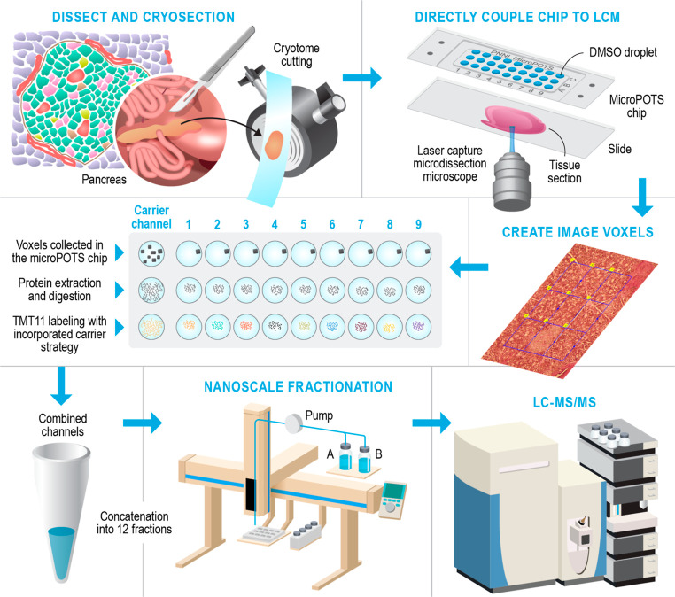
There is increasing interest in developing in-depth proteomic approaches for mapping tissue heterogeneity in a cell-type-specific manner to better understand and predict the function of complex biological systems such as human organs. Existing spatially resolved proteomics technologies cannot provide deep proteome coverage due to limited sensitivity and poor sample recovery. Herein, we seamlessly combined laser capture microdissection with a low-volume sample processing technology that includes a microfluidic device named microPOTS (microdroplet processing in one pot for trace samples), multiplexed isobaric labeling, and a nanoflow peptide fractionation approach. The integrated workflow allowed us to maximize proteome coverage of laser-isolated tissue samples containing nanogram levels of proteins. We demonstrated that the deep spatial proteomics platform can quantify more than 5000 unique proteins from a small-sized human pancreatic tissue pixel (∼60,000 μm2) and differentiate unique protein abundance patterns in pancreas. Furthermore, the use of the microPOTS chip eliminated the requirement for advanced microfabrication capabilities and specialized nanoliter liquid handling equipment, making it more accessible to proteomic laboratories.
Conflict of interest statement
The authors declare no competing financial interest.
Figures
Update of
-
Coupling microdroplet-based sample preparation, multiplexed isobaric labeling, and nanoflow peptide fractionation for deep proteome profiling of tissue microenvironment.bioRxiv [Preprint]. 2023 Mar 13:2023.03.13.531822. doi: 10.1101/2023.03.13.531822. bioRxiv. 2023. Update in: Anal Chem. 2024 Aug 13;96(32):12973-12982. doi: 10.1021/acs.analchem.4c00523. PMID: 36993277 Free PMC article. Updated. Preprint.
References
-
- Zhu Y.; Dou M.; Piehowski P. D.; Liang Y.; Wang F.; Chu R. K.; Chrisler W. B.; Smith J. N.; Schwarz K. C.; Shen Y.; et al. Spatially Resolved Proteome Mapping of Laser Capture Microdissected Tissue with Automated Sample Transfer to Nanodroplets. Mol. Cell. Proteomics 2018, 17 (9), 1864–1874. 10.1074/mcp.TIR118.000686. - DOI - PMC - PubMed
-
- Yi L.; Tsai C. F.; Dirice E.; Swensen A. C.; Chen J.; Shi T.; Gritsenko M. A.; Chu R. K.; Piehowski P. D.; Smith R. D.; et al. Boosting to Amplify Signal with Isobaric Labeling (BASIL) Strategy for Comprehensive Quantitative Phosphoproteomic Characterization of Small Populations of Cells. Anal. Chem. 2019, 91 (9), 5794–5801. 10.1021/acs.analchem.9b00024. - DOI - PMC - PubMed
MeSH terms
Substances
Grants and funding
LinkOut - more resources
Full Text Sources
