Review
. 2024 Oct 3;45(10):1403-1412.
doi: 10.3174/ajnr.A8299.

Myelographic Techniques for the Localization of CSF-Venous Fistulas: Updates in 2024

Affiliations
Review

Myelographic Techniques for the Localization of CSF-Venous Fistulas: Updates in 2024

Ajay A Madhavan et al. AJNR Am J Neuroradiol. .

Abstract

CSF-venous fistulas (CVFs) are a common cause of spontaneous intracranial hypotension. Despite their relatively frequent occurrence, they can be exceedingly difficult to detect on imaging. Since the initial description of CVFs in 2014, the recognition and diagnosis of this type of CSF leak has continually increased. As a result of multi-institutional efforts, a wide spectrum of imaging modalities and specialized techniques for CVF detection is now available. It is important for radiologists to be familiar with the multitude of available techniques, because each has unique advantages and drawbacks. In this article, we review the spectrum of imaging modalities available for the detection of CVFs, explain the advantages and disadvantages of each, provide typical imaging examples, and discuss provocative maneuvers that may improve the conspicuity of CVFs. Discussed modalities include conventional CT myelography, dynamic myelography, digital subtraction myelography, conebeam CT myelography, decubitus CT myelography by using conventional energy-integrating detector scanners, decubitus photon counting CT myelography, and intrathecal gadolinium MR myelography. Additional topics to be discussed include optimal patient positioning, respiratory techniques, and intrathecal pressure augmentation.


Figures

FIG 1.
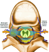
FIG 1.
Illustration of the venous anatomy relevant to CVF imaging. CVFs most commonly arise in association with meningeal diverticula. The internal vertebral venous plexus is an often overlooked drainage pathway that can be particularly subtle on imaging.
FIG 2.
FIG 2.
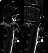
CVFs seen on conventional CT myelography and dynamic fluoroscopic myelography. Axial (A) and coronal (B) images from a conventional supine CT myelogram demonstrate subtle opacification of the internal epidural venous plexus (A and B, arrow), compatible with a CVF, which was subsequently confirmed on decubitus digital subtraction myelography (not shown). In a different patient, 2 AP images spaced 20 seconds apart from a left lateral decubitus dynamic myelogram (C and D) demonstrate a left T8 CVF involving several paraspinal veins (C, arrows), as well as the basivertebral venous plexus (D, arrows). The temporal resolution conferred by dynamic myelography is helpful to characterize the full extent of venous drainage.
FIG 3.
FIG 3.
Left T4 CSF-venous fistula seen on digital subtraction myelography and CB-CTM. Left lateral decubitus digital subtraction myelogram (A) demonstrates curvilinear venous opacification adjacent to a left T4 diverticulum (A, arrows), compatible with a CVF. High-resolution CBCT was performed next after centering the flat panel detector over the level of T4, with imaging performed during active injection of 4 mL Omnipaque 300. Axial (B, D, and E) and sagittal (C) reformatted CBCT images demonstrate a definitive left T4 CVF involving the paraspinal segmental vein (B and C, arrows), the hemiazygous vein (C, dashed arrows), the internal epidural venous plexus (D, arrows), and small lateral and paraspinal muscular venous branches (E, arrows).
FIG 4.
FIG 4.
Typical appearance of CVFs on single-day bilateral decubitus CTM in 2 different patients (A and B versus C and D). In both patients, right lateral decubitus CTM was performed initially with injection of 5 mL Omnipaque 300 (not shown). Subsequently, the spinal needle was removed, the patients were rotated to the left decubitus position, and myelography was repeated with another 5 mL Omnipaque 300 after placement of a new spinal needle. In the first patient, axial (A) and coronal (B) images demonstrate a left T7 CVF involving the paraspinal segmental vein (A and B, solid arrows) and a lateral venous branch (A, dashed arrow). In the second patient, whose myelogram was performed on photon-counting CT, coronal 0.2 mm images (C and D) demonstrate a left T10 CVF involving the internal epidural venous plexus (C and D, solid arrows) and the intervertebral vein (D, dashed arrow). Detection of venous opacification immediately adjacent to meningeal diverticula often requires high spatial resolution.
FIG 5.
FIG 5.
Advantage of high spatial resolution on PC-CTM for detection of a subtle CVF. Coronal and sagittal T3D 0.2 mm images (A and B) demonstrate a curvilinear opacification involving the right T2 intervertebral vein (A and B, solid arrows), clearly separate from the contrast-filled meningeal diverticulum (A–D, dashed arrows). The fistula is not visible when the images are reconstructed at 0.4 mm, because the spatial resolution is insufficient to discriminate the vein from the meningeal diverticulum. In some cases, high spatial resolution is necessary to confidently visualize subtle CVFs, particularly those adjacent to other high-attenuation structures.
FIG 6.
FIG 6.
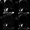
Benefit of high spatial resolution with a sharp kernel reconstruction and denoising to detect a subtle right T8 internal epidural CVF on PC-CTM. Four images from a right lateral decubitus PC-CTM are shown, all at the same section, timepoint, and window/level setting. Images were reconstructed at 0.4 mm with a smooth Br56 kernel (A), 0.2 mm with a smooth Br56 kernel (B), 0.2 mm with a sharp Qr89 kernel (C), and 0.2 mm with a sharp Qr89 kernel and denoising (D). A subtle CVF involving the ventral internal epidural venous plexus (A–D, solid arrows) is best seen and distinguished from the posterior vertebral body cortex (A–D, dashed arrows) on the denoised 0.2 mm sharp Qr89 kernel image.
FIG 7.
FIG 7.
Right T1 CVF associated with a paraspinal venous malformation, demonstrating the complementary benefits of high spatial resolution, 40 keV VMIs, and high temporal resolution on PC-CTM. Axial T2-weighted MR imaging demonstrates a T2 hyperintense venous malformation (A, solid arrows), which extended from C7–T2. Right lateral decubitus DSM shows a small right T1 meningeal diverticulum (B, solid arrow), but no evidence of adjacent venous opacification. Right lateral decubitus PC-CTM was performed next. Axial 0.2 mm T3D (C) from this PC-CTM demonstrates a right T1 CVF (C, solid arrows). A 0.4 mm 40 keV (D) image obtained 8 seconds later shows that the vein has washed away (D, solid arrow), with new subtle opacification of the venous malformation (D, dashed arrow). 40 keV images are especially helpful for detecting subtle areas of contrast opacification such as this.
FIG 8.
FIG 8.
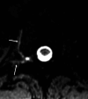
T1-weighted image from an GdM demonstrating a right T7 CVF involving the paraspinal segmental vein and lateral branches (arrows).
FIG 9.
FIG 9.
Benefit of resisted inspiration for the detection of a subtle right T11 CVF. Adjacent coronal images from a right lateral decubitus PC-CTM at 2 different time points (A and B versus C and D) obtained with resisted inspiration through a narrow syringe (A and B) and during slow inspiration through the mouth and nose (C and D) are shown. A clear right T11 CVF involving the internal epidural venous plexus is apparent during resisted inspiration (A and B, arrows) and completely occult during slow inspiration (C and D).

References

    1. Schievink WI, Maya MM, Jean-Pierre S, et al. A classification system of spontaneous spinal CSF leaks. Neurology 2016;87:673–79 10.1212/WNL.0000000000002986 - DOI - PubMed
    1. Liaw V, McCreary M, Friedman DI. Quality of life in patients with confirmed and suspected spinal CSF leaks. Neurology 2023;101:e2411–22 10.1212/WNL.0000000000207763 - DOI - PMC - PubMed
    1. Dobrocky T, Grunder L, Breiding PS, et al. Assessing spinal cerebrospinal fluid leaks in spontaneous intracranial hypotension with a scoring system based on brain magnetic resonance imaging findings. JAMA Neurol 2019;76:580–87 10.1001/jamaneurol.2018.4921 - DOI - PMC - PubMed
    1. Benson JC, Madhavan AA, Mark IT, et al. Likelihood of discovering a CSF leak based on intracranial MRI findings in patients without a spinal longitudinal extradural collection: a new probabilistic scoring system. AJNR Am J Neuroradiol 2023;44:1339–44 10.3174/ajnr.A8030 - DOI - PMC - PubMed
    1. Schievink WI, Maya M, Prasad RS, et al. Spontaneous spinal cerebrospinal fluid-venous fistulas in patients with orthostatic headaches and normal conventional brain and spine imaging. Headache 2021;61:387–91 10.1111/head.14048 - DOI - PubMed

MeSH terms

LinkOut - more resources