Review
. 2024 Aug 1;15(8):556.
doi: 10.1038/s41419-024-06939-5.

Oxidative cell death in cancer: mechanisms and therapeutic opportunities

Affiliations
Review

Oxidative cell death in cancer: mechanisms and therapeutic opportunities

Xiaoqin An et al. Cell Death Dis. .

Abstract

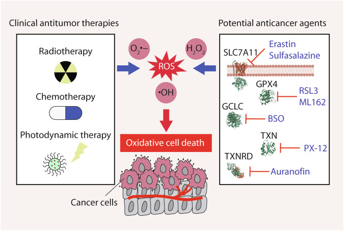
Reactive oxygen species (ROS) are highly reactive oxygen-containing molecules generated as natural byproducts during cellular processes, including metabolism. Under normal conditions, ROS play crucial roles in diverse cellular functions, including cell signaling and immune responses. However, a disturbance in the balance between ROS production and cellular antioxidant defenses can lead to an excessive ROS buildup, causing oxidative stress. This stress damages essential cellular components, including lipids, proteins, and DNA, potentially culminating in oxidative cell death. This form of cell death can take various forms, such as ferroptosis, apoptosis, necroptosis, pyroptosis, paraptosis, parthanatos, and oxeiptosis, each displaying distinct genetic, biochemical, and signaling characteristics. The investigation of oxidative cell death holds promise for the development of pharmacological agents that are used to prevent tumorigenesis or treat established cancer. Specifically, targeting key antioxidant proteins, such as SLC7A11, GCLC, GPX4, TXN, and TXNRD, represents an emerging approach for inducing oxidative cell death in cancer cells. This review provides a comprehensive summary of recent progress, opportunities, and challenges in targeting oxidative cell death for cancer therapy.


Conflict of interest statement

The authors declare no competing interests.

Figures

Fig. 1
Fig. 1. Overview of ROS generation and elimination.
Reactive oxygen species (ROS) are labile oxygen-containing molecules primarily generated by the mitochondrial electron transport chain (ETC), peroxisomes, NADPH oxidase (NOX), lipoxygenase (ALOX), cyclooxygenases (COXs), and cytochrome P450s (CYPs). ROS elimination is facilitated by antioxidant systems, encompassing enzymatic antioxidants (e.g., SOD, CAT, GPX, and the thioredoxin [TXN]-thioredoxin reductase [TXNRD] system) and non-enzymatic antioxidants (e.g., glutathione [GSH], vitamins or analogs, selenium, and metabolites such as bilirubin and melatonin). Superoxide dismutase (SOD) transforms O2•− into H2O2, subsequently reduced to H2O by catalase (CAT), glutathione peroxidase (GPX), or peroxiredoxins (PRDX). Among these, ALOX significantly contributes to lipid peroxidation, while GPX4, a selenocysteine-containing enzyme, quenches lipid peroxides. Central to the antioxidant network, TXNRD—a pivotal selenoprotein antioxidant—donates electrons to the TXN–PRDX axis. Moreover, the transcription factor NRF2 prominently regulates the antioxidant system, orchestrating the expression of genes crucial to antioxidant defense mechanisms.
Fig. 2
Fig. 2. The role of ROS in ferroptosis.
Ferroptosis, an iron-dependent form of cell death, is initiated by ROS-mediated lipid hydroperoxides. ROS primarily stem from the mitochondrial electron transport chain (ETC), NADPH oxidase (NOX), and Fe2+-mediated Fenton reactions. The transporter system xc relies on SLC7A11, a key subunit, to uptake extracellular cystine, a rate-limiting substrate for glutathione (GSH) synthesis. GSH, in turn, cofunctions with GPX4, a critical enzyme quenching lipid peroxidation, and aids in generating antioxidant hydropersulfides (RSSH) from cysteine. GSH-independent ferroptosis suppressors—FSP1 and DHODH—participate in CoQ10 to CoQH2 conversion, alongside FSP1’s roles in vitamin K reduction and membrane repair. The pivotal transcription factor NRF2 orchestrates gene expression to counteract ferroptosis. Fatty acids influence ferroptosis, with polyunsaturated fatty acids (PUFA) promoting it and monounsaturated fatty acids (MUFA) inhibiting it. Enzymes like ACSL4, LPCAT3, and SOAT1 mediate PUFA-CoA formation and subsequent esterification, while 4-hydroxynonenal (4HNE) from lipid hydroperoxides can activate cellular damage through the NOX pathway. Aldehyde dehydrogenase (ALDH) clears 4HNE, limiting ferroptosis. Iron’s import via transferrin (TF) and transferrin receptor (TFRC), as well as its export by SLC40A1/ferroportin, tightly regulates ferroptosis. Ferritinophagy, the autophagic degradation of ferritin, increases cytoplasmic Fe2+ levels, triggering ROS generation. Copper ions, along with iron, significantly contribute to initiating lipid peroxidation.
Fig. 3
Fig. 3. The role of ROS in apoptosis.
Apoptotic pathways can be classified into two categories: extrinsic apoptotic pathways triggered by cell death receptors, and intrinsic apoptotic pathways involving mitochondria. Mitochondria serve as the primary intracellular source of ROS, emanating from electron leakage within the respiratory electron transport chain (ETC). The activation of mitogen-activated protein kinase 14 (MAPK14/p38) or ER stress by ROS influences this balance through anti-apoptotic BCL2 and pro-apoptotic BAX, which results in the release of apoptotic molecules, such as cytochrome c (CYCS). CYCS’s release into the cytoplasm activates initiator caspase 9 (CASP9). ROS may increase the expression of the tumor suppressor protein TP53, which fosters apoptosis not only through the transcriptional regulation of apoptosis-related genes, but also by translocating to the mitochondria. Mitochondrial TP53 interacts with BCL2 family proteins and amplifies mitochondrial membrane permeability independent of transcriptional mechanisms. In addition, ROS is involved in the extrinsic apoptotic pathway through enhancing the expression of both FAS and FASL genes. Eventually, the sequential activation of executor caspase 3 (CASP3) by CASP8 in the extrinsic pathways or CASP9 in the apoptotic pathways initiates apoptosis. On the contrary, NRF2 triggers the transcription of downstream antioxidant genes, effectively neutralizing ROS and mitigating apoptosis.
Fig. 4
Fig. 4. The role of ROS in necroptosis.
Necroptosis is a regulated cell death orchestrated by receptor-interacting serine/threonine kinase 1 (RIPK1), receptor-interacting serine/threonine kinase 3 (RIPK3), and mixed lineage kinase domain-like pseudokinase (MLKL). Activation of receptors (e.g., TNFR1, TLR3, TLR4, and IFNAR1) prompts the recruitment of RHIM-containing proteins like RIPK1, TRIF, and ZBP1, and subsequent necrosome on the cytoplasmic side. Necroptosis is suppressed by caspase 8 (CASP8), and simultaneous treatment with TNF and caspase inhibitors can activate it. Subsequently, necrosomes form involving RIPK3 and MLKL in response to activation cues, which drives MLKL phosphorylation, oligomerization, and translocation to the plasma membrane for pore formation. ROS triggers RIPK1 autophosphorylation and MLKL activation. RIPK3 can also elevate ROS by stimulating mitochondrial metabolism and NADPH oxidase (NOX) activity. It enhances energy metabolism and mitochondrial ROS production through interactions with metabolic enzymes like pyruvate dehydrogenase complex (PDH), glutamate-ammonia ligase (GLUL), glutamate dehydrogenase 1 (GLUD1), and glycogen phosphorylase L (PYGL). Conversely, NRF2 induces transcription of antioxidant genes, mitigating ROS and ameliorating necroptosis.
Fig. 5
Fig. 5. The role of ROS in pyroptosis.
Pyroptosis is a mode of cell death marked by inflammasome activation and inflammation-associated caspases. Upon sensing damage-associated molecular pattern molecules (DAMPs) or pathogen-associated molecular pattern molecules (PAMPs), absent in melanoma 2 inflammasome (AIM2) and NLR family pyrin domain-containing 3 (NLRP3) inflammasomes assemble, which leads to caspase 1 (CASP1) activation. Alternatively, direct binding of lipopolysaccharide (LPS) to caspase 11 (CASP11) triggers CASP11 activation. Activated CASP1 or CASP11 cleaves gasdermin D (GSDMD), while CASP3 cleaves gasdermin E (GSDME), generating the pore-forming proteins N-GSDMD or N-GSDME, which induces cell death. ROS act as an upstream signal for NLRP3 inflammasome activation by upregulating pyroptosis-related genes like NLRP3 and CASP1. ROS or lipid peroxidation can also enhance GSDMD cleavage and CASP1 activation. Iron-induced ROS production activates caspase 3 (CASP3) via mitochondrial translocase of outer mitochondrial membrane 20 (TOMM20), triggering pyroptosis. Moreover, macrophage stimulating 1 (MST1) plays a role in pyroptosis regulation through promoting the production of ROS. In contrast, the pivotal regulator NRF2 curbs pyroptosis by reducing intracellular ROS levels.
Fig. 6
Fig. 6. The role of ROS in paraptosis.
Paraptosis is characterized by cytoplasmic vacuolation, resulting from extensive dilation of the endoplasmic reticulum (ER) and mitochondria. Vacuole formation in paraptosis necessitates the activation of mitogen-activated protein kinases (MAPKs). The onset of paraptosis is driven by ROS generation, initiating ER stress and Ca2+ overload. In contrast, thioredoxin reductase 1 (TXNRD1) critically curtails paraptosis by diminishing ROS production. Additionally, the interplay between mitochondria-associated ER membranes (MAMs) and the coordination of Ca2+ flux from the ER to mitochondria stand out as pivotal factors in inducing oxidative metabolic stress during paraptotic cell death.
Fig. 7
Fig. 7. The role of ROS in parthanatos and oxeiptosis.
a Parthanatos is a distinctive programmed cell death process driven by the enzymatic activity of poly(ADP-ribose) polymerase 1 (PARP1). This ribosyltransferase enzyme is crucial for DNA repair, detecting single- and double-strand DNA breaks and aiding in repair machinery recruitment. Oxidative stress acts as a central trigger, causing widespread DNA damage and excessive PARP1 activation. The resultant surplus of poly(ADP-ribose) (PAR) molecules leads to the liberation of apoptosis-inducing factor mitochondria-associated 1 (AIFM1) from mitochondria. AIFM1 then complexes with macrophage migration inhibitory factor (MIF), instigating parthanatos by orchestrating chromatin condensation and DNA fragmentation. The parthanatos process is influenced by mitogen-activated protein kinase 8 (MAPK8/JNK), AKT, and endoplasmic reticulum (ER) stress, which modulate intracellular ROS levels. b Oxeiptosis is distinguished by a notable ROS buildup, initiated via the kelch-like ECH-associated protein 1 (KEAP1)–PGAM family member 5-mitochondrial serine/threonine protein phosphatase (PGAM5)–apoptosis-inducing factor mitochondria-associated 1 (AIFM1/AIF) signaling cascade. KEAP1 acts as an inherent inhibitor of NRF2, leading to its proteasomal degradation. Under low ROS levels-induced oxidative stress, KEAP1 oxidizes and dissociates from NRF2, allowing NRF2 to translocate into the nucleus, thereby activating the transcription of numerous protective antioxidant genes. However, high ROS levels disrupt KEAP1’s interaction with another partner, PGAM5. This causes PGAM5 to relocate to the mitochondria, where it dephosphorylates AIFM1 and activates AIFM1, inducing oxeiptosis. The OTU deubiquitinase 1 (OTUD1) binds to and suppresses KEAP1’s ubiquitination, consequently inhibiting ROS-triggered oxeiptosis.
Fig. 8
Fig. 8. Strategies to inducing oxidative cell death in cancer.
Approaches that promote the generation of ROS to trigger oxidative cell death hold great promise in anti-cancer therapeutics. Conventional anti-tumor treatments such as radiotherapy, chemotherapy, and photodynamic therapy, can leverage the elevation of ROS levels within cancer cells, leading to damage to the malignancy. Furthermore, potential anti-cancer agents targeting specific components of the antioxidant system, such as SLC7A11, GCLC, GPX4, TXN, and TXNRD, have the potential to selectively eliminate cancer cells.

References

    1. Galluzzi L, Vitale I, Aaronson SA, Abrams JM, Adam D, Agostinis P, et al. Molecular mechanisms of cell death: recommendations of the Nomenclature Committee on Cell Death 2018. Cell Death Differ. 2018;25:486–541. 10.1038/s41418-017-0012-4 - DOI - PMC - PubMed
    1. Tang D, Kang R, Berghe TV, Vandenabeele P, Kroemer G. The molecular machinery of regulated cell death. Cell Res. 2019;29:347–64. 10.1038/s41422-019-0164-5 - DOI - PMC - PubMed
    1. Tsvetkov P, Coy S, Petrova B, Dreishpoon M, Verma A, Abdusamad M, et al. Copper induces cell death by targeting lipoylated TCA cycle proteins. Science. 2022;375:1254–61. 10.1126/science.abf0529 - DOI - PMC - PubMed
    1. Liu X, Nie L, Zhang Y, Yan Y, Wang C, Colic M, et al. Actin cytoskeleton vulnerability to disulfide stress mediates disulfidptosis. Nat Cell Biol. 2023;25:404–14. 10.1038/s41556-023-01091-2 - DOI - PMC - PubMed
    1. Peng F, Liao M, Qin R, Zhu S, Peng C, Fu L, et al. Regulated cell death (RCD) in cancer: key pathways and targeted therapies. Signal Transduct Target Ther. 2022;7:286. 10.1038/s41392-022-01110-y - DOI - PMC - PubMed