

Study on the value of MRI in locating the internal OS of the cervix and influencing factors
- PMID: 39090384
- PMCID: PMC11294610
- DOI: 10.1038/s41598-024-68735-7
Study on the value of MRI in locating the internal OS of the cervix and influencing factors
Abstract
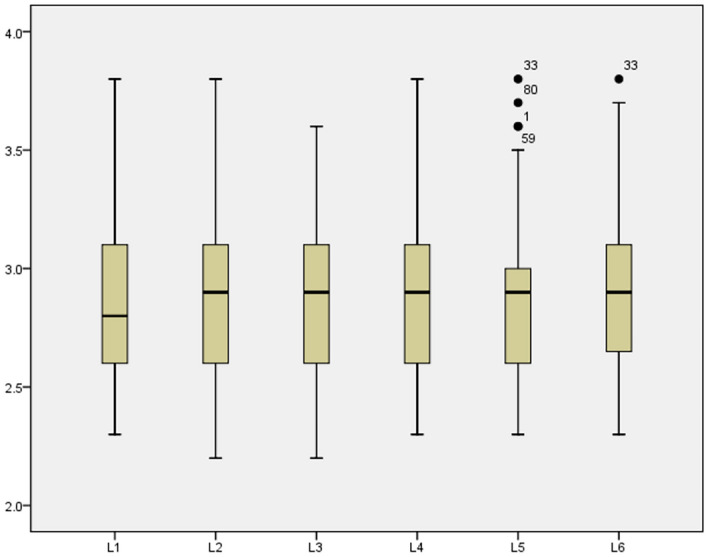
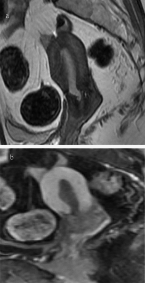
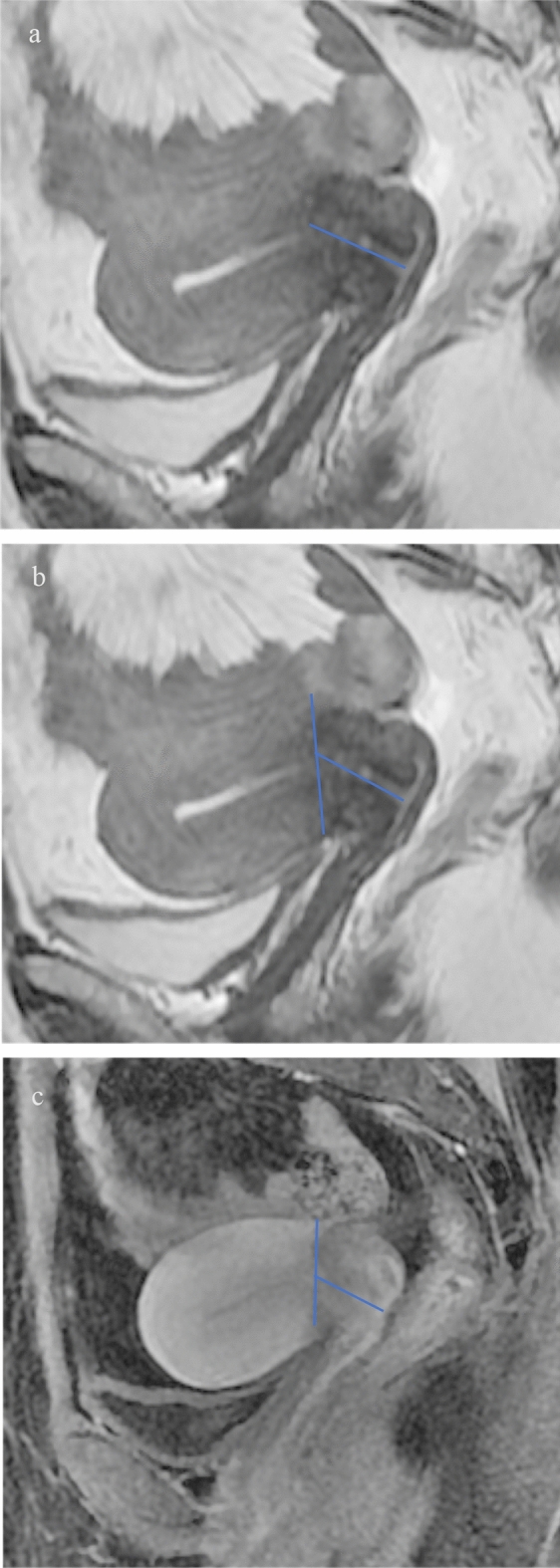
The position of the internal os of the cervix reported in the literature was inconsistent on MRI images. Additionally, the practical impactful data influencing the internal os located by MRI is limited. We aimed to confirm the position of the internal os of the cervix on MRI images, and the influencing factors locating the the internal os by MRI. A single-center retrospective study was conducted. Data from 175 patients who underwent total hysterectomy for stage I endometrial cancer were collected. The internal os of the cervix is positioned as the starting point for measuring the length of the cervix on MRI images. On dynamic contrast-enhanced MRI (DCE-MRI), the section formed by the enhancement difference between the uterus and cervix, and on T2-weighted imaging(T2WI), the section formed by the physiological curvature of the uterus and the low signal intensity of the cervical stroma were used as starting points. The results showed no statistically significant difference compared with the removed uterus specimens (p = 0.208, p = 0.571, p = 0.804). A history of cesarean section(p < 0.001), irregular vaginal bleeding for more than three months(p < 0.001), cervical adenomyosis(p = 0.043), and premenopause(p = 0.001) were not conducive to locating the internal os of the cervix by MRI. Our findings provide valuable information and confirm the position of the internal os of the cervix on MRI images, and the several important infuencing factors. We hope that some patients will benefit from our study.
Keywords: Fertility preservation; Internal Os of cervix; Localization; Magnetic resonance imaging; Surgery.
© 2024. The Author(s).
Conflict of interest statement
The authors declare no competing interests.
Figures
Similar articles
-
Comparison of measurements of the uterus and cervix obtained by magnetic resonance and transabdominal ultrasound imaging to identify the brachytherapy target in patients with cervix cancer.Int J Radiat Oncol Biol Phys. 2014 Mar 15;88(4):860-5. doi: 10.1016/j.ijrobp.2013.12.004. Epub 2014 Jan 22. Int J Radiat Oncol Biol Phys. 2014. PMID: 24462382
-
MRI is highly specific in determining primary cervical versus endometrial cancer when biopsy results are inconclusive.Clin Radiol. 2013 Nov;68(11):1107-13. doi: 10.1016/j.crad.2013.05.095. Epub 2013 Jul 10. Clin Radiol. 2013. PMID: 23849621
-
[MR Imaging of the pelvis in the diagnosis of the endometrium in breast cancer patients in tamoxifen therapy].Rofo. 2006 Mar;178(3):316-23. doi: 10.1055/s-2005-858934. Rofo. 2006. PMID: 16508840 German.
-
MRI of malignant neoplasms of the uterine corpus and cervix.AJR Am J Roentgenol. 2007 Jun;188(6):1577-87. doi: 10.2214/AJR.06.1196. AJR Am J Roentgenol. 2007. PMID: 17515380 Review.
-
Successful management of cervical dysgenesis: Case report and review.Trop Doct. 2023 Apr;53(2):271-275. doi: 10.1177/00494755221149228. Epub 2023 Jan 27. Trop Doct. 2023. PMID: 36705083 Review.
Cited by
-
Multiple Nabothian Cysts and Tunnel Clusters Clinically Mimicking Lobular Endocervical Glandular Hyperplasia: A Case Report.Cureus. 2025 Jan 18;17(1):e77643. doi: 10.7759/cureus.77643. eCollection 2025 Jan. Cureus. 2025. PMID: 39968433 Free PMC article.
