. 2024 Sep;20(9):5861-5888.
doi: 10.1002/alz.13921. Epub 2024 Aug 1.

Primate cerebrospinal fluid CHI3L1 reflects brain TREM2 agonism

Affiliations

Primate cerebrospinal fluid CHI3L1 reflects brain TREM2 agonism

Stephen P Schauer et al. Alzheimers Dement. 2024 Sep.

Abstract

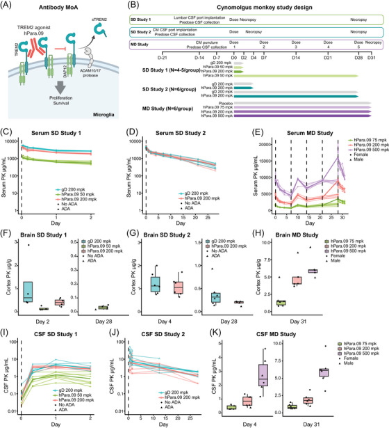
Introduction: Triggering receptor expressed on myeloid cells 2 (TREM2) agonists are being clinically evaluated as disease-modifying therapeutics for Alzheimer's disease. Clinically translatable pharmacodynamic (PD) biomarkers are needed to confirm drug activity and select the appropriate therapeutic dose in clinical trials.

Methods: We conducted multi-omic analyses on paired non-human primate brain and cerebrospinal fluid (CSF), and stimulation of human induced pluripotent stem cell-derived microglia cultures after TREM2 agonist treatment, followed by validation of candidate fluid PD biomarkers using immunoassays. We immunostained microglia to characterize proliferation and clustering.

Results: We report CSF soluble TREM2 (sTREM2) and CSF chitinase-3-like protein 1 (CHI3L1/YKL-40) as PD biomarkers for the TREM2 agonist hPara.09. The respective reduction of sTREM2 and elevation of CHI3L1 in brain and CSF after TREM2 agonist treatment correlated with transient microglia proliferation and clustering.

Discussion: CSF CHI3L1 and sTREM2 reflect microglial TREM2 agonism and can be used as clinical PD biomarkers to monitor TREM2 activity in the brain.

Highlights: CSF soluble triggering receptor expressed on myeloid cells 2 (sTREM2) reflects brain target engagement for a novel TREM2 agonist, hPara.09. CSF chitinase-3-like protein 1 reflects microglial TREM2 agonism. Both can be used as clinical fluid biomarkers to monitor TREM2 activity in brain.

Keywords: Alzheimer's disease; cerebrospinal fluid; chitinase‐3‐like protein 1; microglia; pharmacodynamic biomarker; triggering receptor expressed on myeloid cells 2.


Conflict of interest statement

All authors are current or prior employees and stakeholders of Genentech, Inc. or Hoffman‐La Roche. The authors have no additional financial interests. Author disclosures are available in the supporting information.

Figures

FIGURE 1
FIGURE 1
PK assessments of hPara.09 in two single‐dose studies and one multiple‐dose study in cynomolgus monkeys. A, Mechanism of action of hPara.09: The TREM2 agonist binds to TREM2 in a region spanning the ADAM10/17 cleavage site, which stabilizes the receptor and promotes intracellular signaling. Created with BioRender.com. B, Single‐ and multiple‐dose study designs to evaluate the PK and PD properties of hPara.09 in cynomolgus monkey. SD Study 1 (n = 4–5/group, all females): animals received a single dose of 200 mg/kg gD, 50 mg/kg hPara.09, or 200 mg/kg hPara.09. CSF/plasma samples were collected at pre‐dose and multiple post‐dose time points; brains were collected on days 2 and 28 and multi‐omic discovery and targeted approaches were applied. SD Study 2 (n = 6/group, all females): animals received a single dose of 200 mg/kg gD or 200 mg/kg hPara.09. Pre‐ and post‐dose CSF/plasma samples were collected every 12 hours; brains were collected on days 4 and 28. Brain and CSF multi‐omic discovery and targeted approaches were applied at different post‐dose time points. MD Study (n = 6/group, 3 males, 3 females): animals received 5 weekly doses of placebo or hPara.09 at 75, 200, and 500 mg/kg. CSF/plasma samples were collected at pre‐dose and multiple post‐dose time points; brains were collected on day 31 (3 days after the last dose). A different CSF collection method was used across studies (see Methods). C‐E, Per subject (regular lines) and median (bold line) concentration (μg/mL) of hPara.09 or gD in serum in SD study 1 (C for days 0–2, see Figure S1A for days 0–28), SD Study 2 (D), and MD Study (E). F‐H, Box plots of brain (cortex) hPara.09 concentrations (μg/g) in SD Study 1 (F), SD Study 2 (G), and MD Study (H). I‐K, Per subject (regular lines) and median (bold line) concentration (μg/mL) of hPara.09 in CSF in SD Study 1 (I for days 0–2, see Figure S1B for days 0–28. Animal 4509 was excluded from analysis due to CSF blood contamination), SD Study 2 (J), and MD Study (K). Circles represent ADA‐negative animals for SD Studies 1 and 2 (C, D, F, G, I, J) and female animals for MD study (E, H, K). Triangles represent ADA‐positive animals for SD Studies 1 and 2 (C, D, F, G, I, J) and male animals for MD Study (E, H, K). Vertical dashed lines represent dosing. Missing values were above or below the quantitation limit. ADA, anti‐drug antibody; CM, cisterna magna; CSF, cerebrospinal fluid; gD, glycoprotein D; MD, multiple dose; PD, pharmacodynamic; PK, pharmacokinetic; SD, single dose; TREM2, triggering receptor expressed on myeloid cells 2.
FIGURE 2
FIGURE 2
hPara.09 treatment reduces brain, CSF, and plasma sTREM2 in cynomolgus monkeys. A‐C, Box plots of brain (cortex) sTREM2 concentrations (ng/g) from SD Study 1 on days 2 and 28 after gD 200 mg/kg or hPara.09 (50 and 200 mg/kg) (A), from SD Study 2 on days 4 and 28 after 200 mg/kg gD or 200 mg/kg hPara.09 (B), and from the MD Study on day 31 after placebo or hPara.09 (75, 200, or 500 mg/kg) (C). Dashed line in (C) refers to the LLOQ = 1.03 ng/g, values below LLOQ were imputed (0.5 * 1.03 ng/g = 0.515). *P < 0.05, Kruskal–Wallis (SD Study 1), **P < 0.01, Mann–Whitney (SD Study 2), no statistical test was performed on MD Study as all hPara.09 groups contained imputed values. D‐I, Per subject (regular lines) and median (bold line) CSF (D‐F) or plasma (G‐I) sTREM2 change from baseline after 200 mg/kg gD or hPara.09 (50 and 200 mg/kg) in SD study 1 (D, G), after 200 mg/kg gD or hPara.09 in SD study 2 (E, H), and after placebo or hPara.09 (75, 200, or 500 mg/kg) in MD study (F, I). Circles represent ADA‐negative animals for SD Studies 1 and 2 (A, B, D, E, G, H) and female animals for MD Study (C, F, I). Triangles represent ADA‐positive animals for SD Studies 1 and 2 (A, B, D, E, G, H) and male animals for MD Study (C, F, I). Vertical dashed lines represent dosing, horizontal dashed lines represent the baseline. ADA, anti‐drug antibody; CSF, cerebrospinal fluid; gD, glycoprotein D; LLOQ, lower limit of quantitation; MD, multiple dose; SD, single dose; sTREM2, soluble triggering receptor expressed on myeloid cells 2.
FIGURE 3
FIGURE 3
hPara.09 treatment induces microglial clustering and proliferation in cynomolgus monkeys. A‐B, SD Study 1 cyno brain IBA1 (green) and MKI67 (red) immunostaining 2 days post dose from 200 mg/kg hPara.09 (A) and average number of IBA1+/MKI67+ co‐localized cells from the superior temporal gyrus 2 days post dose from 200 mg/kg gD or hPara.09 (50 mg/kg and 200 mg/kg) (B). Arrows indicate IBA1+/MKI67+ co‐stained cells, scale bar = 100 μm (A); *P < 0.05, one‐way ANOVA on log10‐transformed data (B). C‐H, IBA1 immunohistochemistry staining in cyno brain from SD Study 2, 4 days post dose 200 mg/kg gD or 200 mg/kg hPara.09 (C‐E) and MD Study 31 days after 5 weekly doses of placebo or 200 mg/kg hPara.09 (F‐H). Representative IBA1 images from hemibrain (C, F), with red box denoting superior temporal gyrus images shown in (D, G), and quantification of IBA1 cluster density in hemi‐brains (E, H). Arrows indicate IBA1+ clusters (D, G); **P < 0.01, ***P < 0.001, Kruskal–Wallis followed by Dunn multiple comparisons test; scale bar in (C, F) = 5 mm; scale bar in (D, G) = 100 μm. I, Correlations between CSF sTREM2 levels normalized to baseline and IBA1 microglia cluster density (counts per mm2) in SD study 1 (day 2; Rho = −0.64, P = 0.025), SD study 2 (day 4; Rho = −0.78, P = 0.005), and the MD study (day 31; Rho = − 0.72, P < 0.001). ANOVA, analysis of variance; CSF, cerebrospinal fluid; gD, glycoprotein D; IBA1, ionized calcium‐binding adapter molecule 1; MD, multiple dose; MKI67, marker of proliferation Ki67; SD, single dose; sTREM2, soluble triggering receptor expressed on myeloid cells 2.
FIGURE 4
FIGURE 4
Identification of several PD biomarkers of hPara.09 treatment and TREM2 agonism in cynomolgus monkey by RNA‐seq and proteomics. Ai, Volcano plot showing differentially expressed genes in brain with 200 mg/kg hPara.09 compared to 200 mg/kg gD on day 2 in SD Study 1. Log2 fold changes (hPara.09 200 mg/kg vs. gD 200 mg/kg) are plotted against the FDR (‐log10 transformed). mRNAs significantly regulated by hPara.09 are represented in red (upregulated) or blue (downregulated); FDR < 0.05, log2(1.5) log2 fold change cut‐off. Aii, Gene expression heatmap and Ingenuity Canonical Pathway terms of selected genes. The heatmap represents the Z scored gene expression of the differentially regulated mRNAs identified in (Ai) in the brains of animals treated with gD 200 mg/kg or hPara.09 (50 mg/kg and 200 mg/kg). n = 4 animals per treatment group. B, Volcano plot showing CSF proteins differentially regulated from baseline in response to hPara.09 on day 4 post dose in SD Study 2. Log2 fold changes (hPara.09 200 mg/kg day 4 vs. day −1) are plotted against the FDR (‐log10 transformed). Proteins significantly regulated by hPara.09 dosing are represented in red (upregulated) or blue (downregulated); FDR < 0.1; log2(1.5) log2 fold change cut‐off; n = 12 animals per treatment group. C, Volcano plot showing proteins differentially regulated in iPSC‐MG cultured on hPara.09 compared to gD. Log2 fold changes (hPara.09 vs. gD) are plotted against the FDR (‐log10 transformed). Proteins significantly regulated by hPara.09 dosing are represented in red (upregulated) or blue (downregulated); FDR < 0.05; log2(1.5) log2 fold change cut‐off; n = 3 experimental replicates from cells of three differentiation batches. For all volcano plots, the dashed horizontal and vertical lines represent the FDR and log2 fold change cut‐offs, respectively. CSF, cerebrospinal fluid; gD, glycoprotein D; FDR, false discovery rate; iPSC‐MG, induced pluripotent stem cell microglia; PD, pharmacodynamic; TREM2, triggering receptor expressed on myeloid cells 2.
FIGURE 5
FIGURE 5
hPara.09 increases brain CHI3L1 and sCSF1R in cynomolgus monkeys. A‐D, Box plots of brain (cortex) CHI3L1 (μg/g) (A, B) and sCSF1R (ng/g) (C, D) concentrations at day 4 and day 28 after gD or hPara.09 200 mg/kg in SD Study 2 (A, C) and on day 31 after placebo or hPara.09 (75, 200, or 500 mg/kg) in MD Study (B, D). Circles represent ADA‐negative animals for SD study 2 (A, C) and female animals for MD Study (B, D). Triangles represent ADA‐positive animals for SD study 2 (A, C) and male animals for MD Study (B, D). *P < 0.05, **P < 0.01, Kruskal–Wallis followed by Dunn multiple comparisons test. E, F, Correlations between brain (cortex) CHI3L1 (E) and sCSF1R (F) concentrations and microglial cluster density (counts per mm2) on day 31 after five administrations of placebo or hPara.09 (75, 200, or 500 mg/kg) in the MD Study. Rho = 0.74, P = 5.296e‐05 (E); Rho = 0.58, P = 0.003 (F). ADA, anti‐drug antibody; CHI3L1, chitinase‐3‐like protein 1; CSF1R, colony stimulating factor 1 receptor; gD, glycoprotein D; LLOQ, lower limit of quantitation; MD, multiple dose; SD, single dose.
FIGURE 6
FIGURE 6
hPara.09 increases CSF CHI3L1 across multiple cynomolgus monkey studies. A‐D, CSF CHI3L1 changes from baseline over the entire study (A, C) or on selected post‐dose days (B, D) after gD or hPara.09 200 mg/kg in SD Study 2 (A, B) and after placebo or hPara.09 (75, 200, or 500 mg/kg) in MD Study (C, D). Outlier animal 23 was removed for analysis from MD Study (high CSF responder, 500 mg/kg hPara.09). E, F, Correlation between CSF CHI3L1 changes from baseline and microglial cluster density (counts per mm2) on post‐dose day 31 (E) or CSF sTREM2 changes from baseline on post‐dose day 4 (circle) and day 31 (triangle) (F) in the MD Study. Rho = 0.72, P = 0.0001 (E); Rho = −0.64, P = 6.57e‐06 (F). G‐I, CSF sCSF1R changes from baseline over the entire study (G, I) or on selected post‐dose days (H) after gD or hPara.09 200 mg/kg in SD Study 2, and after placebo or hPara.09 (75, 200, or 500 mg/kg) in MD Study. J, K, CSF IL1RN changes from baseline over the entire study (J) or on selected post‐dose days (K) after placebo or hPara.09 (75, 200, or 500 mg/kg) in MD Study. L, M, Correlation between CSF IL1RN changes from baseline and microglial cluster density (counts per mm2) on post‐dose day 31 (L) or CSF sTREM2 changes from baseline on post‐dose day 4 (circle) and day 31 (triangle) (M) in the MD Study. Rho = 0.61, P = 0.002 (L); Rho = −0.54, P = 0.0004 (M). Vertical dashed lines represent dosing, horizontal dashed line represents the baseline (A, C, G, I, J). Vertical dashed line represents 50% reduction in CSF sTREM2, horizontal dashed line represents the baseline (F, M). On the line plots, regular and bold lines represent per subject and median changes from baseline, respectively (A, C, G, I, J). On the box plots, circles represent ADA‐negative animals for SD Study 2 (B, H) and female animals for MD Study (D, K). Triangles represent ADA‐positive animals for SD Study 2 (B, H) and male animals for MD Study (D, K); *P < 0.05, **P < 0.01, Mann–Whitney (SD Study 2) and Kruskal–Wallis followed by Dunn multiple comparisons test (MD Study). ADA, anti‐drug antibody; CHI3L1, chitinase‐3‐like protein 1; sCSF1R, soluble colony stimulating factor 1 receptor; gD, glycoprotein D; IL1RN, interleukin 1 receptor antagonist; MD, multiple dose; SD, single dose; sTREM2, soluble triggering receptor expressed on myeloid cells 2.

References

    1. Wightman DP, Jansen IE, Savage JE, et al. A genome‐wide association study with 1,126,563 individuals identifies new risk loci for Alzheimer's disease. Nat Genet. 2021;53:1276‐1282. doi:10.1038/s41588-021-00921-z - DOI - PMC - PubMed
    1. Bryois J, Calini D, Macnair W, et al. Cell‐type‐specific cis‐eQTLs in eight human brain cell types identify novel risk genes for psychiatric and neurological disorders. Nat Neurosci. 2022;25:1104‐1112. doi:10.1038/s41593-022-01128-z - DOI - PubMed
    1. Bohlen CJ, Friedman BA, Dejanovic B, Sheng M. Microglia in brain development, homeostasis, and neurodegeneration. Annu Rev Genet. 2019;53:1‐26. doi:10.1146/annurev-genet-112618-043515 - DOI - PubMed
    1. Guerreiro R, Wojtas A, Bras J, et al. TREM2 variants in Alzheimer's disease. N Engl J Med. 2013;368:117‐127. doi:10.1056/nejmoa1211851 - DOI - PMC - PubMed
    1. Thorlakur J, Hreinn S, Stacy S, et al. Variant of TREM2 associated with the risk of Alzheimer's disease. N Engl J Med. 2013;368:107‐116. doi:10.1056/nejmoa1211103 - DOI - PMC - PubMed

MeSH terms

Grants and funding