. 2024 Oct;15(5):1953-1964.
doi: 10.1002/jcsm.13544. Epub 2024 Aug 2.

l-Carnitine relieves cachexia-related skeletal muscle fibrosis by inducing deltex E3 ubiquitin ligase 3L to negatively regulate the Runx2/COL1A1 axis

Affiliations

l-Carnitine relieves cachexia-related skeletal muscle fibrosis by inducing deltex E3 ubiquitin ligase 3L to negatively regulate the Runx2/COL1A1 axis

Zongliang Lu et al. J Cachexia Sarcopenia Muscle. 2024 Oct.

Abstract

Background: Cancer cachexia-induced skeletal muscle fibrosis (SMF) impairs muscle regeneration, alters the muscle structure and function, reduces the efficacy of anticancer drugs, diminishes the patient's quality of life and shortens overall survival. RUNX family transcription factor 2 (Runx2), a transcription factor, and collagen type I alpha 1 chain (COL1A1), the principal constituent of SMF, have been linked previously, with Runx2 shown to directly modulate COL1A1 mRNA levels. l-Carnitine, a marker of cancer cachexia, can alleviate fibrosis in liver and kidney models; however, its role in cancer cachexia-associated fibrosis and the involvement of Runx2 in the process remain unexplored.

Methods: Female C57 mice (48 weeks old) were inoculated subcutaneously with MC38 cells to establish a cancer cachexia model. A 5 mg/kg dose of l-carnitine or an equivalent volume of water was administered for 14 days via oral gavage, followed by assessments of muscle function (grip strength) and fibrosis. To elucidate the interplay between the deltex E3 ubiquitin ligase 3L(DTX3L)/Runx2/COL1A1 axis and fibrosis in transforming growth factor beta 1-stimulated NIH/3T3 cells, a suite of molecular techniques, including quantitative real-time PCR, western blot analysis, co-immunoprecipitation, molecular docking, immunofluorescence and Duolink assays, were used. The relevance of the DTX3L/Runx2/COL1A1 axis in the gastrocnemius was also explored in the in vivo model.

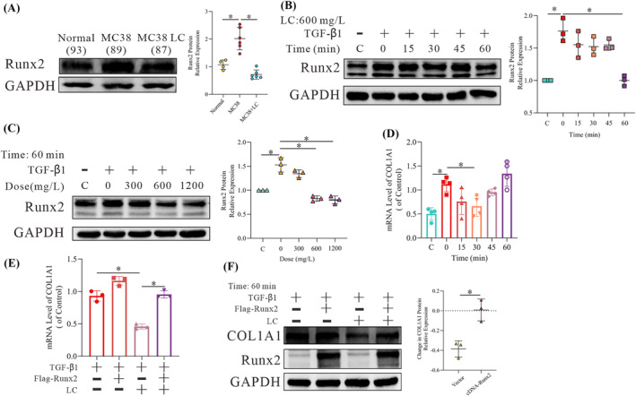
Results: l-Carnitine supplementation reduced cancer cachexia-induced declines in grip strength (>88.2%, P < 0.05) and the collagen fibre area within the gastrocnemius (>57.9%, P < 0.05). At the 5 mg/kg dose, l-carnitine also suppressed COL1A1 and alpha-smooth muscle actin (α-SMA) protein expression, which are markers of SMF and myofibroblasts. Analyses of the TRRUST database indicated that Runx2 regulates both COL1A1 and COL1A2. In vitro, l-carnitine diminished Runx2 protein levels and promoted its ubiquitination. Overexpression of Runx2 abolished the effects of l-carnitine on COL1A1 and α-SMA. Co-immunoprecipitation, molecular docking, immunofluorescence and Duolink assays confirmed an interaction between DTX3L and Runx2, with l-carnitine enhancing this interaction to promote Runx2 ubiquitination. l-Carnitine supplementation restored DTX3L levels to those observed under non-cachectic conditions, both in vitro and in vivo. Knockdown of DTX3L abolished the effects of l-carnitine on Runx2, COL1A1 and α-SMA in vitro. The expression of DTX3L was negatively correlated with the levels of Runx2 and COL1A1 in untreated NIH/3T3 cells.

Conclusions: This study revealed a previously unrecognized link between Runx2 and DTX3L in SMF and demonstrated that l-carnitine exerted a significant therapeutic impact on cancer cachexia-associated SMF, potentially through the upregulation of DTX3L.

Keywords: DTX3L; Runx2; cancer cachexia; l‐carnitine; skeletal muscle fibrosis.


Conflict of interest statement

The authors declare that they have no known competing financial interests or personal relationships that could have appeared to influence the work reported in this paper.

Figures

Figure 1
Figure 1
l‐Carnitine (LC) relieves cachexia‐induced skeletal muscle fibrosis (SMF). (A) Changes in the mRNA expression of members of the collagen family in the gastrocnemius muscles of mice with cancer cachexia identified by RNA sequencing in CT26 tumour‐bearing nude mice (n = 3, 3). (B) Grip strength assessment in cachectic mice with and without LC treatment (5 mg/kg). The bar graph shows the muscle strength (grip strength) determined by a muscle strength metre (n = 4, 6, 6). (C) The collagenous fibre area in cachectic mice with and without LC intervention. The bar graph shows the SMF level in each group (n = 4, 6, 6). (D) Western blot (WB) analysis of the collagen type I alpha 1 chain (COL1A1) expression in the gastrocnemius. The bar graph shows the relative COL1A1 protein expression (n = 4, 6, 6). (E, F) WB analysis of the COL1A1 expression in NIH/3T3 cells following exposure to 10 ng/mL transforming growth factor beta 1 (TGF‐β1) for 24 h and after subsequent treatment with 600 mg/L LC for different amounts of time or with different concentrations of LC for 60 min. The dot chart shows the relative COL1A1 protein expression. Representative images are shown, and images were assessed using the Image Lab software. The data are shown as the means ± SEM (n = 3). A one‐way analysis of variance (B–F) followed by the least significant difference test was used to compare data among groups (*P < 0.05).
Figure 2
Figure 2
l‐Carnitine (LC) downregulates RUNX family transcription factor 2 (Runx2) to improve fibrosis. (A) Western blot (WB) analysis of Runx2 expression in the gastrocnemius of experimental mice. The bar graph shows the relative Runx2 protein expression (n = 4, 6, 6). (B–D) WB and qPCR analysis of the Runx2 and collagen type I alpha 1 chain (COL1A1) expression in NIH/3T3 cells following exposure to 10 ng/mL transforming growth factor beta 1 (TGF‐β1) for 24 h and after subsequent treatment with 600 mg/L LC for different amounts of time or with different concentrations of LC for 60 min. The bar graph shows the relative Runx2 protein and COL1A1 mRNA expression (n = 3). (E, F) WB and qPCR analysis of the Runx2 and COL1A1 expression in NIH/3T3 cells transfected with Flag‐Runx2 for 24 h following exposure to 10 ng/mL TGF‐β1 for 24 h and after subsequent treatment with LC for 30 min, 60 min or 6 h. The dot chart shows the relative COL1A1 protein and mRNA levels (n = 3). Representative images are shown, and images were analysed using the Image Lab software (A–F). The data are shown as the means ± SEM. A one‐way analysis of variance (A–E) followed by the least significant difference test or t‐test (F) was used to compare data among groups (*P < 0.05).
Figure 3
Figure 3
l‐Carnitine (LC) promotes RUNX family transcription factor 2 (Runx2) ubiquitination. (A) qPCR analysis of the Runx2 expression in NIH/3T3 cells following exposure to 10 ng/mL transforming growth factor beta 1 (TGF‐β1) for 24 h and after subsequent treatment with 600 mg/L LC for different amounts of time. The bar graph shows the Runx2 mRNA expression (n = 3). (B) Western blot (WB) analysis of the Runx2 expression in NIH/3T3 cells following exposure to 10 ng/mL TGF‐β1 for 24 h in the culture medium (CM) or 600 mg/L LC for 5 min and treatment with 15 mg/L cycloheximide (CHX) for different times. Representative images are shown, and images were analysed using the Image Lab software. A one‐way analysis of variance was used to compare data among the groups (*P < 0.05) (n = 3). (C) WB analysis of the Runx2 expression in NIH/3T3 cells following exposure to 10 ng/mL TGF‐β1 for 24 h and treatment with 10 μM/mL MG132 for 12 h, followed by subsequent treatment with 600 mg/L LC for 1 h. (D) WB analysis of ub‐Runx2 expression in NIH/3T3 cells transfected with Flag‐Runx2 for 24 h following exposure to 10 ng/mL TGF‐β1 for 12 h and after treatment with 10 μM/mL MG132 for 12 h and 600 mg/L LC for 15 min based on the enrichment of Runx2 by immunoprecipitation.
Figure 4
Figure 4
l‐Carnitine (LC) relieves fibrosis via deltex E3 ubiquitin ligase 3L (DTX3L). (A) Western blot (WB) analysis of RUNX family transcription factor 2 (Runx2) and DTX3L in HEK 293 cells after treatment with a Flag‐Runx2 and GFP‐DTX3L plasmid for 24 h after enrichment for Flag or GFP by immunoprecipitation. (B) HEK 293 cells were transfected with Flag‐Runx2 and GFP‐DTX3L for 48 h and then stained with Flag and Cy3 antibodies and counterstained with DAPI to determine whether Runx2 and DTX3L were co‐localized within the cell. Representative images are shown. Arrows indicate the co‐localization of Runx2 and DTX3L. (C) Duolink analysis of the spatial distance between Flag‐Runx2 and GFP‐DTX3L. (D) WB analysis of Runx2 and DTX3L in NIH/3T3 cells after treatment with Flag‐Runx2 for 24 h following exposure to 10 ng/mL transforming growth factor beta 1 (TGF‐β1) for 24 h and 600 mg/L LC for 15 min after enrichment for Flag by immunoprecipitation. (E) WB analysis of the Runx2 expression in NIH/3T3 cells transfected with siRNA‐DTX3L for 24 h following exposure to 10 ng/mL TGF‐β1 for 24 h and after subsequent treatment with LC for 60 min. Representative images are shown, and images were analysed using the Image Lab software. The data are shown as the means ± SEM (n = 3). A t‐test was used to compare data between groups (*P < 0.05). (F) WB analysis of the ub‐Runx2 expression in NIH/3T3 cells after treatment with Flag‐Runx2 and siRNA‐DTX3L for 24 h following exposure to 10 ng/mL TGF‐β1 for 12 h and treatment with 10 μM/mL MG132 for 12 h and 600 mg/L of LC for 15 min after the enrichment of Flag by immunoprecipitation (n = 3).
Figure 5
Figure 5
l‐Carnitine (LC) relieves fibrosis through the deltex E3 ubiquitin ligase 3L (DTX3L)/RUNX family transcription factor 2 (Runx2) axis. (A) Western blot (WB) analysis of DTX3L expression in the gastrocnemius. The dot chart shows the relative DTX3L protein expression (n = 4, 6, 6). (B, C) WB analysis of the DTX3L expression in NIH/3T3 cells following exposure to 10 ng/mL transforming growth factor beta 1 (TGF‐β1) for 24 h and after subsequent treatment with 600 mg/L LC for different amounts of time or with different concentrations of LC for 60 min. The dot chart shows the relative protein expression of DTX3L (n = 3). (D) WB and qPCR analysis of the DTX3L and collagen type I alpha 1 chain (COL1A1) expression in NIH/3T3 cells transfected with siRNA‐DTX3L for 24 h following exposure to 10 ng/mL TGF‐β1 for 24 h and 600 mg/L LC for 30 min, 60 min or 6 h. The bar graph and dot chart show the relative COL1A1 protein and mRNA expression (n = 3). (E) WB and qPCR analysis of DTX3L, Runx2 and COL1A1 expression in NIH/3T3 cells transfected with Flag‐Runx2 and either empty vector or GFP‐DTX3L plasmid for 24 h following exposure to 10 ng/mL TGF‐β1 for 24 h and 600 mg/L LC for 30 min, 60 min or 6 h. The bar graph and dot chart show the relative COL1A1 protein and mRNA expression (n = 3). Representative images are shown, and images were analysed using the Image Lab software. The data are shown as the means ± SEM (n = 3). A one‐way analysis of variance (A–E) followed by the least significant difference test or t‐test (D, E) was used to compare data among groups (*P < 0.05).
Figure 6
Figure 6
Transforming growth factor beta 1 (TGF‐β1) disturbs the deltex E3 ubiquitin ligase 3L (DTX3L)/RUNX family transcription factor 2 (Runx2) axis. (A, B) Western blot (WB) analysis of the Runx2, collagen type I alpha 1 chain (COL1A1) and alpha‐smooth muscle actin (α‐SMA) expression in NIH/3T3 cells transfected with GFP‐DTX3L or siRNA‐DTX3L for 48 h (n = 3). (C, D) WB analysis of the Runx2, COL1A1 and α‐SMA expression in NIH/3T3 cells transfected with GFP‐DTX3L or siRNA‐DTX3L for 24 h, followed by treatment with 10 ng/mL TGF‐β1 for 24 h (n = 3). (E) A schematic representation of how l‐carnitine promotes DTX3L to induce Runx2 ubiquitination to relieve cachexia‐related skeletal muscle fibrosis.

References

    1. Fearon K, Strasser F, Anker SD, Bosaeus I, Bruera E, Fainsinger RL, et al. Definition and classification of cancer cachexia: an international consensus. Lancet Oncol 2011;12:489–495. - PubMed
    1. Mahdy M. Skeletal muscle fibrosis: an overview. Cell Tissue Res 2019;375:575–588. - PubMed
    1. Judge SM, Nosacka RL, Delitto D, Gerber MH, Cameron ME, Trevino JG, et al. Skeletal muscle fibrosis in pancreatic cancer patients with respect to survival. JNCI Cancer Spectr 2018;2:pky043. - PMC - PubMed
    1. Mochamat Cuhls H, Marinova M, Kaasa S, Stieber C, Conrad R, et al. A systematic review on the role of vitamins, minerals, proteins, and other supplements for the treatment of cachexia in cancer: a European Palliative Care Research Centre cachexia project. J Cachexia Sarcopenia Muscle 2017;8:25–39. - PMC - PubMed
    1. Takagi A, Hawke P, Tokuda S, Toda T, Higashizono K, Nagai E, et al. Serum carnitine as a biomarker of sarcopenia and nutritional status in preoperative gastrointestinal cancer patients. J Cachexia Sarcopenia Muscle 2022;13:287–295. - PMC - PubMed