. 2024 Jul 18:15:1425374.
doi: 10.3389/fimmu.2024.1425374. eCollection 2024.

Tetanus-diphtheria vaccine can prime SARS-CoV-2 cross-reactive T cells

Affiliations

Tetanus-diphtheria vaccine can prime SARS-CoV-2 cross-reactive T cells

Sara Alonso Fernandez et al. Front Immunol. .

Abstract

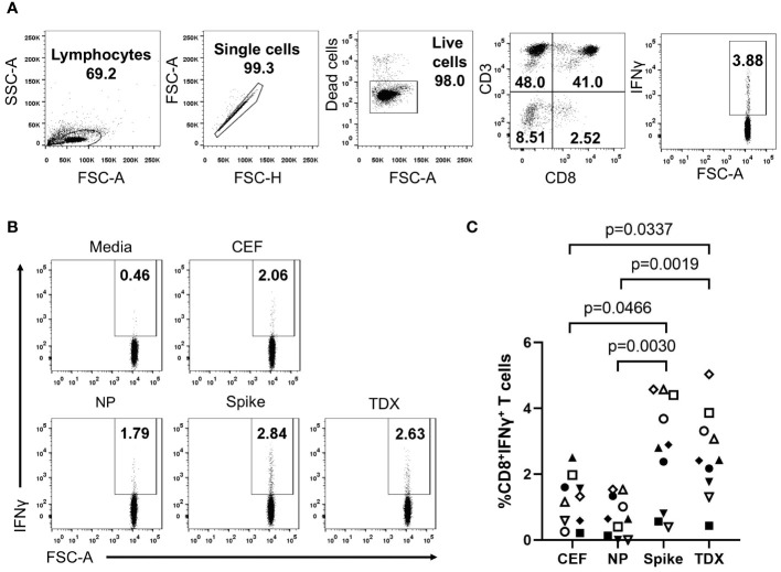
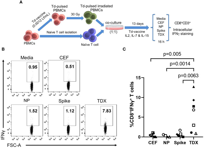
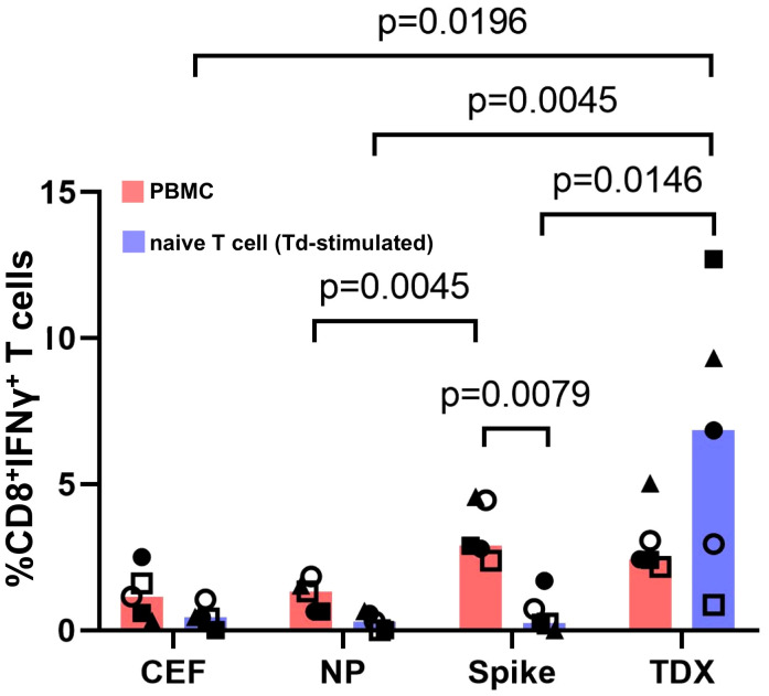
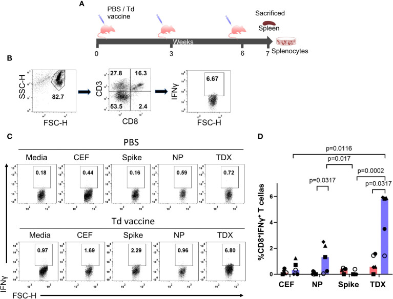
Vaccines containing tetanus-diphtheria antigens have been postulated to induce cross-reactive immunity to severe acute respiratory syndrome coronavirus 2 (SARS-CoV-2), which could protect against coronavirus disease (COVID-19). In this work, we investigated the capacity of Tetanus-diphtheria (Td) vaccine to prime existing T cell immunity to SARS-CoV-2. To that end, we first collected known SARS-CoV-2 specific CD8+ T cell epitopes targeted during the course of SARS-CoV-2 infection in humans and identified as potentially cross-reactive with Td vaccine those sharing similarity with tetanus-diphtheria vaccine antigens, as judged by Levenshtein edit distances (≤ 20% edits per epitope sequence). As a result, we selected 25 potentially cross-reactive SARS-CoV-2 specific CD8+ T cell epitopes with high population coverage that were assembled into a synthetic peptide pool (TDX pool). Using peripheral blood mononuclear cells, we first determined by intracellular IFNγ staining assays existing CD8+ T cell recall responses to the TDX pool and to other peptide pools, including overlapping peptide pools covering SARS-CoV-2 Spike protein and Nucleocapsid phosphoprotein (NP). In the studied subjects, CD8+ T cell recall responses to Spike and TDX peptide pools were dominant and comparable, while recall responses to NP peptide pool were less frequent and weaker. Subsequently, we studied responses to the same peptides using antigen-inexperienced naive T cells primed/stimulated in vitro with Td vaccine. Priming stimulations were carried out by co-culturing naive T cells with autologous irradiated peripheral mononuclear cells in the presence of Td vaccine, IL-2, IL-7 and IL-15. Interestingly, naive CD8+ T cells stimulated/primed with Td vaccine responded strongly and specifically to the TDX pool, not to other SARS-CoV-2 peptide pools. Finally, we show that Td-immunization of C57BL/6J mice elicited T cells cross-reactive with the TDX pool. Collectively, our findings support that tetanus-diphtheria vaccines can prime SARS-CoV-2 cross-reactive T cells and likely contribute to shape the T cell responses to the virus.

Keywords: COVID-19; SARS-CoV-2; T cell cross-reactivity; epitope; tetanus-diphtheria toxoid vaccines.


Conflict of interest statement

The authors declare that the research was conducted in the absence of any commercial or financial relationships that could be construed as a potential conflict of interest. The author(s) declared that they were an editorial board member of Frontiers, at the time of submission. This had no impact on the peer review process and the final decision.

Figures

Figure 1
Figure 1
Existing T cell responses to SARS-CoV-2 peptide pools. PBMCs from 10 healthy subjects were stimulated with SARS-CoV-2 peptide pools (Spike, NP and TDX), CEF pool or media (complete RPMI with 0.3% DMSO) during 16 hours and CD8+ T cell responses detected by intracellular IFNγ staining assays (A) Gating strategy for the detection of intracellular IFNγ expression within the CD8+ T cell population by flow cytometry using a representative PBMC sample stimulated with the TDX peptide pool. IFNγ+CD3+CD8+ cells were identified after the following steps: a) Adequate adjustment of the gate of the lymphocytes using the light scatter parameters (FSC and SSC-A), b) Exclusion of doublets with the identification of singlets improving the accuracy of the analysis, c) Selection of the viable lymphocytes, d) Identification of the CD3+CD8+ cell subset and e) IFNγ+ cells within the CD3+CD8+ cells. IFNγ+ gate was set after FMO stainings with PHA-L stimulation (see Supplementary Figure S1 ). (B) Representative dot plot showing IFNγ+ cells on CD3+CD8+ gated cells in response to the different stimuli (Media, CEF, Spike, NP and TDX). Percentage of IFNγ+ cells are indicated (C) Graph depicting the percentage of CD8+ T cells expressing IFNγ (y-axis) in response to peptide pools (x-axis) after subtracting response to media (n = 10). Individual values are plotted. Statistically significant differences were obtained by applying Kruskal-Wallis tests. Significant differences are indicated and p-values shown.
Figure 2
Figure 2
Cross-reactive responses of naive T cells stimulated with Td vaccine to SARS-CoV-2 peptide pools. (A) Experimental design to stimulate/prime T cells against Td vaccine. PBMCs were pulsed with Td vaccine (0.0012 Lf/ml of diphtheria toxoid) and homogenously irradiated at 30 Gy. Td-pulsed irradiated PBMCs were co-cultured with autologous naive T cells (ratio 1:1) for 13 days in the presence of IL-2, IL-7, IL-15 and Td vaccine (details in Methods). Subsequently, T cell responses to peptide pools (TDX pool, Spike pool, NP pool and CEF pool) and media (0.3% DMSO) were determined by intracellular IFNγ staining assays. IFNγ positive gate was set after FMO staining with PHA stimulation. (B) Representative dot plot showing the IFNγ+CD8+ T cells after stimulation with the relevant peptide pools (C) Percentage of CD8+ T cells expressing IFNγ after subtracting value from control media. Individual values were plotted (n = 7, except for NP with n = 5). Statistically significant differences were obtained by applying Kruskal-Wallis tests. Significant differences are indicated and p-values shown.
Figure 3
Figure 3
Responses to SARS-CoV-2 peptide pools from matched PBMCs and Td-stimulated native T cells. T cell responses to SARS-CoV-2 peptide pools (NP, Spike and TDX) and CEF pool determined using PBMCs (pink) and Td-stimulated naive T cells (blue) from the same five subjects. PBMCs and Td-stimulated naive T cells were incubated with peptide pools or media for 16 hours and responses were detected by intracellular IFNγ staining assays. All responses (value from media control subtracted) are plotted and bars represent median values. Statistically significant differences between conditions are indicated and p-values shown. Kruskal-Wallis tests were used for comparing responses to peptide pools from PBMC recall and Td-stimulated naive T cells. Wilcoxon signed-rank tests were carried out to compare T cell responses between PBMCs and Td-stimulated naive T cells in the same individuals to the same peptide pools.
Figure 4
Figure 4
Cross-reactive T cell responses to SARS-CoV-2 peptide pools in Td-vaccinated mice. (A) Immunization schedule. Mice were immunized intramuscularly (IM) at 3-week intervals with Td vaccine (0.1 Lf diphtheria toxoid)(Td-vaccine group, n = 5) or PBS (PBS control group, n=5). Mice were sacrificed seven days after the last immunization, spleens were collected and splenocytes prepared and responses to SARS-CoV-2 peptide pools (NP, Spike and TDX), CEF pool and media (complete RPMI with 0.3% DMSO) after 36-hour stimulations determined by intracellular IFNγ staining assays (B) Gating strategy for intracellular IFNγ detection within the CD8+ T cell population by flow cytometry using mouse splenocytes stimulated with the TDX peptide pool. IFNγ+CD3+CD8+ cells were identified after the following steps: a) Adequate selection of cells using the light scatter parameters (FSC, SSC), b) Selection of CD3+CD8+ cells, c) Selection of IFNγ+ cells within the CD3+CD8+ cells. IFNγ+ gate set after FMO stainings with PHA-L stimulation (see Supplementary Figure S1 ) (C) Representative dot plot showing IFNγ+CD8+ T cells responding to SARS-CoV-2 peptide pools (TDX, Spike and NP), CEF pool or media in both, PBS and Td-vaccine groups. (D) Percentage of CD8+ T cells producing IFNγ (value from control media subtracted) in both, PBS control group (pink) and Td-vaccine group (blue). All values are plotted and bars represent median values. Statistically significant differences between responses are shown with p-values. Kruskal-Wallis tests were used for comparing T cell responses to peptide pools in Td-vaccinated and control mice. Mann-Whitney U tests were carried out to compare T cell responses to the same peptides (CEF, NP, Spike and TDX pools) between Td-vaccinated mice and control mice.

Similar articles

Cited by

References

    1. Viruses, CSGotICoTo . The species Severe acute respiratory syndrome-related coronavirus: classifying 2019-nCoV and naming it SARS-CoV-2. Nat Microbiol. (2020) 5:536–44. doi: 10.1038/s41564-020-0695-z - DOI - PMC - PubMed
    1. Barbour V. COVID-19: no longer a global health emergency, now a long term challenge. Med J Aust. (2023) 218:437. doi: 10.5694/mja5692.51976 - DOI - PubMed
    1. Yewdell JW. Individuals cannot rely on COVID-19 herd immunity: Durable immunity to viral disease is limited to viruses with obligate viremic spread. PloS Pathog. (2021) 17:e1009509. doi: 10.1001371/journal.ppat.1009509 - DOI - PMC - PubMed
    1. Ballesteros-Sanabria L, Pelaez-Prestel HF, Ras-Carmona A, Reche PA. Resilience of spike-specific immunity induced by COVID-19 vaccines against SARS-coV-2 variants. Biomedicines. (2022) 10:996. doi: 10.3390/biomedicines10050996 - DOI - PMC - PubMed
    1. Li R, Tian J, Yang F, Lv L, Yu J, Sun G, et al. . Clinical characteristics of 225 patients with COVID-19 in a tertiary Hospital near Wuhan, China. J Clin Virol. (2020) 127:104363. doi: 10.1016/j.jcv.2020.104363 - DOI - PMC - PubMed