[Preprint]. 2025 Apr 22:2024.07.17.603924.
doi: 10.1101/2024.07.17.603924.

An enhancer-AAV toolbox to target and manipulate distinct interneuron subtypes

Elisabetta Furlanis  1   2   3 Min Dai  1   2   3 Brenda Leyva Garcia  1   2   3 Thien Tran  1   2 Josselyn Vergara  1   2 Ana Pereira  1   2 Bram L Gorissen  1   2 Sara Wills  1   2 Anna Vlachos  4 Ariel Hairston  1 Deepanjali Dwivedi  1   2 Sarah Du  1   2 Justin McMahon  1   2 Shuhan Huang  1 Annunziato Morabito  1 Arenski Vazquez  5 Soyoun Kim  5 Anthony T Lee  6   7 Edward F Chang  6   7 Taha Razzaq  8 Ahmed Qazi  8 Geoffrey Vargish  4 Xiaoqing Yuan  4 Adam Caccavano  4 Steven Hunt  4 Ramesh Chittajallu  4 Nadiya McLean  4 Lauren Hewitt  4 Emily Paranzino  4 Haley Rice  4 Alex C Cummins  9 Anya Plotnikova  9 Arya Mohanty  9 Anne Claire Tangen  9 Jung Hoon Shin  9 Reza Azadi  9 Mark A G Eldridge  9 Veronica A Alvarez  9 Bruno B Averbeck  9 Mansour Alyahyay  10 Tania Reyes Vallejo  10 Mohammed Soheib  10 Lucas G Vattino  11   12 Cathryn P MacGregor  11   12 Carolina Piletti Chatain  13   14 Emmie Banks  15 Viktor Janos Olah  15 Shovan Naskar  16 Sophie Hill  17 Sophie Liebergall  17 Rohan Badiani  1   2 Lili Hyde  1   2 Ella Hanley  1   2 Qing Xu  10   18 Kathryn C Allaway  1   2 Ethan M Goldberg  17 Matthew J M Rowan  15 Tomasz J Nowakowski  6   7 Soohyun Lee  16 Emilia Favuzzi  13   14 Pascal S Kaeser  1 Lucas Sjulson  5   19 Renata Batista-Brito  5   19 Anne E Takesian  11   12 Leena A Ibrahim  10 Asim Iqbal  8 Kenneth A Pelkey  4 Chris J McBain  4 Jordane Dimidschstein  2 Gord Fishell  1   2 Yating Wang  1   2
Affiliations

An enhancer-AAV toolbox to target and manipulate distinct interneuron subtypes

Elisabetta Furlanis et al. bioRxiv. .

Update in

  • An enhancer-AAV toolbox to target and manipulate distinct interneuron subtypes.
    Furlanis E, Dai M, Leyva Garcia B, Tran T, Vergara J, Pereira A, Gorissen BL, Wills S, Vlachos A, Hairston A, Dwivedi D, Du S, McMahon J, Huang S, Morabito A, Vazquez A, Kim S, Lee AT, Chang EF, Razzaq T, Qazi A, Vargish G, Yuan X, Caccavano A, Hunt S, Chittajallu R, McLean N, Hewitt L, Paranzino E, Rice H, Cummins AC, Plotnikova A, Mohanty A, Tangen AC, Shin JH, Azadi R, Eldridge MAG, Alvarez VA, Averbeck BB, Alyahyay M, Vallejo TR, Soheib M, Vattino LG, MacGregor CP, Chatain CP, Banks E, Olah VJ, Naskar S, Hill S, Liebergall S, Badiani R, Hyde L, Hanley E, Xu Q, Allaway KC, Goldberg EM, Rowan MJM, Nowakowski TJ, Lee S, Favuzzi E, Kaeser PS, Sjulson L, Batista-Brito R, Takesian AE, Ibrahim LA, Iqbal A, Pelkey KA, McBain CJ, Dimidschstein J, Fishell G, Wang Y. Furlanis E, et al. Neuron. 2025 May 21;113(10):1525-1547.e15. doi: 10.1016/j.neuron.2025.05.002. Neuron. 2025. PMID: 40403705

Abstract

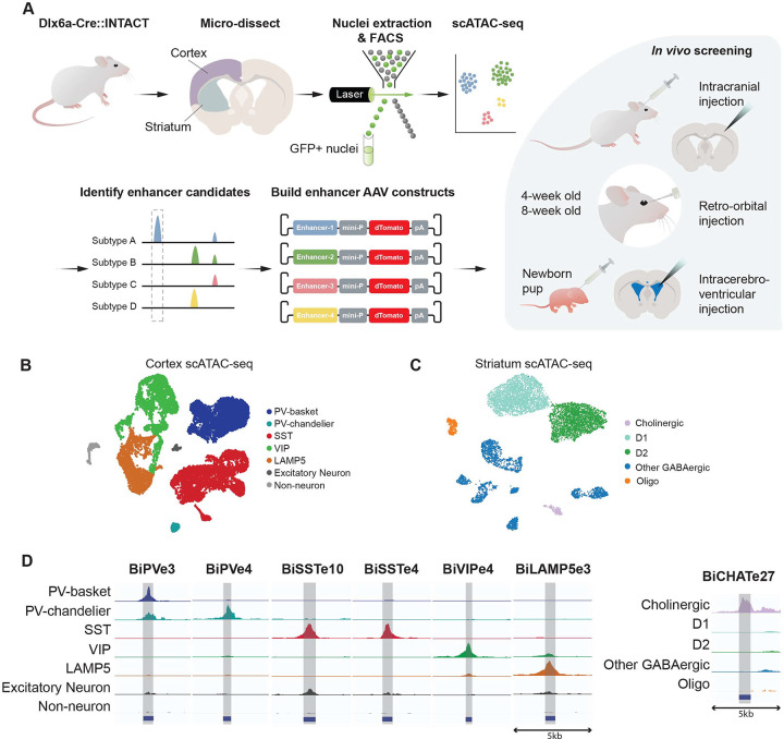
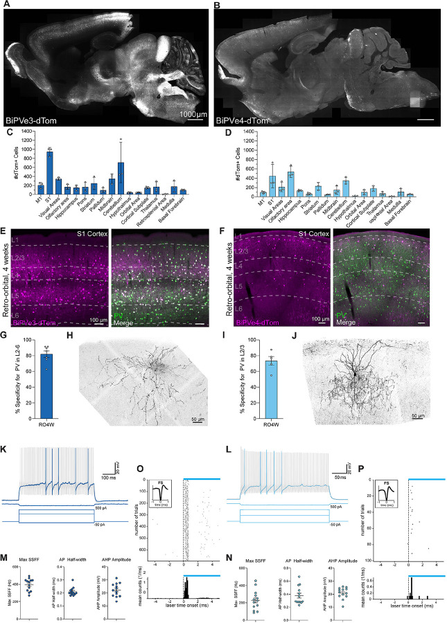
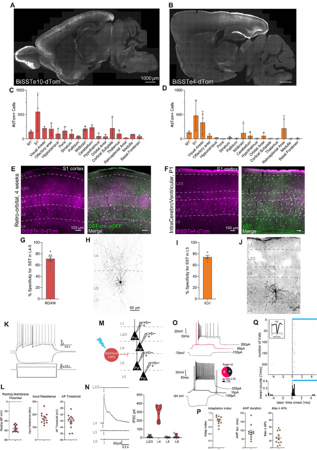
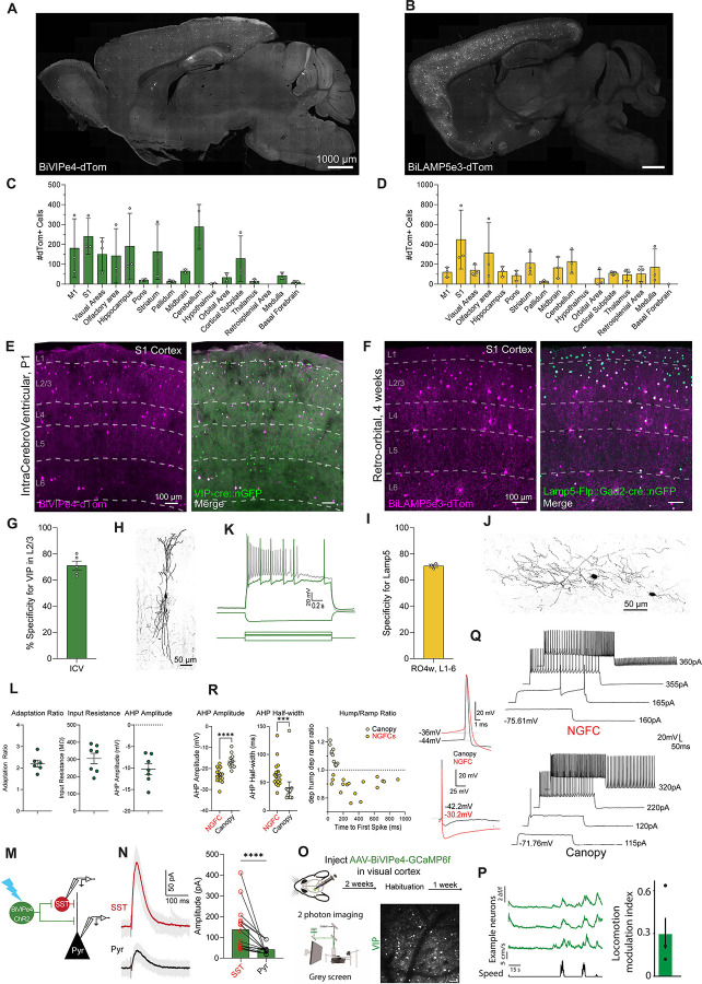
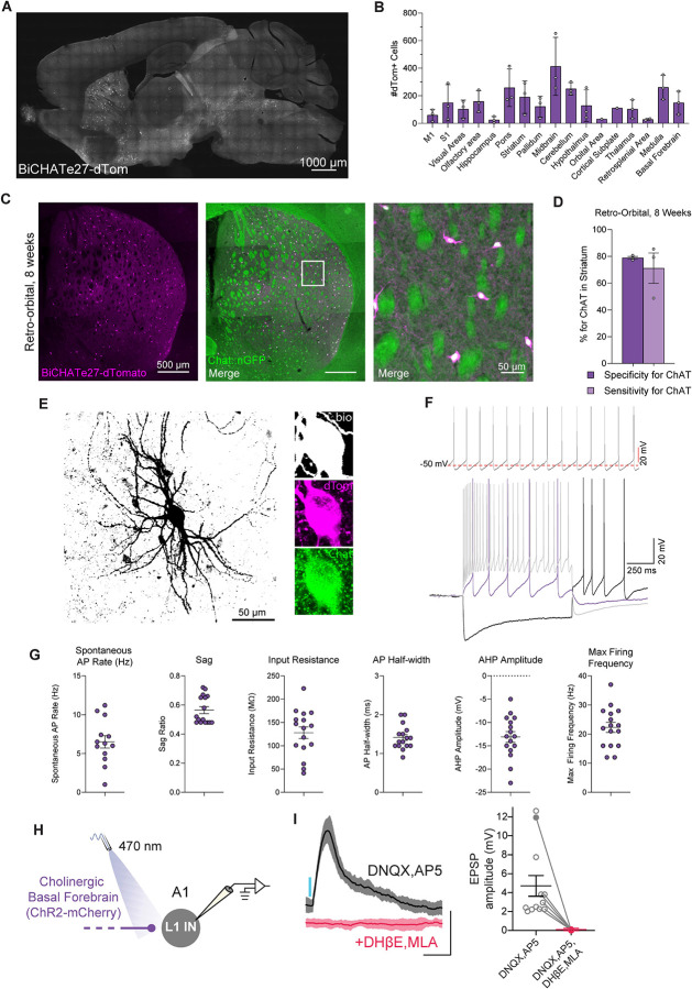
In recent years, we and others have identified a number of enhancers that, when incorporated into rAAV vectors, can restrict the transgene expression to particular neuronal populations. Yet, viral tools to access and manipulate specific neuronal subtypes are still limited. Here, we performed systematic analysis of single cell genomic data to identify enhancer candidates for each of the telencephalic interneuron subtypes. We established a set of enhancer-AAV tools that are highly specific for distinct cortical interneuron populations and striatal cholinergic interneurons. These enhancers, when used in the context of different effectors, can target (fluorescent proteins), observe activity (GCaMP) and manipulate (opto-genetics) specific neuronal subtypes. We also validated our enhancer-AAV tools across species. Thus, we provide the field with a powerful set of tools to study neural circuits and functions and to develop precise and targeted therapy.


Conflict of interest statement

DECLARATION OF INTERESTS G.F. is a founder of Regel Therapeutics, which has no competing interests with the present manuscript. G.F. is an advisor for Neuron and Annual Review of Neuroscience. J.D. and K.A. are employees of Regel Therapeutics and J.D. is also a founder. A.I. is the founder of Tibbling Technologies. Patents are pending on all enhancers present in this manuscript. For BiCHATe27 and BiSSTe10 G.F. and J.D hold this patent. For the remainder of enhancer patents, they are held by G.F., M.D. and Y.W.

Figures

Figure 1.
Figure 1.. Strategies to identify and characterize enhancer-based AAVs tools with cell type specificity
(A) A schematic illustrating the identification and testing of cell type-specific enhancers in rAAV constructs. (B and C) UMAP projections of single cell ATAC-seq data from mouse cortex (B) and striatum (C). Cells are colored by annotated cell types. (D) Chromatin accessibility pattern of the 7 top enhancers, visualized as normalized genome browser tracks representing the aggregated signals of cells from different cell types..
Figure 2.
Figure 2.. Enhancer-based viral targeting of PV neuron subtypes in mice
(A and B) Representative para-sagittal sections showing the distribution of dTomato-positive cells resulting from expression controlled by the BiPVe3 (A) or BiPVe4 (B) enhancers within the mouse brain, three weeks after retro-orbital AAV injection at 4 weeks of 2.0E+11 total vg/mouse. Scale bar is indicated in the figure. (C and D) Bar graphs showing the number of dTomato-positive cells under the control of the BiPVe3 (C) or BiPVe4 (D) enhancers in the brain areas indicated. Data from 5 sagittal sections from 5 different medio-lateral coordinates were collected, to cover the majority of brain regions; each individual datapoint represents a distinct biological replicate (N=3 mice). Error bars represent standard error of the mean (SEM). (E and F) Representative images showing the expression of dTomato reporter-expressing cells (in magenta) under the control of the BiPVe3 (E) or BiPVe4 (F) enhancers, and parvalbumin (Pvalb)-positive cells (in green) in the primary somatosensory cortex (S1) of the mouse brain, following retro-orbital AAV injection at 4 weeks. dTomato-positive cells co-expressing the PV marker (merge) are labeled in white. Cortical layers (L1–6) and scale bars are indicated in the figure. (G and I) Bar graphs showing the percentage specificity of BiPVe3 (G) or BiPVe4 (I) in targeting PV-positive neurons in the indicated layers. N=7 and 6 mice, respectively. Error bars represent SEM. RO4w= retro-orbital injection at 4 weeks of age (H and J) Representative confocal stack of a biocytin-filled cell expressing BiPVe3-dTomato (H) or BiPVe4-GFP (J) in L2/3 of primary somatosensory cortex, displaying characteristic PV basket (H) and chandelier (J) cell morphology. Scale bar is indicated in the figure. (K and L) Representative traces from current clamp recordings of cells expressing (K) BiPVe3-dTomato or (L) BiPVe4-GFP in L2/3 primary somatosensory cortex in response to 600 ms (K) or 300 ms (L) square wave current injections. In both cases, rAAV transduced cells show classical, non-accommodating, fast-spiking firing behavior. (M and N) BiPVe3-dTomato and BiPVe4-dTomato expressing cells in L2/3 display characteristic electrophysiologic properties of fast-spiking PV-positive interneurons, including a high maximum steady state firing frequency (SSFF), a narrow action potential (AP) halfwidth, and large afterhyperpolarization (AHP) (n = 13 cells from N = 4 mice for BiPVe3; n = 13 cells from N = 2 mice for BiPVe4: 4 cells from V1, 5 cells from A1 and 4 cells from S1). Error bars represent SEM. (O and P) Example of fast-spiking cell showing successful optogenetic in vivo activation across trials of cells transduced with with rAAV-BiPVe3-ChR2 (O) and rAAV-BiPVe4-ChR2 (P) viruses.
Figure 3.
Figure 3.. Enhancer-based viral targeting of SST neuron subtypes in mice
(A and B) Representative para-sagittal sections showing the distribution of dTomato-positive cells resulting from expression controlled by the BiSSTe10 (A) or BiSSTe4 (B) enhancers within the mouse brain, three weeks after retro-orbital AAV injection at 4 weeks of 2.0E+11 total vp/mouse (A) or ICV injections of 1.0E+10 total vp/pup at P1, respectively. Scale bar is indicated in the figure. (C and D) Bar graphs showing the number of dTomato-positive cells under the control of the BiSSTe10 (C) or BiSSTe4 (D) enhancers in the brain areas indicated. Data from 5 sagittal sections from 5 different medio-lateral coordinates were collected, to cover the majority of brain regions. Each individual datapoint represents a distinct biological replicate; N=3 mice (D). Error bars represent SEM. (E and F) Representative images showing the expression of dTomato reporter-expressing cells (in magenta) under the control of the BiSSTe10 (E) or BiSSTe4 (F) enhancers, and somatostatin (SST)-positive cells (in green) in the primary somatosensory cortex (S1) of the mouse brain, following retro-orbital AAV injection at 4 weeks or ICV injections at P1, respectively. dTomato-positive cells co-expressing the SST marker (merge) are labeled in white. Cortical layers (L1–6) and scale bars are indicated in the figure. (G and I) Bar graphs showing the percentage specificity of BiSSTe10 (G) or BiSSTe4 (I) in targeting SST-positive neurons across the layers indicated. N=7 and 4, respectively. Error bars represent SEM. RO4w= retro-orbital injection at 4 weeks of age, ICV=intracerebroventricular. (H and J) Representative confocal stack of a biocytin-filled cell expressing BiSSTe10-dTomato (H) following RO 4 weeks AAV injection or BiSSTe4-dTomato following ICV injection (J) in S1, displaying characteristic morphology. Scale bar is indicated in the figure. (K) Representative traces of voltage responses to 500 ms step current injection in current-clamp whole-cell configuration of BiSSTe10-dTomato positive cells in L4/5 primary somatosensory cortex, following RO 4 weeks AAV injections. (L) Dot plots showing three intrinsic physiological properties of BiSSTe10-dTomato positive cells. n = 13 cells from N = 2 mice. Error bars represent SEM. (M) Schematic representation of testing the synaptic connection from BiSSTe10-positive to pyramidal (Pyr) neurons located in different cortical layers. AAV-BiSSTe10-ChR-mCherry was injected by RO at 4 weeks and activity was recorded in Pyr neurons in S1. (N) left panel: Example traces of photo-stimulation-evoked postsynaptic currents recorded from pyramidal neurons in the indicated layers at 0 mV under voltage-clamp configuration. Experiments were performed in the presence of TTX and 4AP. Blue bar indicates ChR2 photo-stimulation (470 nm, 1 ms). right panel:Violin plot showing IPSCs responses of pyramidal neurons from the indicated layers, following light stimulation of BiSSTe10-dTomato cells. Data highlight a very selective inhibition from BiSSTe10-mCherry-positive cells to L4 pyramidal neurons. n = 30 cells (7 for L2/3, 9 for L4, 7 for L5 and 7 for L6), from N = 4 mice.(O) Top: Representative traces of a fast adapting (non LTS) (top panel) and low threshold spiking (LTS) (bottom panel) recorded from BiSSTe4-tdtomato positive neurons in L5 of S1. LTS=low threshold spikers. (P) Dot plots representing three recorded intrinsic properties of BiSSTe4 cells. Graphs are presented as mean and SEM. n=10 from N=3 mice. (Q) Example of regular-spiking cell showing successful optogenetic in vivo activation across trials of cells transduced with rAAV-BiSSTe4-ChR2.
Figure 4.
Figure 4.. Enhancer-based viral targeting of CGE-derived cINs in mice
(A and B) Representative para-sagittal sections showing the distribution of dTomato-positive cells resulting from expression controlled by the BiVIPe4 (A) or BiLAMP5e3 (B) enhancers within the mouse brain, three weeks after ICV injections of 1.0E+10 total vg/pup at P1 (A) or retro-orbital AAV injection at 4 weeks of 2.0E+11 total vg/mouse (B) respectively. Scale bar is indicated in the figure. (C and D) Bar graphs showing the number of dTomato-positive cells under the control of the BiVIPe4 (C) or BiLAMP5e3 (D) enhancers in the brain areas indicated. Data from 5 sagittal sections from 5 different medio-lateral coordinates were collected, to cover the majority of brain regions. Each individual datapoint represents a distinct biological replicate; N32 mice. Error bars represent SEM. (E and F) Representative images showing the expression of dTomato-expressing cells (in magenta) under the control of the BiVIPe4 (E) or BiLAMP5e3 (F) enhancers, and vasoactive intestinal peptide (Vip)-positive cells (E) or the genetically-expressed GFP reporter under the control of Lamp5-Flp and Gad2-cre (F) (in green) in the primary somatosensory cortex (S1) of the mouse brain, following ICV injections at P1 (E) or retro-orbital AAV injection at 4 weeks (F), respectively. dTomato-positive cells co-expressing the marker (merge) are labeled in white. Cortical layers (L1–6) and scale bars are indicated in the figure. (G and I) Bar graphs showing the percentage specificity of BiVIPe4 (G) or BiLAMP5e3 (I) in targeting VIP- or LAMP5-positive neurons in L2/3 (G) or across all cortical layers (I). N=5 and N=3 mice, respectively. Error bars represent SEM. (H and J) Representative morphological reconstructions of neurobiotin-filled BiVIPe4-mCherry-positive neuron in L2/3 (H) and a BiLAMP5e3-dTomato (J) neuron in L1 of S1. Scale bar is indicated. (K) Representative traces of voltage responses to 800 ms step current injection in current-clamp whole-cell configuration of BiVIPe4-mCherry-positive cells. Scale bar is indicated. (L) Summary plots showing the intrinsic physiological properties of BiVIPe4-mCherry--positive cells. n = 7 cells from N = 2 mice for BiVIPe4. Error bars represent SEM. (M) Schematic representation of testing the synaptic connection from BiVIPe4-positive cells to SST-positive and pyramidal (Pyr) neurons. AAV-BiVIPe4-ChR-mCherry was injected at P0 into GIN-GFP mice and photo-evoked activity was recorded in SST and Pyr neurons in S1. (N, left panel) Example traces of photo-stimulation-evoked postsynaptic currents recorded from an SST cell and a pyramidal neuron at 0 mV under voltage-clamp configuration. Gray traces depict individual sweeps, and solid traces the average of these sweeps. Blue bar indicates ChR2 photo-stimulation (470 nm, 3 ms). (right panel): Histogram showing the amplitude of postsynaptic response of SST and simultaneously recorded nearby pyramidal neurons upon optogenetic stimulation of BiVIPe4-labeled cells. n=16 SST and pyramidal cells from N=4 mice. Error bars represent SEM. (O) Imaging locomotion responses of BiVIPe4 targeted neurons in the visual cortex. Mice were injected with AAV-BiVIPe4-GCaMP6f in the visual cortex. After operation recovery, mice were habituated to head-fixation and were able to run freely on a belt treadmill. Following habituation, spontaneous activity in VIP interneurons was acquired while the mice were presented with a gray screen. Bottom left: Set up for two-photon imaging of VIP interneurons in the visual cortex of awake-behaving mice. Bottom right: An example image plane in L2/3 showing GCaMP6f-expressing neurons. (P) Traces of three example neurons of three neurons (orange) in relation to running speed (black). Right panel: Mean of the locomotion modulation index (LMI; LMI = (RL–RM) / (RL+RM)). Individual points represent the mean LMI for each mouse (N= 3 mice). (Q) Left panel: Representative traces of action potentials recorded from L1 NGFC and Canopy interneurons. The top panel shows action potentials aligned to the maximum amplitude, while the bottom panel aligned to their action potential threshold. Right panel: Two examples of an NGFC and a Canopy cell responding to a 1-second increasing current injection. Differences in electrophysiological properties can be observed at subthreshold, threshold, and suprathreshold current steps. (R) Summary plots showing three intrinsic physiological properties of BiLAMP5e3-dTomato-positive cells. n=17 cells for NGFCs and n=11 cells for Canopy, from N=5 mice. Error bars represent SEM.***p<0.001 ****=p<0.0001 Mann Whitney test.
Figure 5.
Figure 5.. Enhancer-based viral targeting of CHAT-positive neurons in mice
(A) Representative para-sagittal sections showing the distribution of dTomato-positive cells resulting from expression controlled by the BiCHATe27 enhancer within the mouse brain, three weeks after RO injection of 2.0E+11 total vg/mouse at 4 weeks. Scale bar is indicated in the figure. (B) Histograms showing the number of dTomato-positive cells under the control of the BiCHATe27 enhancer in the brain areas indicated. Data from 5 sagittal sections from 5 different medio-lateral coordinates were collected, to cover the majority of brain regions. Each individual datapoint represents a distinct biological replicate; N=3 mice. Error bars represent SEM. (C) Representative image showing the expression of dTomato reporter (in magenta) under the control of the BiCHATe27 enhancer, and the Chat-cre dependent expression of GFP reporter (in green) in striatum of the mouse brain, following retro-orbital AAV injection at 8 weeks. The dTomato-positive cells co-expressing the marker (merge) are labeled in white. Right panel: zoomed-in image from the left panel. Scale bars are indicated in the figure. (D) Bar graphs showing the percentage specificity and sensitivity of the BiCHATe27 enhancer in targeting CHAT-positive neurons in the striatum. N=3 mice. Error bars represent SEM. (E) Biocytin filled reconstruction of BiCHATe27 infected cell recorded in mouse striatum. Inset: Reconstructed cell body in biocytin (white), BiCHATe27-dTomato (magenta), and CHAT immunostaining (green). Scale bar is indicated in the figure. (F) Top panel: Example trace of spontaneous action potential firing pattern during cell-attached patch clamp of BiCHATe27-dTomato infected cell. Bottom panel: Intrinsic firing properties recorded from BiCHATe27-dTomato infected cell, electrophysiological membrane and action potential firing responses at hyperpolarized (black), threshold (red), and maximum firing depolarization (gray) current steps. (G) Intrinsic membrane and firing properties of BiCHATe27 infected cells recorded in mouse striatum. n = 15–16 cells across N=3 mice. Error bars represent SEM. (H) Schematic of whole-cell current clamp recordings in layer 1 cortical interneurons (L1 INs) of primary auditory cortex (A1) in response to optogenetic stimulation of BiCHATe27-ChR2-mCherry axons. (I) Left panel: Representative (mean ± SD) excitatory postsynaptic potentials (EPSPs) from a L1 IN in response to a 5 ms blue light pulse delivered with a 470 nm LED (~14 mW/mm2, 0.1 Hz). Responses were recorded in the presence of 20 μM DNQX and 50 μM AP5, AMPA-R and NMDA-R blockers, respectively, and eliminated in the presence of nAChR blockers 10 μM DHβE and 10 μM MLA (red traces). Scale bars: 5 mV, 200 ms. Right panel: Average EPSP amplitude in the presence of DNQX and AP5 (n = 12 cells) and in the presence of DNQX, AP5, DHβE and MLA (n = 6 cells), N=4 mice. Error bar represents SEM. Filled circle corresponds to the representative cell shown in the left panel.
Figure 6.
Figure 6.. Validation of enhancer-based viral tools in non-human primates and human tissue
Validation of (A) BiPVe3, (B) BiPVe4, (C) BiSSTe10, (D) BiLAMP5e3, (E) BiCHATe27, (F) BiSSTe4, and (G) BiVIPe4 in rhesus macaque. (A-Gi) Overview images of reporter expression (magenta) colocalized with immunohistochemical (IHC) staining of target molecular markers (green). The scale bar is indicated in the figure. nNOS: neuronal nitric oxide synthase, CR: calretinin, HPC: hippocampus. (A-Gii) Percentage specificity of rAAV transduced cells for the target cell type-specific markers. BiPVe3: 12 sections from 1 animal, BiPVe4: 25 sections from 1 animal, BiSSTe10: 8 sections from 2 animals, BiLAMP5e3: 13 sections from 2 animals, BiCHATe27: 20 sections from 2 animals, BiSSTe4: 5 sections from 1 animal, BiVIPe4: 5 sections from 2 animals. Error bars represent SEM. (A-Eiii) Biocytin filled cell-reconstructions of rAAV transduced cells. (Inset) Reconstructed cell body in biocytin (white) and reporter expression (magenta). (A-Eiv) Intrinsic firing properties recorded from rAAV transduced cells. (Left) Electrophysiological membrane and action potential firing responses at hyperpolarized (black), threshold (red), and maximum firing depolarization (gray) current steps. (Right) Spike shape and accommodation of rAAV transduced cells depicting initial (black), intermediate (gray), and final (red) spikes during maximum firing trains. Inset in Div shows persistent firing of BiLAMP5 cells after the end of depolarizing pulse. (H) Validation of BiVIPe4 enhancer activity in human tissue. Representative images of human tissue sections transduced with AAV-BiVIPe4-ChR2-mCherry, co-stained with Calretinin (CR). The scale bar is indicated in the figure.

References

    1. Yao Z., van Velthoven C.T.J., Nguyen T.N., Goldy J., Sedeno-Cortes A.E., Baftizadeh F., Bertagnolli D., Casper T., Chiang M., Crichton K., et al. (2021). A taxonomy of transcriptomic cell types across the isocortex and hippocampal formation. Cell 184, 32223241 e26. 10.1016/j.cell.2021.04.021. - DOI - PMC - PubMed
    1. Tasic B., Yao Z., Graybuck L.T., Smith K.A., Nguyen T.N., Bertagnolli D., Goldy J., Garren E., Economo M.N., Viswanathan S., et al. (2018). Shared and distinct transcriptomic cell types across neocortical areas. Nature 563, 72–78. 10.1038/s41586-018-0654-5. - DOI - PMC - PubMed
    1. Gouwens N.W., Sorensen S.A., Baftizadeh F., Budzillo A., Lee B.R., Jarsky T., Alfiler L., Baker K., Barkan E., Berry K., et al. (2020). Integrated Morphoelectric and Transcriptomic Classification of Cortical GABAergic Cells. Cell 183, 935–953 e19. 10.1016/j.cell.2020.09.057. - DOI - PMC - PubMed
    1. Wu S.J., Sevier E., Dwivedi D., Saldi G.A., Hairston A., Yu S., Abbott L., Choi D.H., Sherer M., Qiu Y., et al. (2023). Cortical somatostatin interneuron subtypes form celltype-specific circuits. Neuron 111, 2675–2692 e9. 10.1016/j.neuron.2023.05.032. - DOI - PMC - PubMed
    1. Dimidschstein J., Chen Q., Tremblay R., Rogers S.L., Saldi G.A., Guo L., Xu Q., Liu R., Lu C., Chu J., et al. (2016). A viral strategy for targeting and manipulating interneurons across vertebrate species. Nat Neurosci 19, 1743–1749. 10.1038/nn.4430. - DOI - PMC - PubMed

Publication types

LinkOut - more resources