. 2024 Jul 18:14:1384499.
doi: 10.3389/fonc.2024.1384499. eCollection 2024.

3D bioprinted CRC model brings to light the replication necessity of an oncolytic vaccinia virus encoding FCU1 gene to exert an efficient anti-tumoral activity

Affiliations

3D bioprinted CRC model brings to light the replication necessity of an oncolytic vaccinia virus encoding FCU1 gene to exert an efficient anti-tumoral activity

Christophe A Marquette et al. Front Oncol. .

Abstract

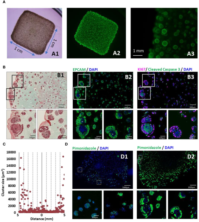
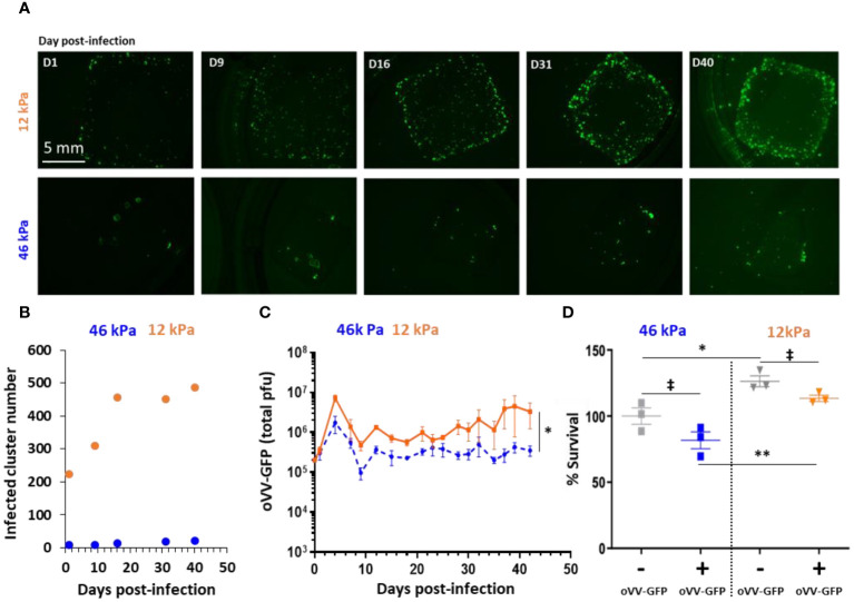
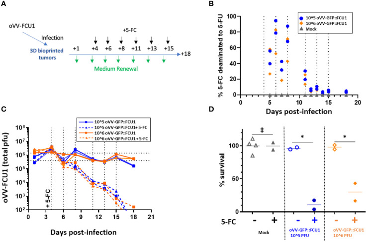
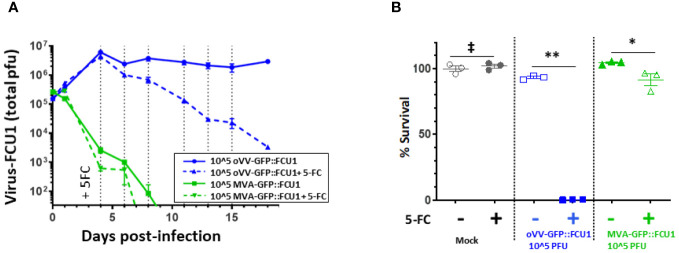
The oncolytic virus represents a promising therapeutic strategy involving the targeted replication of viruses to eliminate cancer cells, while preserving healthy ones. Despite ongoing clinical trials, this approach encounters significant challenges. This study delves into the interaction between an oncolytic virus and extracellular matrix mimics (ECM mimics). A three-dimensional colorectal cancer model, enriched with ECM mimics through bioprinting, was subjected to infection by an oncolytic virus derived from the vaccinia virus (oVV). The investigation revealed prolonged expression and sustained oVV production. However, the absence of a significant antitumor effect suggested that the virus's progression toward non-infected tumoral clusters was hindered by the ECM mimics. Effective elimination of tumoral cells was achieved by introducing an oVV expressing FCU1 (an enzyme converting the prodrug 5-FC into the chemotherapeutic compound 5-FU) alongside 5-FC. Notably, this efficacy was absent when using a non-replicative vaccinia virus expressing FCU1. Our findings underscore then the crucial role of oVV proliferation in a complex ECM mimics. Its proliferation facilitates payload expression and generates a bystander effect to eradicate tumors. Additionally, this study emphasizes the utility of 3D bioprinting for assessing ECM mimics impact on oVV and demonstrates how enhancing oVV capabilities allows overcoming these barriers. This showcases the potential of 3D bioprinting technology in designing purpose-fit models for such investigations.

Keywords: bioprinting; colorectal (colon) cancer; hydrogel; oncovirus; tumor.


Conflict of interest statement

Author AS, CE, MN, BM, PE, J-MB, EQ and CZ were employed by company Transgene SA. The remaining authors declare that the research was conducted in the absence of any commercial or financial relationships that could be construed as a potential conflict of interest.

Figures

Figure 1
Figure 1
3D-bioprinted tumor models characterization. (A) 35 days after 3D printing, models were stained with calcein AM and were observed in bright field (A1), under fluorescent light to detect live cells (A2) and at higher magnification (A3). (B) Models were fixed, paraffin embedded and sliced. Consecutive sections were analyzed by Masson’s trichrome staining (B1), EPCAM (green) (B2), and immunofluorescent staining against KI67 (purple) and cleaved caspase 3 (green) (B3). (C) Cluster size distribution along 10 mm CRC model histological sections, quantified using imageJ software and particle analysis. (D) Hypoxia was monitored by short incubation with pimonidazole followed by specific immunofluorescent staining at 10 days (D1) or 35 days (D2) post-printing. Nucleus were counter-stained with DAPI (Blue).
Figure 2
Figure 2
Stiffness of 3D bioprinted tumor model impacts infection and proliferation of oncolytic Vaccinia Virus. 3D bioprinted tumor models with different matrix stiffness were infected 40 days post-printing by 10^5 PFU of oVV expressing eGFP. (A) Virus expression was monitored by fluorescent microscopy during at least 40 day post-infection. Matrix with stiffness of 46 kPa (top) or 12 kPa (bottom) were assessed. (B) Numbers of infected-clusters by oVV-GFP were quantified using imageJ software and particle analysis. (C) Following infection, supernatant was renewed every 2–3 days and oVV presents in the supernatant was titrated. The orange line and the dashed blue line represented the mean of the total PFU ± SEM (n=3) for the model with 12 kPa and 46 kPa stiffness respectively. (D) At 42 days post-infection, the viability of the models was determined using Celltiter Blue assay. The 100% viability was based on mock-treated models. Symbols represent individual models, and horizontal lines indicate the mean ± SEM (n=3). ‡: non-significant ANOVA test (p>0.1). *: significant ANOVA test (p<0.02). **: significant ANOVA test (p<0.002).
Figure 3
Figure 3
oVV expressing GFP::FCU1 as payload demonstrates efficient antitumor cytotoxicity. (A) Schematic depicting the experimental procedure to evaluate the antitumor efficiency of oVV-GFP::FCU1. 3D bioprinted-tumor models were infected with 10^6 or 10^5 PFU of oVV-GFP::FCU1, the following day input virus was removed and new medium added. Then, 4 days post infection 5-FC (1mM) was added in new medium. Every 2/3 days medium with 5-FC was renewed. Experiment was stopped 18 days post-infection and model viability was measured. As control, same procedure was applied with medium without 5-FC to infected and non-infected models (for each condition with infected models (n=2), mock models + 5-FC (n=2) and mock models (n=4). (B) Concentration of 5-FC and 5-FU in model supernatants was determined by high-performance liquid chromatography. The results are presented as percent of 5-FU generated from 5-FC with each symbol representing an individual model The percent of 5-FC/5-FU conversion was defined in medium before the medium was renew. (C) Virus in the supernatant was quantified by PFU titration. Each line shows the total PFU measured for individual models. Dotted vertical line indicated the addition of new medium with 5-FC (D) Viability of the models was determined at 18 days post-infection using Celltiter Blue assay. The 100% viability was based on mock-treated models. Symbols represent individual models, and the horizontal line indicates the mean. ‡: non-significant ANOVA test (p>0.9). *: significant ANOVA test (p<0.02).
Figure 4
Figure 4
oVV-GFP::FCU1 replication is needed for efficient antitumoral and bystander effect. (A) 3D bioprinted-tumor models were infected by 10^5 PFU of oVV-GFP::FCU1 or MVA-GFP::FCU1 as described in Figure 3A . 5-FC (1mM) was added 4 days post infection and every 2/3 days medium was renewed with 5-FC. Virus in the supernatant was quantified by PFU titration. Results are represented as the mean of the total PFU ± SEM (n=3). As a control, identical protocol was carried over without the addition of 5-FC. Dotted vertical line indicated the addition of new medium with 5-FC (B) 18 post-infection the viability of the models was determined using Celltiter Blue assay. The 100% viability was based on mock-treated models. Symbols represent individual model, and horizontal line indicates the mean ± SEM (n=3). ‡: non-significant ANOVA test (p>0.9). **: significant ANOVA test (p<0.002). *: significant ANOVA test (p<0.02).

Similar articles

References

    1. Davis LE, Shalin SC, Tackett AJ. Current state of melanoma diagnosis and treatment. Cancer Biol Ther. (2019) 20:1366–79. doi: 10.1080/15384047.2019.1640032 - DOI - PMC - PubMed
    1. Wang M, Herbst RS, Boshoff C. Toward personalized treatment approaches for non-small-cell lung cancer. Nat Med. (2021) 27:1345–56. doi: 10.1038/s41591-021-01450-2 - DOI - PubMed
    1. Dagogo-Jack I, Shaw AT. Tumour heterogeneity and resistance to cancer therapies. Nat Rev Clin Oncol. (2018) 15:81–94. doi: 10.1038/nrclinonc.2017.166 - DOI - PubMed
    1. Drost J, Clevers H. Organoids in cancer research. Nat Rev Cancer. (2018) 18:407–18. doi: 10.1038/s41568-018-0007-6 - DOI - PubMed
    1. Neufeld L, Yeini E, Pozzi S, Satchi-Fainaro R. 3D bioprinted cancer models: from basic biology to drug development. Nat Rev Cancer. (2022) 22:679–92. doi: 10.1038/s41568-022-00514-w - DOI - PubMed

LinkOut - more resources