Review
. 2024 Jun 4;4(5):100343.
doi: 10.1016/j.bpsgos.2024.100343. eCollection 2024 Sep.

Using Organoids to Model Sex Differences in the Human Brain

Collaborators, Affiliations
Review

Using Organoids to Model Sex Differences in the Human Brain

Adam Pavlinek et al. Biol Psychiatry Glob Open Sci. .

Abstract

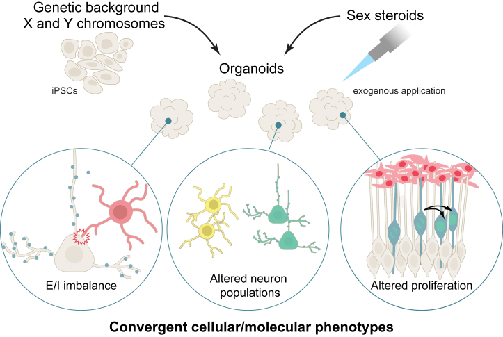
Sex differences are widespread during neurodevelopment and play a role in neuropsychiatric conditions such as autism, which is more prevalent in males than females. In humans, males have been shown to have larger brain volumes than females with development of the hippocampus and amygdala showing prominent sex differences. Mechanistically, sex steroids and sex chromosomes drive these differences in brain development, which seem to peak during prenatal and pubertal stages. Animal models have played a crucial role in understanding sex differences, but the study of human sex differences requires an experimental model that can recapitulate complex genetic traits. To fill this gap, human induced pluripotent stem cell-derived brain organoids are now being used to study how complex genetic traits influence prenatal brain development. For example, brain organoids from individuals with autism and individuals with X chromosome-linked Rett syndrome and fragile X syndrome have revealed prenatal differences in cell proliferation, a measure of brain volume differences, and excitatory-inhibitory imbalances. Brain organoids have also revealed increased neurogenesis of excitatory neurons due to androgens. However, despite growing interest in using brain organoids, several key challenges remain that affect its validity as a model system. In this review, we discuss how sex steroids and the sex chromosomes each contribute to sex differences in brain development. Then, we examine the role of X chromosome inactivation as a factor that drives sex differences. Finally, we discuss the combined challenges of modeling X chromosome inactivation and limitations of brain organoids that need to be taken into consideration when studying sex differences.

Keywords: Autism; Brain organoids; Sex chromosomes; Sex differences; Steroids; X chromosome inactivation.

Plain language summary

Sex differences are a contributing factor in neuropsychiatric conditions such as autism, which is more prevalent in males. Sex differences occur through interactions between sex steroid hormones such as estrogen and testosterone and sex chromosomes (chrX and chrY). Human stem cell–derived brain organoids are laboratory models that mimic brain development. For example, in individuals with neurodevelopmental conditions, brain organoids have revealed an imbalance of neuron populations compared with neurotypical individuals. In this review, we discuss sex steroid and sex chromosome influences on brain development and challenges of this model that need to be taken into account when studying sex differences.


Figures

Figure 1
Figure 1
Timeline of human brain development from conception to birth showing human brain morphology over developmental time (top), predicted testosterone dynamics (middle), and key events in sex-biased development (bottom). The box shows an axial section with examples of regions with growth trajectory (blue, male-biased; purple, female-biased) sex differences found in fetal MRI (141). The insular cortex is an example of a focal region that was larger in males than in females. The sex chromosomes begin exerting sex-specific effects soon after conception. Testosterone is given here as a classic example of a steroid sex hormone with levels that differ by sex. Fetal testosterone increases after the differentiation of the testes in males. Male testosterone is thought to peak between GW14 and GW18 and then remain relatively consistent (141). Studies have generally reported higher male testosterone, and some studies have reported increasing testosterone in females over time (142). GW, gestational week; MRI, magnetic resonance imaging.
Figure 2
Figure 2
Sex steroid hormones from various sources (top left) and sex chromosome effects (bottom left) give rise to phenotypic sex differences at the cellular level. Note that although we show the placenta in the context of hormones, the placenta can influence sex differences both through sex steroid hormones or sex chromosome complement. Sex steroid hormones in the fetus can originate from the maternal adrenal gland, the placenta, and from the fetal adrenal gland. After entering the fetal circulatory system, hormones can cross the blood-brain barrier to affect brain development. In males, the testes develop and are a source of androgens. DHEA, DHEAS, A4, E2 are examples of key steroid sex hormones. Aromatase (bottom right) can aromatize testosterone to E2. The sex chromosomes (bottom left) can be a source of gene expression differences. In males, there is 1 active X chromosome and the Y chromosome. The SRY gene on the Y chromosome is responsible for testes development. Expression of other Y genes (purple) that have diverged from their X chromosome homologs in evolution can be a source of sex differences. Females have 1 Xa and 1 Xi. Sex-specific gene expression can originate from genes that are expressed from the Xi (escapees) or from modulation of gene expression on the Xa by the Xi. Finally, X-chromosomal mosaicism in females results in allelic diversity (bottom). The placenta is one example of a tissue where sex chromosome complement has an important role in sex differences. Examples of how various cellular and molecular brain sex differences may arise are shown (right). Exposure to steroid hormones can have rapid cellular effects mediated by rapid signaling cascades or canonical gene expression effects through nuclear signaling. Sex chromosome gene expression effects combined with gene expression in response to steroid hormone signaling can modulate regional receptor expression and steroid signaling pathways, resulting in further sex-specific cellular responses. The combined action of the sex chromosomes and steroid hormones can establish sex-biased gene expression, gene regulatory networks, and epigenetic modifications. One example of epigenetic modifications is demethylation, such as by the KDM5C and KDM6A demethylases, which are discussed. A4, androstenedione; AR, androgen receptor; DHEA, dehydroepiandrosterone; DHEAS, dehydroepiandrosterone sulfate; E2, estradiol; ESR, estrogen receptor; GPER1, G protein–coupled estrogen receptor 1; Xa, active X chromosome; Xi, inactive X chromosome; Xm, X maternal; Xp, X paternal.
Figure 3
Figure 3
Typical chrX states in female human embryo development. Human genome activation and expression from the X chromosomes occurs by the 8-cell stage (far left). In the preimplantation embryo (left), gene expression occurs from both of the X chromosomes, which are coated with the long noncoding RNAs XIST and XACT. Gene expression is modulated through chrX dampening. Sometime after implantation (middle), random XCI occurs. Gene expression becomes restricted to only Xa, and Xa is coated with XACT. Xi is coated with XIST, and gene expression is silenced. In somatic cells, Xi remains silenced except for escapee genes. Random XCI is preserved in somatic cells (box), resulting in X-chromosomal mosaicism in females, where a subset of cells will express the maternal allele of a gene, and the remaining cells will express the paternal alleles of a gene. chrX, X chromosome; G, gene; M, maternal; P, paternal; Xa, active chrX; XCI, X chromosome inactivation; Xi, inactive chrX.
Figure 4
Figure 4
Examples of convergent cellular and molecular neuropsychiatric condition phenotypes revealed by studies using human iPSC-derived brain organoids: E/I imbalance, altered cell proliferation, and phenotypes that affect specific neuron classes. Future organoid studies can combine the study of genetic background, sex chromosome complement, and exogeneous steroid hormone application. E/I, excitation/inhibition; iPSC, induced pluripotent stem cell.

References

    1. Dewing P., Shi T., Horvath S., Vilain E. Sexually dimorphic gene expression in mouse brain precedes gonadal differentiation. Brain Res Mol Brain Res. 2003;118:82–90. - PubMed
    1. De Vries G.J., Rissman E.F., Simerly R.B., Yang L.Y., Scordalakes E.M., Auger C.J., et al. A model system for study of sex chromosome effects on sexually dimorphic neural and behavioral traits. J Neurosci. 2002;22:9005–9014. - PMC - PubMed
    1. Wierman M.E. Sex steroid effects at target tissues: Mechanisms of action. Adv Physiol Educ. 2007;31:26–33. - PubMed
    1. Alexander G.M., Wilcox T., Woods R. Sex differences in infants’ visual interest in toys. Arch Sex Behav. 2009;38:427–433. - PubMed
    1. Arnold A.P., Chen X. What does the “four core genotypes” mouse model tell us about sex differences in the brain and other tissues? Front Neuroendocrinol. 2009;30:1–9. - PMC - PubMed

LinkOut - more resources