. 2024 Jul 18:17:1415207.
doi: 10.3389/fnmol.2024.1415207. eCollection 2024.

Drosophila larval motor patterning relies on regulated alternative splicing of Dscam2

Affiliations

Drosophila larval motor patterning relies on regulated alternative splicing of Dscam2

G Lorenzo Odierna et al. Front Mol Neurosci. .

Abstract

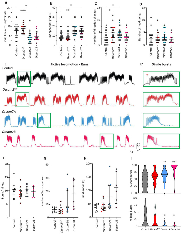
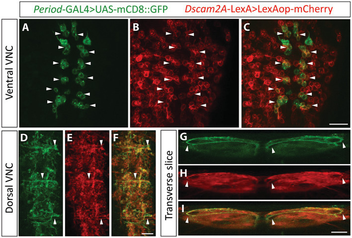
Recent studies capitalizing on the newly complete nanometer-resolution Drosophila larval connectome have made significant advances in identifying the structural basis of motor patterning. However, the molecular mechanisms utilized by neurons to wire these circuits remain poorly understood. In this study we explore how cell-specific expression of two Dscam2 isoforms, which mediate isoform-specific homophilic binding, contributes to motor patterning and output of Drosophila larvae. Ablating Dscam2 isoform diversity resulted in impaired locomotion. Electrophysiological assessment at the neuromuscular junction during fictive locomotion indicated that this behavioral defect was largely caused by weaker bouts of motor neuron activity. Morphological analyses of single motor neurons using MultiColour FlpOut revealed severe errors in dendrite arborization and assessment of cholinergic and GABAergic projections to the motor domain revealed altered morphology of interneuron processes. Loss of Dscam2 did not affect locomotor output, motor neuron activation or dendrite targeting. Our findings thus suggest that locomotor circuit phenotypes arise specifically from inappropriate Dscam2 interactions between premotor interneurons and motor neurons when they express the same isoform. Indeed, we report here that first-order premotor interneurons express Dscam2A. Since motor neurons express Dscam2B, our results provide evidence that Dscam2 isoform expression alternates between synaptic partners in the nerve cord. Our study demonstrates the importance of cell-specific alternative splicing in establishing the circuitry that underlies neuromotor patterning without inducing unwanted intercellular interactions.

Keywords: A02; Drosophila; Dscam2; alternative splicing; dendrite targeting; fictive locomotion; looper; motor neuron.


Conflict of interest statement

The authors declare that the research was conducted in the absence of any commercial or financial relationships that could be construed as a potential conflict of interest.

Figures

Figure 1
Figure 1
Locomotor and fictive locomotor defects following loss of Dscam2 isoform diversity. (A–D) Motor behavior assessment of larvae lacking Dscam2 (Dscam2null) and larvae with ablated Dscam2 alternative splicing, expressing only isoform A (Dscam2A) or only isoform B (Dscam2B). Quantification of the number of grid lines crossed by larvae in 60 s (A) and time spent within a single grid (B) revealed that Dscam2A and Dscam2B larvae had a locomotor defect relative to controls. Number of direction changes (C) was measured by counting the number of times larvae performed a head swing during forward linear locomotion and continued along the new path set by the head swing. Number of head swings (D) was measured by counting the number of times larvae performed a head swing while immobile. Groups analyzed using a Kruskal-Wallis test with Dunn’s multiple comparison, *p < 0.05, **p < 0.01, ***p < 0.001. N = number of larvae assessed; Control = 23, Dscam2null = 16, Dscam2A = 25, Dscam2B = 17. Error bars show 95% confidence interval. (E,E′) Representative traces of spontaneous compound EJPs recorded from larvae performing fictive locomotion. Bursts, which occur via sequential summing of EJPs, occur in a rhythmic fashion. Green insets show individual bursts within the runs (E′). Red arrowhead shows a single EJP. Control = black, Dscam2null = red, Dscam2A = blue, Dscam2B = magenta. (F–H) Quantification of burst parameters within individual runs, where a run was defined by more than 3 consecutive bursts (within 10 s of each other). The frequency of bursts (F), average number of bursts per run (G) and average duration of individual runs (H) is not statistically different between any of the measured genotypes. (I,J) Quantification of the composition of runs after separating bursts into short (<75 EJPs), medium (75–150 EJPs) and long (>150 EJPs) categories. Dscam2A and Dscam2B larvae displayed a statistically significant increase in the prevalence of short bursts (I) and a statistically significant decrease in the prevalence of long bursts (J). Groups analyzed using a Kruskal-Wallis test with Dunn’s multiple comparison, **p < 0.01, ****p < 0.0001. N = number of larvae assessed; Control = 11, Dscam2null = 11, Dscam2A = 9, Dscam2B = 6. Error bars show 95% confidence interval. Lines in violin plots represent median (unbroken line) and quartiles (broken lines).
Figure 2
Figure 2
Dscam2 protein is expressed in the ventral nerve cord. (A–F) Representative images of immunostaining against Bruchpilot (Brp) and Dscam2 in the 3rd instar larval ventral nerve cord (VNC). Brp immunoreactivity (red) identifies the VNC neuropil (A, max-projected z-stack; A′, z-stack transverse reslice). Dscam2 immunoreactivity (green) using an antibody directed against the cytoplasmic region reveals protein localization to the VNC neuropil (B,C, max-projected z-stack; B′,C′, z-stack transverse reslice). Loss of signal in larvae lacking Dscam2 (Dscam2null) confirms the specificity of the Dscam2 antibody (D–F′). Scale bars in panels (A–F) are 50 μm. Scale bars in panels (A′–F′) are 20 μm. (G,G′) V5 immunoreactivity (green) in larvae harboring a V5-tagged Dscam2 (BAC-Dscam2-V5). A single optical slice is shown in panel (G) and a single optical transverse reslice is shown in panel (G′). Scale bar in panel (G) is 20 μm. Scale bar in panel (G′) is 10 μm. (H–J) GFP fluorescence in the VNC of larvae expressing Dscam2::GFP-FLAG from the endogenous Dscam2 locus (CRISPR knock-in). A single optical slice is shown in panel (H) and a single optical transverse reslice is shown in panel (H′). Color-coded insets highlight GFP in presumed axon bundles containing sensory afferent and motor efferent axons (I, white arrowheads in yellow inset) and in commissural fascicles that contain axonal and dendritic neurites (J, white arrowheads in red inset). Scale bar in panel (H) is 20 μm. Scale bar in panel (H′) is 10 μm.
Figure 3
Figure 3
Looper premotor interneurons express Dscam2A. (A–C) Single optical slice from the ventral cortex of the nerve cord in larvae expressing Period-directed GFP (A, Period-GAL4 > UAS-mCD8::GFP) and Dscam2A-directed mCherry (B, Dscam2A-LexA>LexAop-mCherry). Merged image in panel (C) shows that multiple Period-positive interneurons express Dscam2A (white arrowheads). Note that the ventral cortex of the nerve cord is where the cell bodies of period-positive median segmental interneurons (PMSIs, otherwise known as “loopers”) reside. Scale bar is 20 μm. (D–F) Expression of Period-directed GFP (D) and Dscam2A-directed mCherry (E) in the dorsal region of the VNC neuropil, where looper interneurons form synaptic contacts with motor neurons. Merged image in panel (F) shows colocalization in many neurites arborizing throughout the motor domain. Scale bar is 10 μm. (G–I) Transverse optical slice of the VNC showing the characteristic ventro-dorsal axonal projections of looper interneurons in Period-directed GFP (G, white arrowheads) are also identifiable using Dscam2A-directed mCherry (H, white arrowheads). Merged image in panel (I) shows close overlap between Period-directed GFP and Dscam2A-directed mCherry in presumed looper axons (white arrowheads). Scale bar is 10 μm.
Figure 4
Figure 4
Inappropriate Dscam2 interactions disrupt motor neuron dendrite patterning and inputs to the motor domain. (A–C) Representative example of singly labeled MN6/7-1b neurons in control, Dscam2null and Dscam2B larvae using a MultiColor FlpOut strategy. Yellow arrowheads indicate singly labeled MN6/7-1b motor neurons. Green arrowheads indicate the three distinct dendritic arrays formed by MN6/7-1b. Yellow arrows in panel (A) show the general orientation of MN6/7-1b in the ventral nerve cord; R = Rostral, C = Caudal, M = medial, L = lateral. Scale bar is 10 μm. (D) Quantification of max-projected z-stacks by outlining basal dendritic arbors revealed that the ratio of dorsal-caudal projections was significantly higher in the Dscam2B MN6/7-1b motor neurons compared to control and Dscam2null. Groups analyzed using a Kruskal-Wallis test with Dunn’s multiple comparison, *p < 0.05, ***p < 0.001. N = number of motor neurons analyzed (1 motor neuron per larva); Control = 11, Dscam2null = 8, Dscam2B = 32. Error bars show 95% confidence interval. (E–H″) Representative images of triple labeled ventral nerve cords with OK6-GAL4 > UAS-mCD8::GFP fluorescence, anti-Choline acetyltransferase (ChAT) immunoreactivity and anti-Drosophila Vesicular GABA transporter (dVGAT) immunoreactivity. Dotted outlines show regions with reduced dVGAT processes. Note that in controls and single isoform B larvae these regions are often devoid of ChAT processes as well and overlap with regions of reduced OK6-GAL4 > GFP fluorescence, suggesting they are structural gaps in the neuropil. In Dscam2null and single isoform A larvae, regions of poor dVGAT innervation are often innervated with ChAT processes, showing they are part of the neuropil experiencing reduced GABAergic innervation. White arrowheads show longitudinal ChAT+ve projections that are normally punctate but appear specifically enlarged on the lateral and medial edges of the motor domain in single isoform A and B larvae. Scale bar is 10 μm. (I–L) Quantification of thresholded ChAT and dVGAT processes in the motor domain. The percentage of the motor domain occupied by ChAT processes was not different between all genotypes (I) but Dscam2null and Dscam2A had significantly less motor domain dVGAT processes relative to controls (J). The average ChAT+ve process size was significantly higher in Dscam2A and Dscam2B relative to controls (K) and the average size for dVGAT processes was significantly lower in Dscam2null relative to controls (L). Groups analyzed using a one-way ANOVA with Holm-Sidak’s multiple comparisons test, *p < 0.05, ***p < 0.01. N = number of larvae assessed; Control = 12, Dscam2null = 6, Dscam2A = 20, Dscam2B = 7. Error bars show 95% confidence interval.

Similar articles

References

    1. Ackerman S. D., Perez-Catalan N. A., Freeman M. R., Doe C. Q. (2021). Astrocytes close a motor circuit critical period. Nature 592, 414–420. doi: 10.1038/s41586-021-03441-2, PMID: - DOI - PMC - PubMed
    1. Barclay J. W., Atwood H. L., Robertson M. R. (2002). Impairment of central pattern generation in Drosophila cysteine string protein mutants. J. Comp. Physiol. A 188, 71–78. doi: 10.1007/s00359-002-0279-9 - DOI - PubMed
    1. Berni J., Pulver S. R., Griffith L. C., Bate M. (2012). Autonomous circuitry for substrate exploration in freely moving Drosophila larvae. Curr. Biol. 22, 1861–1870. doi: 10.1016/j.cub.2012.07.048, PMID: - DOI - PMC - PubMed
    1. Crisp S., Evers J. F., Fiala A., Bate M. (2008). The development of motor coordination in Drosophila embryos. Development 135, 3707–3717. doi: 10.1242/dev.026773 - DOI - PMC - PubMed
    1. Fox L. E., Soll D. R., Wu C. F. (2006). Coordination and modulation of locomotion pattern generators in Drosophila larvae: effects of altered biogenic amine levels by the tyramine beta hydroxlyase mutation. J. Neurosci. 26, 1486–1498. doi: 10.1523/jneurosci.4749-05.2006, PMID: - DOI - PMC - PubMed

LinkOut - more resources