. 2024 Jul 18:15:1411652.
doi: 10.3389/fphar.2024.1411652. eCollection 2024.

Phosphodiesterase 7 inhibitor reduces stress-induced behavioral and cytoarchitectural changes in C57BL/6J mice by activating the BDNF/TrkB pathway

Affiliations

Phosphodiesterase 7 inhibitor reduces stress-induced behavioral and cytoarchitectural changes in C57BL/6J mice by activating the BDNF/TrkB pathway

Jiahao Dong et al. Front Pharmacol. .

Abstract

Background: Phosphodiesterase 7 (PDE7) plays a role in neurological function. Increased expression and activity of PDE7 has been detected in several central nervous system diseases. However, the role of PDE7 in regulating stress levels remains unclear. Thus, this study aimed to determine whether and how PDE7 involved in the stress-induced behavioral and neuron morphological changes.

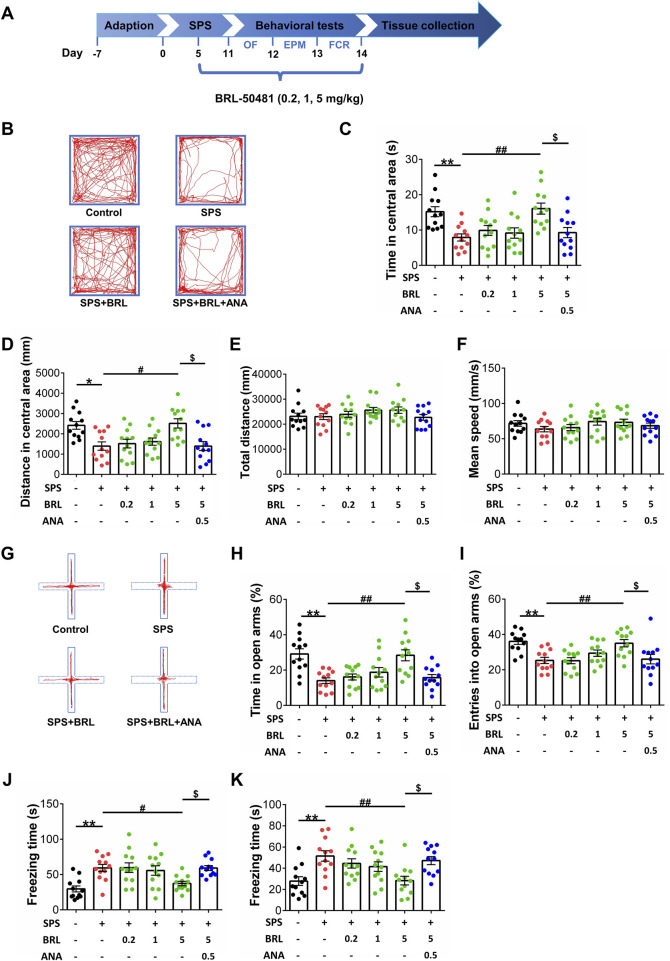
Methods: The single prolonged stress (SPS) was used to build a stress exposure model in C57BL/6 J mice and detected PDE7 activity in hippocampus, amygdala, prefrontal cortex and striatum. Next, three doses (0.2, 1, and 5 mg/kg) of the PDE7 inhibitor BRL-50481 were intraperitoneally administered for 10 days, then behavioral, biochemical, and morphological tests were conducted.

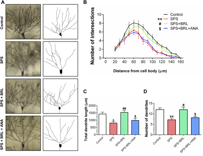
Results: PDE7 activity in hippocampus of mice significantly increased at all times after SPS. BRL-50481 significantly attenuated SPS induced anxiety-like behavior and fear response in both context and cue. In addition, BRL-50481 increased the levels of key molecules in the cAMP signaling pathway which were impaired by SPS. Immunofluorescent staining and Sholl analysis demonstrated that BRL-50481 also restored the nucleus/cytoplasm ratio of hippocampal neurons and improved neuronal plasticity. These effects of BRL-50481 were partially blocked by the TrkB inhibitor ANA-12.

Conclusion: PDE7 inhibitors attenuate stress-induced behavioral changes by protecting the neuron cytoarchitecture and the neuronal plasticity in hippocampus, which is mediated at least partly through the activation of BDNF/TrkB signaling pathway. These results proved that PDE7 is a potential target for treating stress-induced behavioral and physiological abnormalities.

Keywords: cAMP signaling; phosphodiesterase 7; single prolonged stress; stress exposure; synaptic plasticity.


Conflict of interest statement

The authors declare that the research was conducted in the absence of any commercial or financial relationships that could be construed as a potential conflict of interest. The author(s) declared that they were an editorial board member of Frontiers, at the time of submission. This had no impact on the peer review process and the final decision.

Figures

FIGURE 1
FIGURE 1
PDE7 activity in different brain regions of mice at 1, 7, and 14 days after SPS induction. (A) Experimental scheme for measuring cAMP by HPLC. (B) Relative activity of PDE7 in hippocampus, amygdala, prefrontal cortex and striatum of mice. Data are presented as mean ± SEM (n = 6 mice per group). * P < 0.05, ** P < 0.01, *** P < 0.001 vs. control.
FIGURE 2
FIGURE 2
PDE7 inhibitor ameliorated the anxiety-like behaviors in OFT and EPMT as well as the severe fear conditioning response in FCRT after SPS induction. (A) Experimental schedule for the modified single prolonged stress model and behavioral tests. Representative trajectories in OFT (B) and EPMT (G) of each group. Time (C) and distance (D) in the central area of OFT. Mean speed (E) and total distance (F) in OFT. Duration time (H) and entry times (I) on the open arms in EPMT. Freezing time in contextual (J) and cued (K) fear conditioning response. Data were presented as mean ± SEM (n = 12 mice per group). * P < 0.05, ** P < 0.01 vs. control; # P < 0.05, ## P < 0.01 vs. SPS; $ P < 0.05 vs. BRL 5-treated SPS.
FIGURE 3
FIGURE 3
PDE7 inhibitor ameliorated SPS induced BDNF and PSD95 downregulation as well as CREB and TrkB inactivation in hippocampus by increasing cAMP levels. (A) Representative blots of the molecular expression in hippocampal tissue of mice (B) cAMP level in the hippocampal tissue. Relative quantitative ratio of p-CREB/CREB (C) p-TrkB/TrkB (E) in the hippocampal tissue. Quantifications of BDNF (D) PSD95 (F) in the hippocampal tissue. Data are presented as mean ± SEM (n = 5 or 6 mice per group). * P < 0.05, ** P < 0.01 vs. control; # P < 0.05, ## P < 0.01 vs. SPS.
FIGURE 4
FIGURE 4
PDE7 inhibitor ameliorated the increased nucleus/cytoplasm ratio of neurons in hippocampal CA1 induced by SPS. (A) Representative images of the nucleus and cytoplasm of the hippocampal neurons after illustrated with anti-NeuN (green) and DAPI (blue) immunofluorescent staining. Number of hippocampal CA1 neurons (B), and nucleus/cytoplasm ratio of hippocampal CA1 neurons (C). Data were presented as mean ± SEM (n = 24 neurons/4 mice per group). ** P < 0.01 vs. control; ## P < 0.01 vs. SPS; $ P < 0.05 vs. SPS + BRL.
FIGURE 5
FIGURE 5
PDE7 inhibitor ameliorated the impairment of dendritic structure in hippocampal neurons. (A). Representative images of neuronal dendrites labeled with Golgi impregnation. (B). Sholl analysis of the hippocampal neurons revealed the alterations of basal dendritic intersections at distinct distances from soma. The total dendritic length (C), and dendritic numbers (D) of neurons. Data were presented as mean ± SEM (n = 36 neurons/6 mice per group). * P < 0.05, ** P < 0.01 vs. control; # P < 0.05, ## P < 0.01 vs. SPS; $ P < 0.05 vs. SPS + BRL.
FIGURE 6
FIGURE 6
Schematic representation of the mechanism of PDE7 inhibitor improving the behavioral and cytoarchitectural abnormality in SPS mice. PDE7 inhibition reduces severe fear conditioning responses and anxiety-like behavior in SPS mice, meanwhile repairing the neuron damage in hippocampus, and these improvement effects were achieved by the activation of BDNF/TrkB pathway.

References

    1. American Psychiatric Association (2013). Diagnostic and statistical manual of mental disorders. Fifth Edition. American Psychiatric Association. 10.1176/appi.books.9780890425596 - DOI
    1. Azevedo M. F., Faucz F. R., Bimpaki E., Horvath A., Levy I., de Alexandre R. B., et al. (2014). Clinical and molecular genetics of the phosphodiesterases (PDEs). Endocr. Rev. 35, 195–233. 10.1210/er.2013-1053 - DOI - PMC - PubMed
    1. Besnard A., Sahay A. (2016). Adult hippocampal neurogenesis, fear generalization, and stress. Neuropsychopharmacology 41, 24–44. 10.1038/npp.2015.167 - DOI - PMC - PubMed
    1. Blokland A., Heckman P., Vanmierlo T., Schreiber R., Paes D., Prickaerts J. (2019). Phosphodiesterase type 4 inhibition in CNS diseases. Trends Pharmacol. Sci. 40, 971–985. 10.1016/j.tips.2019.10.006 - DOI - PubMed
    1. Bollen E., Prickaerts J. (2012). Phosphodiesterases in neurodegenerative disorders. IUBMB Life 64, 965–970. 10.1002/iub.1104 - DOI - PubMed

LinkOut - more resources