Brown Tumor of the Dorsal Spine With Hemorrhage Causing Acute Neurological Deterioration: A Rare Presentation of Secondary Hyperparathyroidism
- PMID: 39092321
- PMCID: PMC11292460
- DOI: 10.7759/cureus.63645
Brown Tumor of the Dorsal Spine With Hemorrhage Causing Acute Neurological Deterioration: A Rare Presentation of Secondary Hyperparathyroidism
Abstract
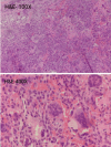
Brown tumor due to secondary hyperparathyroidism in chronic kidney disease is a well-established entity. Brown tumor of the spine with hemorrhage causing acute neurological deficit is a rare entity. A 35-year-old gentleman, with chronic kidney disease (CKD) on dialysis, presented with acute paraplegia and loss of lower limb sensation and bowel and bladder control. Imaging revealed a T8 vertebral body expansile lytic lesion with collapse, exaggerated kyphosis, and cord compression. He underwent an emergency decompressive laminectomy and transpedicular corpectomy of T8, with posterior stabilization. Histopathology revealed lobular clusters of osteoclast-like multinucleated giant cells with background of which was possibly the reason for acute neurological deterioration in this case. Brown tumors of the spine can mimic lytic lesions of the spine like myeloma and metastasis. Suspicion must be raised given in the setting of CKD and hyperparathyroidism. They can present with hemorrhage and acute neurological deficit, which warrants urgent surgical intervention for optimal outcomes.
Keywords: brown tumor; chronic kidney disease; hemorrhage; secondary hyperparathyroidism; spine.
Copyright © 2024, Srinivasan et al.
Conflict of interest statement
Human subjects: Consent was obtained or waived by all participants in this study. Conflicts of interest: In compliance with the ICMJE uniform disclosure form, all authors declare the following: Payment/services info: All authors have declared that no financial support was received from any organization for the submitted work. Financial relationships: All authors have declared that they have no financial relationships at present or within the previous three years with any organizations that might have an interest in the submitted work. Other relationships: All authors have declared that there are no other relationships or activities that could appear to have influenced the submitted work.
Figures


References
-
- Management of brown tumor of spine with primary hyperparathyroidism. A case report and literature review. Hu J, He S, Yang J, Ye C, Yang X, Xiao J. https://www.ncbi.nlm.nih.gov/pmc/articles/PMC6456137/ Medicine (Baltimore) 2019;98:0. - PMC - PubMed
-
- Spinal compression by brown tumor in two patients with chronic kidney allograft failure on maintenance hemodialysis. Gheith O, Ammar H, Akl A, et al. https://pubmed.ncbi.nlm.nih.gov/20622318/ Iran J Kidney Dis. 2010;4:256–259. - PubMed
-
- Brown tumor of the thoracic spine: first manifestation of primary hyperparathyroidism. Sonmez E, Tezcaner T, Coven I, Terzi A. https://www.ijkd.org/index.php/ijkd/article/view/210/210. J Korean Neurosurg Soc. 2015;58:389–392. - PMC - PubMed
-
- Spinal cord compression secondary to brown tumour in a patient on long-term haemodialysis: a case report. Mak KC, Wong YW, Luk KD. J Orthop Surg (Hong Kong) 2009;17:90–95. - PubMed
-
- Primary hyperparathyroidism presenting as spinal cord compression. Shaw MT, Davies M. https://www.ncbi.nlm.nih.gov/pmc/articles/PMC1912178/pdf/brmedj02107-005.... Br Med J. 1968;4:230–231. - PMC - PubMed
Publication types
LinkOut - more resources
Full Text Sources
