. 2024 Sep 5;187(18):4877-4889.e15.
doi: 10.1016/j.cell.2024.07.013. Epub 2024 Aug 1.

Activation of the helper NRC4 immune receptor forms a hexameric resistosome

Affiliations

Activation of the helper NRC4 immune receptor forms a hexameric resistosome

Furong Liu et al. Cell. .

Abstract

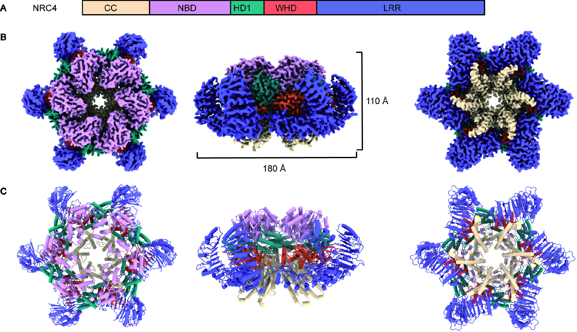
Innate immune responses to microbial pathogens are regulated by intracellular receptors known as nucleotide-binding leucine-rich repeat receptors (NLRs) in both the plant and animal kingdoms. Across plant innate immune systems, "helper" NLRs (hNLRs) work in coordination with "sensor" NLRs (sNLRs) to modulate disease resistance signaling pathways. Activation mechanisms of hNLRs based on structures are unknown. Our research reveals that the hNLR, known as NLR required for cell death 4 (NRC4), assembles into a hexameric resistosome upon activation by the sNLR Bs2 and the pathogenic effector AvrBs2. This conformational change triggers immune responses by facilitating the influx of calcium ions (Ca2+) into the cytosol. The activation mimic alleles of NRC2, NRC3, or NRC4 alone did not induce Ca2+ influx and cell death in animal cells, suggesting that unknown plant-specific factors regulate NRCs' activation in plants. These findings significantly advance our understanding of the regulatory mechanisms governing plant immune responses.

Keywords: ETI; NLR proteins; NRC0; NRC2; NRC3; NRC4 resistosome; PTI; calcium influx; pathogen recognition; plant immunity.


Conflict of interest statement

Declaration of interests B.J.S. is a scientific co-founder of and serves on the board of directors for Mendel Biotechnology. He also serves on the scientific advisory boards of Verinomics and the Sainsbury Laboratory.

Figures

Figure 1.
Figure 1.. Structure of the NRC4 resistosome.
(A) Domain organization of NRC4. The same color code for the domains is used throughout this study, unless specified otherwise. (B) Cryo-EM density map of the hexameric NRC4 structure. (C) The corresponding refined structure model (top, side and bottom views are shown from left to right). Six identical NRC4 protomers are arranged in a wheel-like structure measuring ~180 Å in diameter and 100 Å in height. See also Figure S1, S2, S3 and Table 1.
Figure 2.
Figure 2.. Interfaces in the oligomerization of the NRC4 resistosome.
(A) View of two adjacent protomers in the NRC4 resistosome. Boxes indicate regions of interaction between them, shown in detail in panels B-F. (B-F) Structural details of CC-CC, NBD-HD, WHD-HD1 and LRR-LRR interactions, respectively. Residue labels in both gray and white correspond to two adjacent protomers. (F) ATP is situated in the cleft between the NBD and HD1 domain, interacting exclusively with the NBD despite its proximity to both domains. (G) Hypersensitive response phenotypes of N. benthamiana leaves upon the expression of mutant NRC4 proteins based on the interfaces shown in (B-F). A representative figure from multiple replicates is shown for each case. (H) Protein expression levels of the wild-type NRC4 and the tested mutants in the N. benthamiana leaves were evaluated using SDS-PAGE and subsequent immunoblotting with α-StrepTag II antibody. An InstantBlue® Coomassie-stained gel was used as a loading control (IB, InstantBlue). See also Figure S4.
Figure 3.
Figure 3.. CC-LRR interactions within an NRC4 protomer.
(A) Structure of one NRC4 protomer, with the CC-LRR interaction region indicated by a light blue dashed box. (B) Structural details of the CC-LRR interactions. Three negatively charged residues (E73, D74, and D77) from the CC domain form salt bridges and hydrogen bond interactions with two positively charged residues (R514 and R537) from the LRR domain. (C) Electrostatic surface view of the LRR domain. The position of the conserved R-cluster is indicated by a dashed box. (D) Structure-based sequence alignment encompassing the EDVID motif and R-cluster of ZAR1, Sr35, NRC2, NRC3 and NRC4. Key residues from (B) are highlighted by a solid box, while the additional arginines involved in the CC-LRR interaction of the Sr35 resistosome are highlighted by a dashed box. (E) Hypersensitive response phenotypes of N. benthamiana leaves upon the expression of NRC4 with mutations in the residues from (B). In each case, a representative figure is shown from multiple replicates. (F) Protein expression levels of the wild-type NRC4 and the tested mutants in the N. benthamiana leaves were evaluated using SDS-PAGE and subsequent immunoblotting with α-StrepTag II antibody. An InstantBlue® Coomassie-stained gel was used as a loading control.
Figure 4.
Figure 4.. [Ca2+]cyt dynamics upon the expression of NRC4 variants and indicated treatments.
(A) Time course of [Ca2+]cyt dynamics after infiltration of N. benthamiana leaves expressing the [Ca2+]cyt reporter GCaMP3 with Agrobacterium strains carrying the indicated constructs. NRC4 DV and NRC4 L9E denote NRC4 variants with amino acid substitutions D478V and L9E, respectively. NRC4 L9EDV indicates an NRC4 variant with both mutations. Leaf disc fluorescent (F) intensities of GCaMP3, as indicative of relative [Ca2+]cyt levels, are plotted over a tested time. The activation mimic NRC4 DV, unlike other variants, exhibited a robust increase in [Ca2+]cyt. (B) Relative [Ca2+]cyt levels at indicated time as shown in (A). (C) Time course of [Ca2+]cyt dynamics after expressing indicated constructs. NRG1.1 DV denotes NRG1.1 variant with amino acid substitution D485V, and NRC3 DV denote variant with amino acid substitution D480V. (D) Relative [Ca2+]cyt levels at indicated time (NRC4 DV at 11h, NRG1.1 DV at 13h and NRC3 DV at 16 h) as shown in (C). (E) Time course of [Ca2+]cyt dynamics upon the expression of NRC4 variants co-infiltrated with a PM calcium channel blocker, LaCl3 (2 mM). (F) Relative [Ca2+]cyt levels at indicated times as shown in (E). (G) Time course of [Ca2+]cyt dynamics upon the expression of NRC4 DV co-infiltrated with K+-channel blockers, TEACl (1mM) and CsCl (1mM), an intracellular Ca2+ release blocker RR (1mM), or LaCl3 (1mM). (H) Time required to reach peaks for the relative [Ca2+]cyt levels as shown in (G). (I) Time course of [Ca2+]cyt dynamics, showing additive effects of extracellular Ca2+ (10 mM) on NRC4 DV-mediated Ca2+ influx. (J) Relative [Ca2+]cyt levels at indicated time as shown in (I). Error bars in panels A to F represent standard error (n=6 to 8 discs from 3 independent plants). Experiments were repeated twice with similar phenotypes observed. One way ANOVA comparison among groups for B, D, F, J; or between control with other groups (H). See also Figure S5.
Figure 5.
Figure 5.. Structural analysis of ion permeation in the NRC4 resistosome.
(A) The ion permeation path, calculated by HOLE, for the active NRC4 resistosome is illustrated by purple dots. For clarity, only two protomers positioned diagonally opposite are displayed. (B) A comparison of the corresponding pore radius of NRC4 (blue), Sr35 (green), and ZAR1 (magenta) resistosomes is presented, with the constriction site set as zero. (C) A cross-sectional view of the ion-conducting pore within the NRC4 resistosome, shown as an electrostatic surface.
Figure 6.
Figure 6.. The NRC4 resistosome converges on Ca2+ influx in plant immunity.
The figure illustrates the working model for NRC4 activation. NRC4 initially exists as a homodimer in its resting state. Upon activation by the upstream sNLR Bs2 and the pathogenic effector AvrBs2, NRC4 converts into a hexameric resistosome. The formation of this hexameric resistosome leads to Ca2+ influx in plant immune responses, probably involving other plant-specific factor(s). This mechanism is distinct from previously identified resistosomes such as ZAR1, Sr35 and NRG1, which form calcium channels allowing Ca2+ influx. See also Figure S6.

Update of

References

    1. Ngou BPM, Ding P, and Jones JDG (2022). Thirty years of resistance: Zig-zag through the plant immune system. The Plant cell 34, 1447–1478. 10.1093/plcell/koac041. - DOI - PMC - PubMed
    1. Jones JD, Vance RE, and Dangl JL (2016). Intracellular innate immune surveillance devices in plants and animals. Science 354. 10.1126/science.aaf6395. - DOI - PubMed
    1. Locci F, and Parker JE (2024). Plant NLR immunity activation and execution: a biochemical perspective. Open Biol 14, 230387. 10.1098/rsob.230387. - DOI - PMC - PubMed
    1. Jones JDG, Staskawicz BJ, and Dangl JL (2024). The plant immune system: From discovery to deployment. Cell 187, 2095–2116. 10.1016/j.cell.2024.03.045. - DOI - PubMed
    1. Yuan M, Jiang Z, Bi G, Nomura K, Liu M, Wang Y, Cai B, Zhou JM, He SY, and Xin XF (2021). Pattern-recognition receptors are required for NLR-mediated plant immunity. Nature 592, 105–109. 10.1038/s41586-021-03316-6. - DOI - PMC - PubMed