. 2024 Sep 5;187(18):5010-5028.e24.
doi: 10.1016/j.cell.2024.07.006. Epub 2024 Aug 1.

A replisome-associated histone H3-H4 chaperone required for epigenetic inheritance

Affiliations

A replisome-associated histone H3-H4 chaperone required for epigenetic inheritance

Juntao Yu et al. Cell. .

Abstract

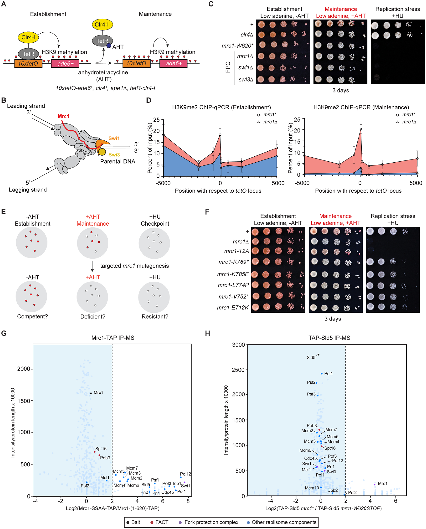
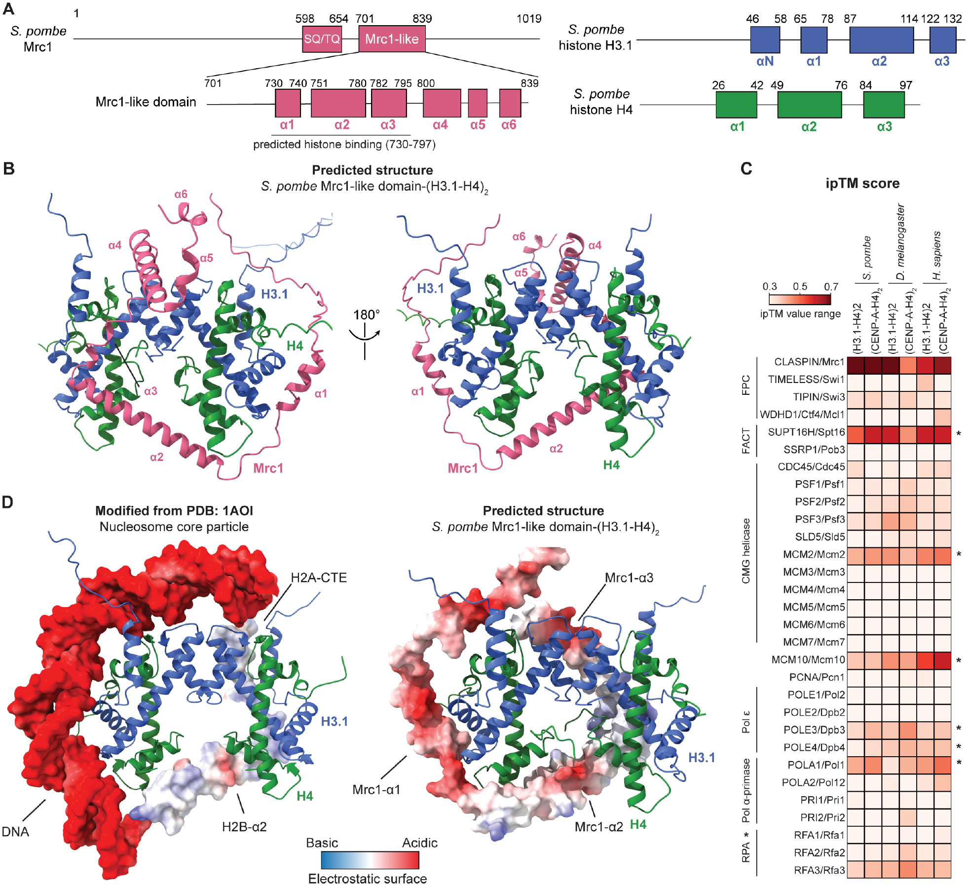
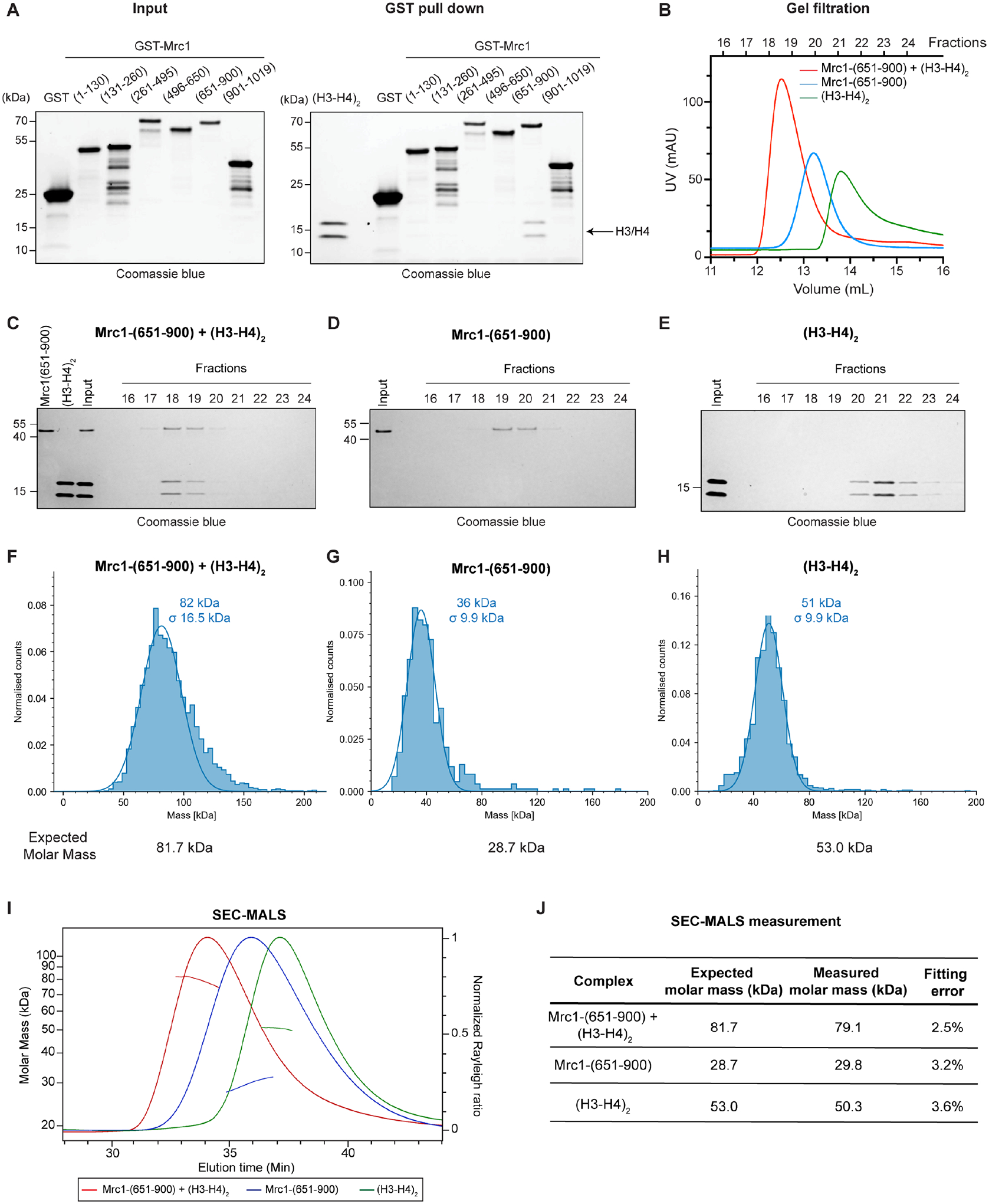
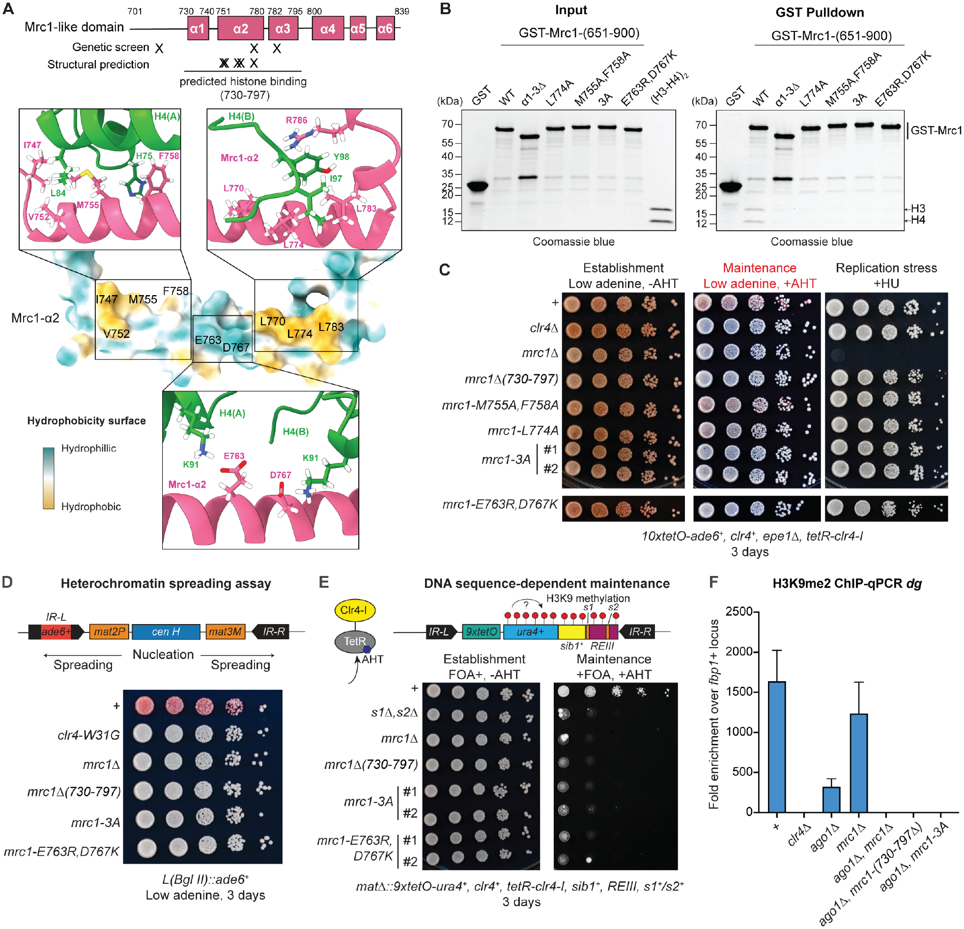
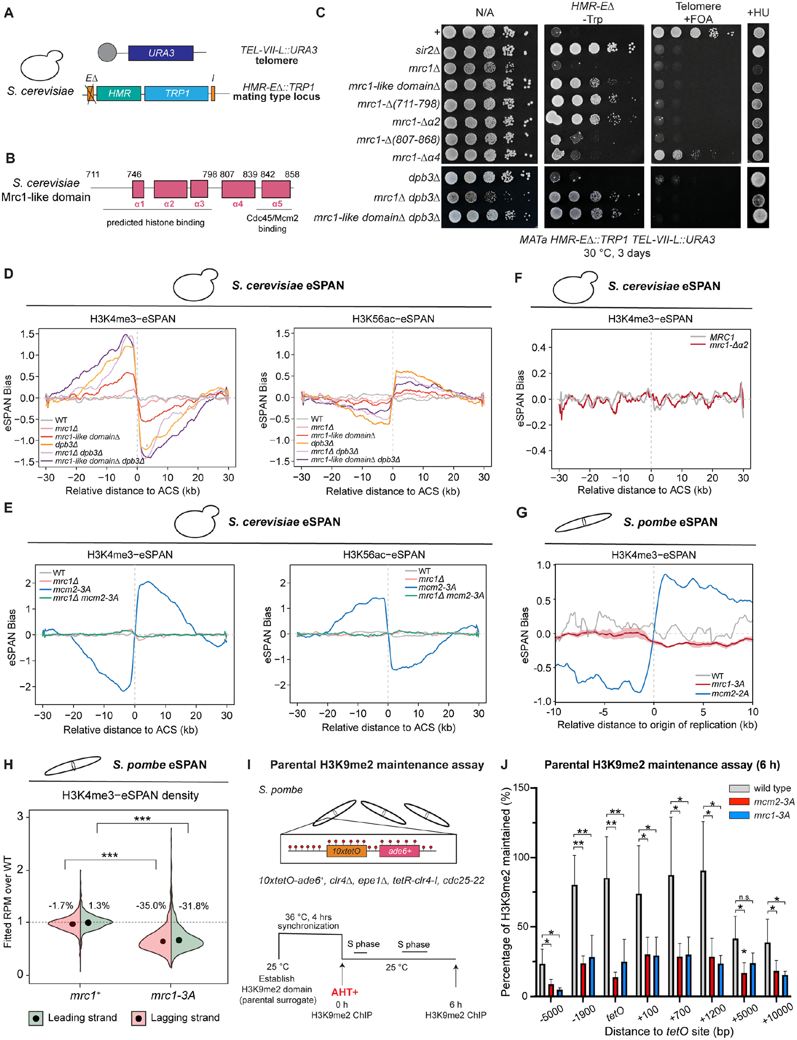
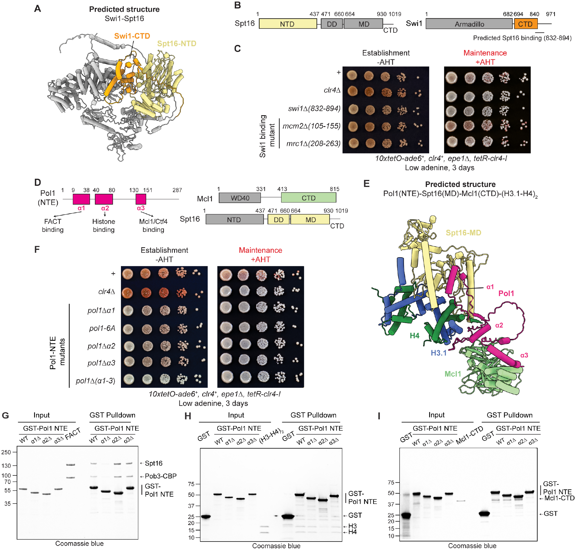
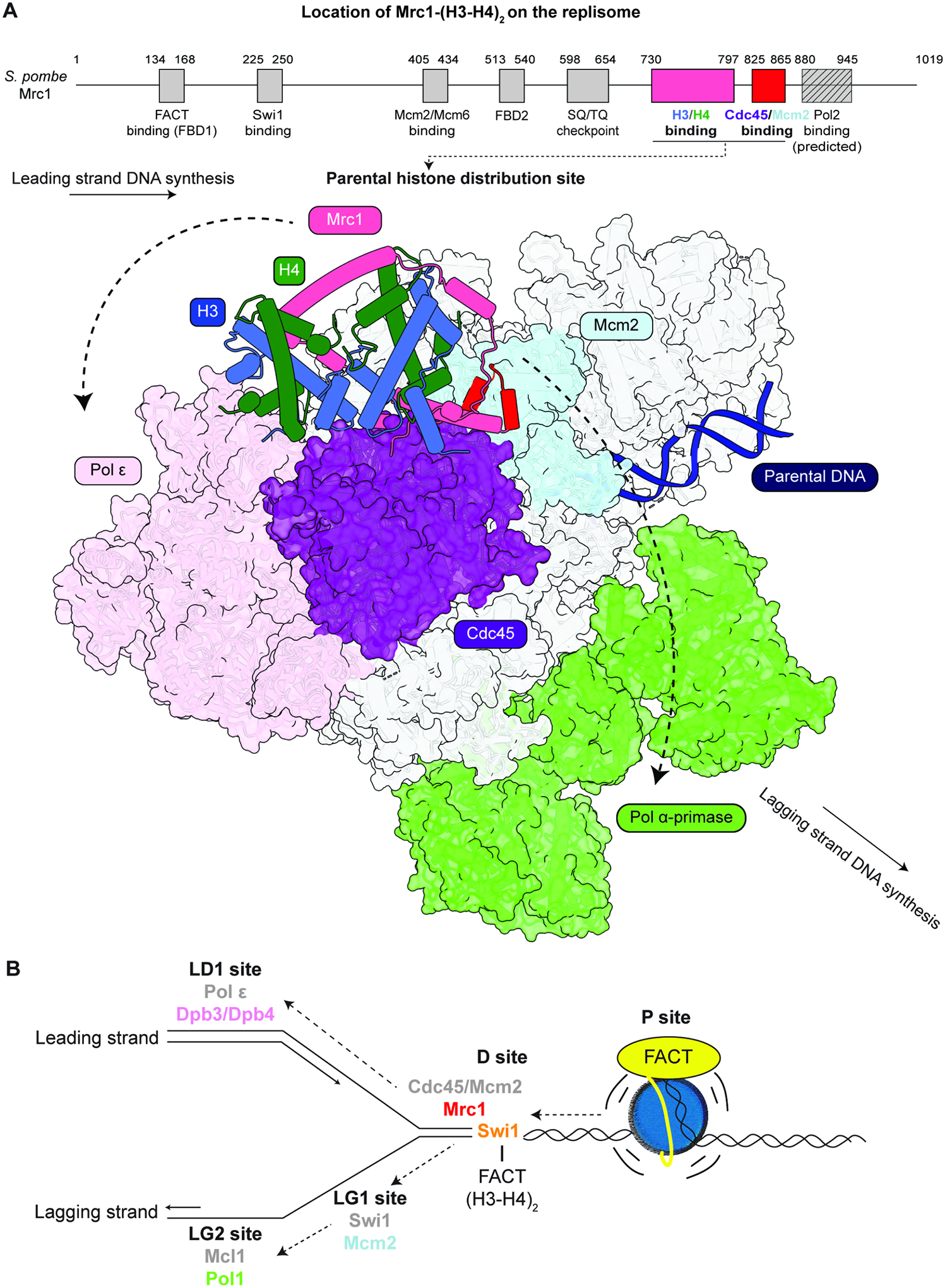
Faithful transfer of parental histones to newly replicated daughter DNA strands is critical for inheritance of epigenetic states. Although replication proteins that facilitate parental histone transfer have been identified, how intact histone H3-H4 tetramers travel from the front to the back of the replication fork remains unknown. Here, we use AlphaFold-Multimer structural predictions combined with biochemical and genetic approaches to identify the Mrc1/CLASPIN subunit of the replisome as a histone chaperone. Mrc1 contains a conserved histone-binding domain that forms a brace around the H3-H4 tetramer mimicking nucleosomal DNA and H2A-H2B histones, is required for heterochromatin inheritance, and promotes parental histone recycling during replication. We further identify binding sites for the FACT histone chaperone in Swi1/TIMELESS and DNA polymerase α that are required for heterochromatin inheritance. We propose that Mrc1, in concert with FACT acting as a mobile co-chaperone, coordinates the distribution of parental histones to newly replicated DNA.

Keywords: CLASPIN; FACT; chromatin replication; epigenetics; fork protection complex; heterochromatin; histone inheritance; parental histone transfer; replisome, Mrc1.


Conflict of interest statement

Declaration of interests The authors declare no competing interests.

Figures

Figure 1.
Figure 1.. The full fork protection complex is required for heterochromatin maintenance.
A) Diagram showing the inducible ectopic heterochromatin system. B) Diagram highlighting the location of the fork protection complex subunits (Swi1, Swi3, Mrc1) on the replisome. C) Heterochromatin maintenance assay testing the roles of subunits of the fork protection complex in epigenetic inheritance. Ten-fold serial dilutions of cells were plated on the indicated growth medium to detect heterochromatin establishment (AHT−) and maintenance (AHT+). Loss of growth on medium containing hydroxyurea (HU+) indicates deficiency in replication checkpoint. * denotes a stop codon. D) H3K9me2 ChIP-qPCR at the 10XtetO-ade6+ locus showing that the H3K9me2 levels in mrc1+ or mrc1Δ cells at the establishment phase (AHT−) and the maintenance phase 24 hours after growth in the presence of AHT. E) Diagram illustrating the gene-targeted random mutagenesis of mrc1+ to isolate mutant cells that are competent for heterochromatin establishment and replication checkpoint but fail to maintain heterochromatin. F) Separation-of-function alleles isolated from the random mutagenesis of mrc1+ that abolish heterochromatin maintenance but not replication checkpoint. G) IP-MS analysis of TAP-tagged heterochromatin maintenance-competent Mrc1-SSAA and mutant Mrc1-(1–620). H) IP-MS of TAP-Sld5 in mrc1+ and mrc1-W620STOP cells. G, H) X-axis, the log2 fold change between wild type and mutant epitope tagged proteins; Y-axis, normalized intensity of proteins associated with the indicated tagged proteins detected by mass spectrometry.
Figure 2.
Figure 2.. AlphaFold-Multimer predictions suggest an interaction interface between the S. pombe Mrc1-like domain and (H3.1-H4)2.
A) The location of the conserved S. pombe Mrc1-like domain and secondary structure features of the Mrc1-like domain predicted by AlphaFold-Multimer. The predicted histone binding domain (amino acid 730 to 797) located within the Mrc1-like domain is indicated at the bottom (left). The structural domains of S. pombe histone H3.1 and H4 (right). B) The front (left) and back (right) views of the predicted structure of S. pombe Mrc1-like domain-(H3.1-H4)2. Mrc1-like domain is colored in pink, and histone H3.1, H4 are colored as blue and green, respectively. C) Heatmap showing the average interface predicted template modeling (ipTM) score of all five predicted models between S. pombe, D. melanogaster and H. sapiens (H3.1-H4)2 or centromere variant (CENP-A-H4)2 (X-axis) against each core replisome component (Y-axis). The ipTM score and the heatmap scale range from 0.3 to 0.7. Asterisk denotes known histone chaperones. D) Comparison of the crystal structure of the nucleosome core particle (PDB: 1AOI) (left) and the predicted structure of Mrc1-like domain-(H3.1-H4)2 (right).
Figure 3.
Figure 3.. S. pombe Mrc1-like domain contains an (H3-H4)2 binding domain
A) In vitro pulldown assays with GST-Mrc1 fragments immobilized on glutathione magnetic beads and (H3-H4)2. B) Chromatogram of purified Mrc1-(651–900), (H3-H4)2, and reconstituted Mrc1-(651–900)/(H3-H4)2 complex on a Superdex 200 increase 10/300 GL gel filtration column. C-E) SDS-PAGE analysis of peak fractions from the gel filtration column showing comigration of Mrc1(651–900) with H3-H4(C), migration of Mrc1-(651–900)(D), and migration of H3-H4(E). F-H) Mass photometry analysis of the measured molecular mass of purified Mrc1-(651–900)-(H3-H4)2 complex(F), Mrc1-(651–900)(G), and (H3-H4)2(H). The measurement of Mrc1-(651–900) is higher than the expected molecular weight, which may be due to the detection limit of 30 kDa for mass photometry. I) SEC-MALS profiles of purified Mrc1-(651–900)-(H3-H4)2 complex, Mrc1-(651–900), and (H3-H4)2. J) Summary of the expected molecular mass and SEC-MALS measured molar mass of purified Mrc1-(651–900)-(H3-H4)2 complex, Mrc1-(651–900), and (H3-H4)2.
Figure 4.
Figure 4.. Mrc1 histone binding activity is required for heterochromatin maintenance in S. pombe.
A) Energy minimized AlphaFold-predicted interaction between Mrc1-α2 and histone H4s. Top, diagram showing the location of Mrc1-α2 and the Mrc1-histone binding domain. Bottom, hydrophobic map of the Mrc1-α2 and detailed predicted interactions between Mrc1-α2 and histone H4. B) In vitro GST pulldown assays showing the effect of hydrophobic (Mrc1-M755A, F758A, L774A) and electrostatic (Mrc1-E763R, D767K) mutations in Mrc1-α2 on histone H3-H4 binding. C) Heterochromatin maintenance assay showing the phenotypes of hydrophobic and electrostatic mutations in mrc1-α2. D) Top, diagram showing the ade6+ reporter gene inserted at the boundary of the mating type locus IR-L (L(BglII)::ade6+). Bottom, silencing assays showing phenotypes of cell carrying Mrc1-histone binding domain mutations in silencing of the ade6+ reporter. E) Top, diagram showing the DNA sequence-dependent heterochromatin maintenance reporter system in S. pombe. Bottom, spotting assay showing the maintenance phenotype of the ura4+ report gene in wildtype cells and cells carrying the indicated mutations. As a control, cells with deletions of Atf1/Pcr1 binding sites (s1Δ,s2Δ) are unable to maintain heterochromatin. F) H3K9me2 ChIP-qPCR analysis of mrc1 mutations in combination of ago1Δ at pericentromere dg repeats. N=3, error bars indicate standard deviations.
Figure 5.
Figure 5.. The histone binding domain of Mrc1 promotes parental histone transfer without affecting transfer symmetry.
A) Diagram illustrating the dual gene silencing reporter systems in S. cerevisiae. B) Diagram of the predicted histone binding domain and Mcm2/Cdc45 interaction region, PDB: 8B9C and AlphaFold prediction (more details are presented in Figure S7J–P), in the Mrc1-like domain of S. cerevisiae Mrc1. C) Growth assays showing the effects of the indicated mutations on silencing and replication stress. D) eSPAN bias of the parental histone surrogate H3K4me3 (left panel) and the new histone surrogate H3K56ac (right panel) distribution around 139 early replicating origins (ACSs) in wild-type (WT), mrc1Δ, mrc1-like domainΔ, dpb3Δ, dpb3Δ mrc1Δ, and dpb3Δ mrc1-like domainΔ S. cerevisiae cells. E) eSPAN bias of parental histones surrogate H3K4me3 (left panel) and the new histone surrogate H3K56ac (right panel) around 139 early ACSs in wild-type (WT), mrc1Δ, mcm2-3A, and mrc1Δ mcm2-3A S. cerevisiae cells. F) eSPAN bias of the parental histone H3K4me3 distribution in MRC1, mrc1-α2Δ S. cerevisiae cells. G) eSPAN bias of parental histone surrogate H3K4me3 distribution around 162 origin of replication in wild-type (WT), mrc1-3A, mcm2-2A S. pombe cells. The shading of the bias line plot is the 95% confidence interval of mean value of at least two biological replicates, which is mean ± 2 folds of the standard error. H) Violin plot showing the average of two biological replicates of S. pombe eSPAN H3K4me3 density on the leading and lagging strand around the replication origin (2.5 kb upstream of replication origin to 2.5 kb downstream of replication origin). The numbers in the figure represent changes of eSPAN density over wild type cells for each strand. *** indicates p-value < 0.001 (two-sample t-test). I) Diagram illustrating a parental H3K9me2 maintenance assay. Top, diagram of the S. pombe reporter system that lacks read-write activity. Bottom, diagram of the designed assay to analyze the maintenance of H3K9me2 in a synchronized cell population after 6 hours after release from cell cycle arrest. J) ChIP-qPCR of parental H3K9me2 in wild-type (WT), mcm2-2A, mrc1-3A cells 6 hours after release from cell cycle arrest. A two-tailed two-sample t-test with unequal variance was used for statistical significant test between wild-type and mutant samples. N=5. *, p-value < 0.05, **, p-value < 0.01, n.s., not significant (p=0.068).
Figure 6.
Figure 6.. Identification of FACT binding sites on the replisome required for heterochromatin maintenance.
A) Predicted structure of Swi1 and FACT subunit Spt16. B) The predicted interacting domains of Spt16 and Swi1 in A are highlighted in yellow and orange, respectively. C) Heterochromatin maintenance assay showing the effects of swi1, mrc1, mcm2 mutations. D) Diagram of regions in the N-terminal extension (NTE) of Pol1 predicted to interact with Spt16, (H3.1-H4)2, and the Mcl1 C-terminal domain (CTD). The predicted interacting domains of Spt16 and Mcl1 in F are highlighted in green and yellow, respectively. E) Predicted structure of Pol1-NTE (α1, α2, and α3) with Spt16-middle domain (MD), (H3.1-H4)2 and Mcl1-CTD). F) Heterochromatin maintenance assay showing the effect of the indicated pol1 mutations. G-I) In vitro GST pull down assays showing the interaction of the indicated GST-Pol1-NTE proteins with purified FACT complex (G), (H3-H4)2 (H), and Mcl1-(CTD)(I).
Figure 7.
Figure 7.. Mrc1 acts as a parental histone distribution site.
A) The predicted location of Mrc1-(H3-H4)2 on the cryo-EM structure of the replisome (PDB: 8B9C and 7QHS). Top, diagram showing indicated regions in the Mrc1 involved in interaction with multiple replisome components, replication checkpoint signaling, and interaction with histones. The predicted Pol2 interacting region was identified by AlphaFold-Multimer and is consistent with previous biochemical results. The newly identified histone binding region is highlighted in pink and the Cdc45/Mcm2(NTD) interacting region is highlighted in red. Bottom, the predicted structure of Mrc1-like domain/(H3-H4)2/Cdc45/Mcm2(NTD) was aligned to the cryo-EM structure (PDB: 8B9C) via the Mrc1-like domain α5 helix. See Figure S7K–P for alignment details. B) Model for DNA replication-coupled directional parental histone transfer with FACT acting as a mobile chaperone. P, Parental site; D, Distribution site; LD1, leading strand site 1; LG1 and LG2, lagging strand sites. See text for details.

References

    1. Margueron R, and Reinberg D (2010). Chromatin structure and the inheritance of epigenetic information. Nat Rev Genet 11, 285–296. nrg2752 [pii] 10.1038/nrg2752. - DOI - PMC - PubMed
    1. Moazed D (2011). Mechanisms for the inheritance of chromatin states. Cell 146, 510–518. 10.1016/j.cell.2011.07.013. - DOI - PMC - PubMed
    1. Allshire RC, and Madhani HD (2018). Ten principles of heterochromatin formation and function. Nat Rev Mol Cell Biol 19, 229–244. 10.1038/nrm.2017.119. - DOI - PMC - PubMed
    1. Serra-Cardona A, and Zhang Z (2018). Replication-Coupled Nucleosome Assembly in the Passage of Epigenetic Information and Cell Identity. Trends Biochem Sci 43, 136–148. 10.1016/j.tibs.2017.12.003. - DOI - PMC - PubMed
    1. Stewart-Morgan KR, Petryk N, and Groth A (2020). Chromatin replication and epigenetic cell memory. Nat Cell Biol 22, 361–371. 10.1038/s41556-020-0487-y. - DOI - PubMed

MeSH terms