. 2024 Aug 20;5(8):101668.
doi: 10.1016/j.xcrm.2024.101668. Epub 2024 Aug 1.

Hybrid immunity to SARS-CoV-2 arises from serological recall of IgG antibodies distinctly imprinted by infection or vaccination

Affiliations

Hybrid immunity to SARS-CoV-2 arises from serological recall of IgG antibodies distinctly imprinted by infection or vaccination

William N Voss et al. Cell Rep Med. .

Abstract

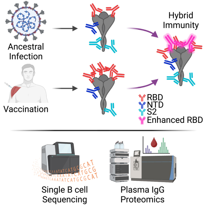
We describe the molecular-level composition of polyclonal immunoglobulin G (IgG) anti-spike antibodies from ancestral severe acute respiratory syndrome coronavirus 2 (SARS-CoV-2) infection, vaccination, or their combination ("hybrid immunity") at monoclonal resolution. Infection primarily triggers S2/N-terminal domain (NTD)-reactive antibodies, whereas vaccination mainly induces anti-receptor-binding domain (RBD) antibodies. This imprint persists after secondary exposures wherein >60% of ensuing hybrid immunity derives from the original IgG pool. Monoclonal constituents of the original IgG pool can increase breadth, affinity, and prevalence upon secondary exposures, as exemplified by the plasma antibody SC27. Following a breakthrough infection, vaccine-induced SC27 gained neutralization breadth and potency against SARS-CoV-2 variants and zoonotic viruses (half-maximal inhibitory concentration [IC50] ∼0.1-1.75 nM) and increased its binding affinity to the protective RBD class 1/4 epitope (dissociation constant [KD] < 5 pM). According to polyclonal escape analysis, SC27-like binding patterns are common in SARS-CoV-2 hybrid immunity. Our findings provide a detailed molecular definition of immunological imprinting and show that vaccination can produce class 1/4 (SC27-like) IgG antibodies circulating in the blood.

Keywords: COVID-19; Ig-seq; SARS-CoV-2; antibody feedback; bNAb; broadly neutralizing monoclonal antibody; cryo-EM; hybrid immunity; immunological imprinting; plasma.


Conflict of interest statement

Declaration of interests R.S.B. is a member of advisory boards for Vaxart, Takeda, and Invivyd and has collaborative projects with Gilead, J&J, and HilleVax, focused on unrelated projects. S.R.L. and R.S.B. are co-inventors of methods and uses of mouse-adapted SARS-CoV-2 viruses (US patent US11225508B1). J.D.B. is on the scientific advisory boards of Apriori Bio, Invivyd, Aerium Therapeutics, and the Vaccine Company. J.D.B. and B.D. are inventors on Fred Hutchinson Cancer Center-licensed patents related to viral deep mutational scanning. R.S.B., G.G., W.N.V., J.J.L., and G.C.I. are inventors on a provisional US patent application for mAb SC27 and other new antibodies described in this manuscript, entitled “Broadly neutralizing human monoclonal antibodies that target the SARS-CoV-2 receptor binding domain (RBD)” (63/491,270).

Figures

None
Graphical abstract
Figure 1
Figure 1
IgG serological recall and hybrid immunity are predetermined by the initial immunological imprint set by infection or vaccination (A) Plasma RBD competition ELISA reveals that mode and order of exposure to SARS-CoV-2 S result in differential persistent antibody orientation, with infection and vaccination imprinting non-RBD and RBD epitopes, respectively. Results are based on two technical replicates. (B) Comparison of post-infection and post-vaccination anti-S plasma IgG repertoire diversity (D80). D80, diversity index 80%: the number of lineages that comprise 80% of the S-reactive plasma IgG repertoire by abundance. Error bars represent 95% CI about the median. (C) Donor P3 plasma IgG repertoire elicited by primary infection (black bars), recalled by subsequent vaccination (light blue bars), and newly elicited by subsequent vaccination (dark blue bars). Each bar represents an individual plasma IgG lineage. Antibody symbols above bars indicate S domain specificity of recombinantly cloned mAbs representative of each lineage. The insert shows anti-S plasma binding titers at each time point, and the UpSet plot below the repertoire bar plot indicates whether the plasma lineage was detected (filled circle) in total B cells, sorted MBCs, and sorted PBs. “Post-V1,” following the first vaccine dose and “Post-V2,” following the second vaccine dose. (D) Donor P25 plasma IgG repertoire elicited by naive vaccination (gray bars), recalled by subsequent BT infection (pink bars), and newly elicited by subsequent vaccination (red bars). Insert and UpSet plot as described in C. (E) Hybrid immunity: proportion of plasma IgG anti-S lineages following secondary exposure, across the cohort, which are recalled from lineages originally imprinted by the initial exposure. The horizontal line between the plots denotes each quartile of the plasma IgG repertoire by relative abundance. Plasma IgG limit of quantitation = 15 ng/mL. Significant differences calculated using the Mann-Whitney U test linked by horizontal lines are indicated by asterisks: ∗∗p < 0.01, ∗∗∗p < 0.001.
Figure 2
Figure 2
Molecular IgG responses: Differential immunological imprinting by infection (S2/NTD) and vaccination (RBD) (A) Representative mAbs from infection-elicited lineages mostly bind non-RBD epitopes (top) whereas those from vaccine-elicited lineages tend to bind RBD epitopes (bottom). (B) Mean VH somatic hypermutation rates are highest in S2-directed lineages, followed by RBD- and then NTD-directed lineages. (C) VH sequences of recalled plasma IgG lineages are significantly more mutated after the second S exposure. (D) Heatmap displaying the origin, S domain specificity, RBD class (when applicable), relative abundance after infection and/or vaccination, binding to Wuhan-Hu-1 and Omicron BA.1, and in vitro neutralization capacity of the representative mAbs cloned and characterized from the donors in this study, each run in duplicate. “Origin” column indicates the time point at which the lineage was first detected (BCR-seq or Ig-seq). Asterisks indicate significant differences calculated using the Mann-Whitney U test: ∗∗p < 0.01, ∗∗∗p < 0.001, ∗∗∗∗p < 0.0001.
Figure 3
Figure 3
Identification of the broad, potent, and protective plasma mAb SC27 (A) Compared with naive vaccination, BT infection elicits a larger fraction of RBD-directed antibodies endowed with greater neutralization potency (Wuhan D614G, left) and breadth (Omicron BA.1, right). (B) Live-virus neutralization assays using RBD-directed plasma mAbs screened against a wide panel of SARS-CoV-2 VOCs as well as zoonotic sarbecoviruses. (C) ELISA binding data for recombinant RBD proteins showing SC27 cross-reactivity across a broad panel of SARS-CoV-2 VOCs (left) and other sarbecoviruses (right). Error bars represent SEM about the mean. (D–G) In vivo prophylactic protection of 12-month-old BALB/c mice against standard intranasal infection challenge dose (103 plaque-forming unit [PFU]) of mouse-adapted (MA10) SARS-CoV-2 (D614G or XBB.1.5). Error bars represent SEM about the mean. (H) Biolayer interferometry sensorgram demonstrating complete inhibition of SARS-CoV-2 Wuhan-Hu-1 S-ACE2 binding by mAb SC27. (I) SPR sensorgram of SC27 Fab binding to stabilized (HexaPro) WA1/2020 and Omicron BA.1 S ECD proteins. All mAb binding and neutralization assays were run in duplicate. Significant differences calculated using the Mann-Whitney U test linked by horizontal lines are indicated by asterisks: ∗p < 0.05, ∗∗p < 0.01, ∗∗∗p < 0.001.
Figure 4
Figure 4
Structural basis for SC27-mediated neutralization (A) Coulomb potential map of SC27 Fab (orange, yellow) bound to the SARS-CoV-2 BA.1 S protein (blue). (B) Coulomb potential map of SC27 Fab bound to the BA.1 RBD, with boxes encompassing interactions by the heavy chain VH (orange) and light chain VL (yellow) with the RBD. (C and D) Zoomed-in view of the interaction between the VH of SC27 (orange) and RBD class 4 epitopic region (blue). (E) Zoomed-in view of the interaction of the VL (yellow) of SC27 and RBD class 1 epitopic region (blue). Blue, nitrogen atoms; red, oxygen atoms; dashed yellow lines, hydrogen bonds. (F) Total escape scores at each site in the XBB.1.5 RBD (top) and key sites targeted in hybrid immunity by human antibodies elicited by SARS-CoV 2003 infection followed by SARS-CoV-2 vaccination (bottom). Aggregated data are available in the Bloom Lab antibody escape calculator at https://jbloomlab.github.io/SARS2-RBD-escape-calc/. (G) Sequence conservation across the RBD-SC27 binding interface (class 1/4 epitope) across an array of SARS-CoV-2 VOCs and zoonotic sarbecoviruses. “+” symbols indicate residues contacted by SC27 via hydrogen bonds.

Update of

Similar articles

Cited by

References

    1. McLean G., Kamil J., Lee B., Moore P., Schulz T.F., Muik A., Sahin U., Türeci Ö., Pather S. The Impact of Evolving SARS-CoV-2 Mutations and Variants on COVID-19 Vaccines. mBio. 2022;13:e0297921. doi: 10.1128/mbio.02979-21. - DOI - PMC - PubMed
    1. McMahan K., Yu J., Mercado N.B., Loos C., Tostanoski L.H., Chandrashekar A., Liu J., Peter L., Atyeo C., Zhu A., et al. Correlates of protection against SARS-CoV-2 in rhesus macaques. Nature. 2021;590:630–634. doi: 10.1038/s41586-020-03041-6. - DOI - PMC - PubMed
    1. Amanat F., Thapa M., Lei T., Ahmed S.M.S., Adelsberg D.C., Carreño J.M., Strohmeier S., Schmitz A.J., Zafar S., Zhou J.Q., et al. SARS-CoV-2 mRNA vaccination induces functionally diverse antibodies to NTD, RBD, and S2. Cell. 2021;184:3936–3948.e10. doi: 10.1016/j.cell.2021.06.005. - DOI - PMC - PubMed
    1. Liu L., Wang P., Nair M.S., Yu J., Rapp M., Wang Q., Luo Y., Chan J.F.-W., Sahi V., Figueroa A., et al. Potent neutralizing antibodies against multiple epitopes on SARS-CoV-2 spike. Nature. 2020;584:450–456. doi: 10.1038/s41586-020-2571-7. - DOI - PubMed
    1. Wang Z., Schmidt F., Weisblum Y., Muecksch F., Barnes C.O., Finkin S., Schaefer-Babajew D., Cipolla M., Gaebler C., Lieberman J.A., et al. mRNA vaccine-elicited antibodies to SARS-CoV-2 and circulating variants. Nature. 2021;592:616–622. doi: 10.1038/s41586-021-03324-6. - DOI - PMC - PubMed

Supplementary concepts