. 2024 Sep;65(9):100612.
doi: 10.1016/j.jlr.2024.100612. Epub 2024 Jul 31.

SCD4 deficiency decreases cardiac steatosis and prevents cardiac remodeling in mice fed a high-fat diet

Affiliations

SCD4 deficiency decreases cardiac steatosis and prevents cardiac remodeling in mice fed a high-fat diet

Marcin Wolosiewicz et al. J Lipid Res. 2024 Sep.

Abstract

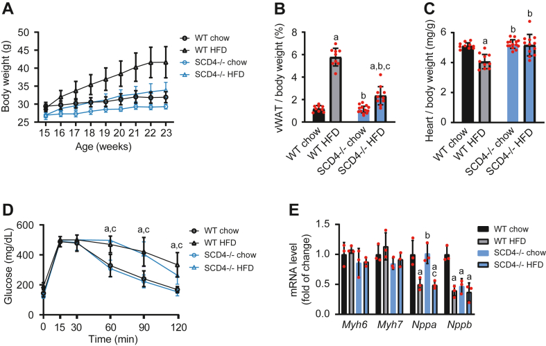
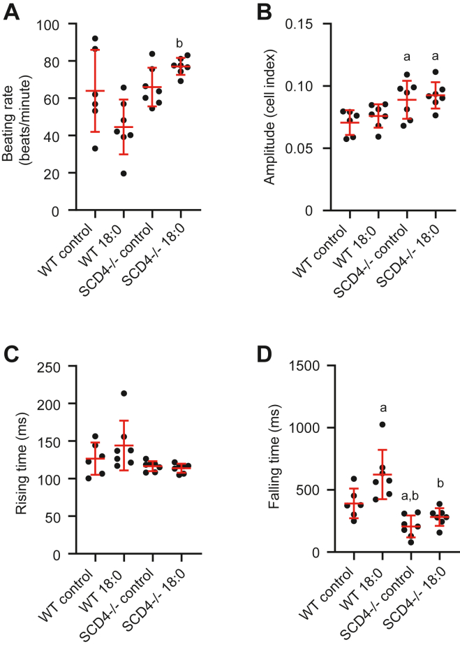
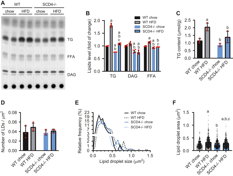
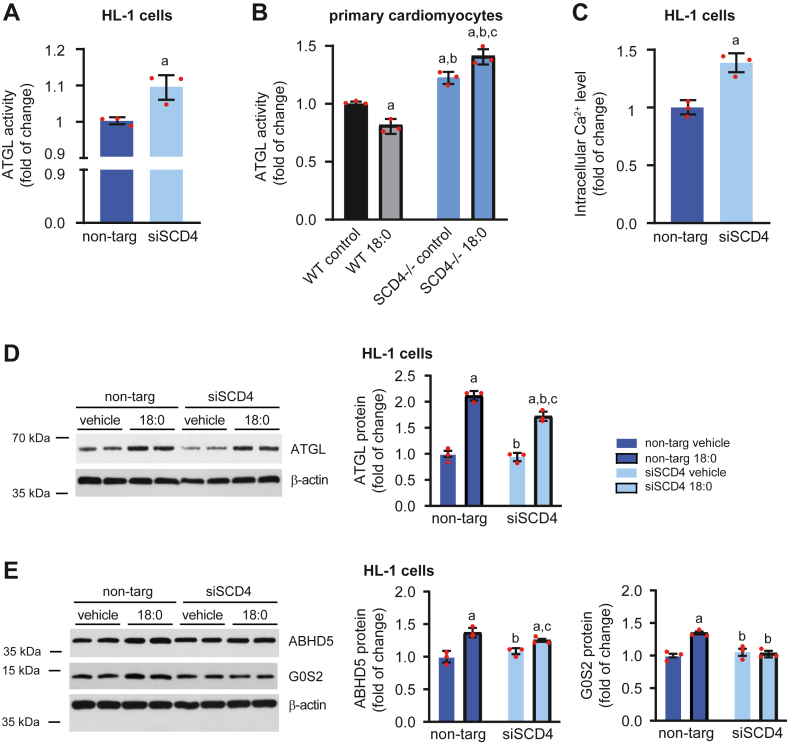
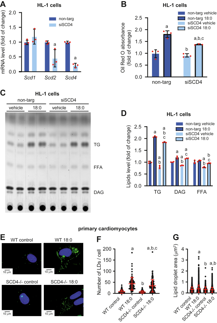
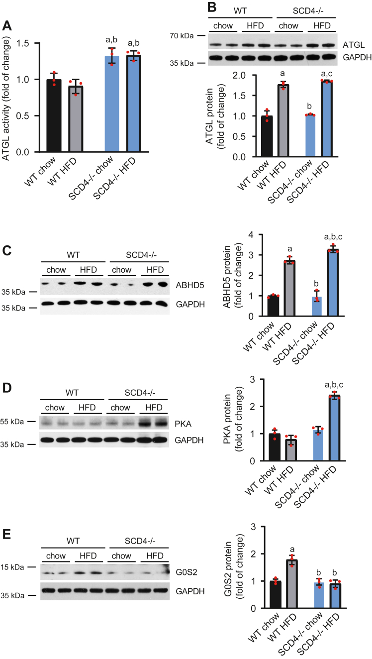
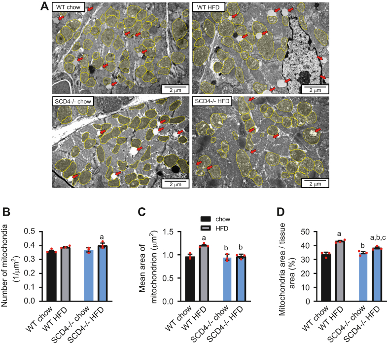
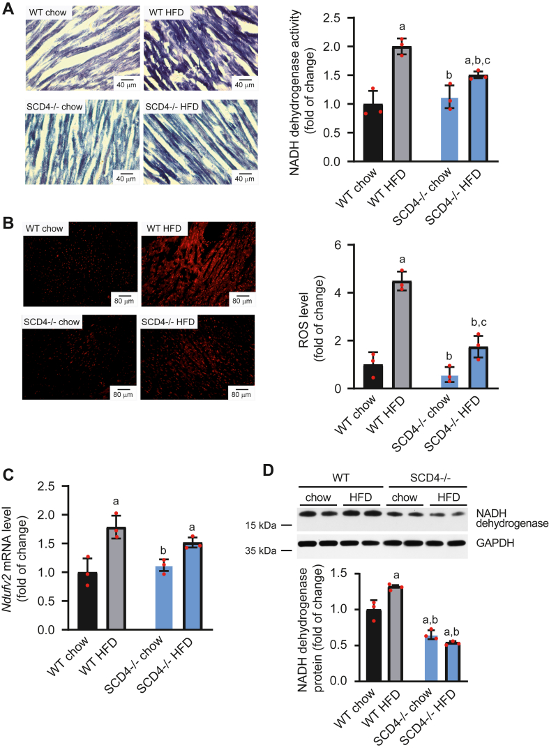
Stearoyl-CoA desaturase (SCD) is a lipogenic enzyme that catalyzes formation of the first double bond in the carbon chain of saturated fatty acids. Four isoforms of SCD have been identified in mice, the most poorly characterized of which is SCD4, which is cardiac-specific. In the present study, we investigated the role of SCD4 in systemic and cardiac metabolism. We used WT and global SCD4 KO mice that were fed standard laboratory chow or a high-fat diet (HFD). SCD4 deficiency reduced body adiposity and decreased hyperinsulinemia and hypercholesterolemia in HFD-fed mice. The loss of SCD4 preserved heart morphology in the HFD condition. Lipid accumulation decreased in the myocardium in SCD4-deficient mice and in HL-1 cardiomyocytes with knocked out Scd4 expression. This was associated with an increase in the rate of lipolysis and, more specifically, adipose triglyceride lipase (ATGL) activity. Possible mechanisms of ATGL activation by SCD4 deficiency include lower protein levels of the ATGL inhibitor G0/G1 switch protein 2 and greater activation by protein kinase A under lipid overload conditions. Moreover, we observed higher intracellular Ca2+ levels in HL-1 cells with silenced Scd4 expression. This may explain the activation of protein kinase A in response to higher Ca2+ levels. Additionally, the loss of SCD4 inhibited mitochondrial enlargement, NADH overactivation, and reactive oxygen species overproduction in the heart in HFD-fed mice. In conclusion, SCD4 deficiency activated lipolysis, resulting in a reduction of cardiac steatosis, prevented the induction of left ventricular hypertrophy, and reduced reactive oxygen species levels in the heart in HFD-fed mice.

Keywords: ATGL; heart; lipid droplets; metabolism; mitochondria.


Conflict of interest statement

Conflict of interest The authors declare that they have no conflicts of interest with the contents of this article.

Figures

Fig. 1
Fig. 1
A: Effect of SCD4 deficiency on body weight in mice. WT and SCD4−/− mice were fed chow or a high-fat diet (HFD). During the 8-week experiment, body weight was monitored weekly. B: Adiposity in mice, calculated as the ratio of visceral white adipose tissue (vWAT) weight to body weight, expressed as a percentage. C: Ratio of heart weight to body weight. D: Glucose tolerance in mice was analyzed at the end of the experiment using an intraperitoneal glucose tolerance test. E: Effect of SCD4 deficiency on the expression of genes that are related to heart dysfunction. mRNA levels of α and β myosin heavy chain (Myh6 and Myh7), atrium natriuretic peptide (Nppa), and B-type natriuretic peptide (Nppb) were analyzed using quantitative real-time PCR and the 2-ΔΔCt method. The data are expressed as the mean ± SD. n = 10–12 mice/group. ap < 0.05, versus WT chow; bp < 0.05, versus WT HFD; cp < 0.05, versus SCD4−/− chow.
Fig. 2
Fig. 2
Effect of SCD4 deficiency on contractile properties of neonatal cardiomyocytes. A: Spontaneous beating rate of primary cardiomyocytes treated with stearic acid. B: Amplitude of cardiomyocyte contraction. C: Rising time (time for the cell index to increase from 10% to 90% of amplitude). D: Falling time (time for the cell index to decrease from 90% to 10% of amplitude). ap < 0.05, versus. WT control; bp < 0.05, versus WT 18:0. The data are expressed as the mean ± SD. n = 6–7 independent measurements.
Fig. 3
Fig. 3
Effect of SCD4 deficiency on myocardial lipid accumulation. A: Representative thin-layer chromatography plate of separated neutral lipids. Lipids were isolated according to the method of Bligh and Dyer (1959). B: After soaking in a mixture of 10% cupric sulfate and 8% phosphoric acid, the plate was burned in at 140°C, and lipids were quantified by densitometry. C: Triglyceride (TG) content was analyzed using gas chromatography-mass spectrometry. D: The number of lipid droplets (LDs) and E: LD size distribution were determined using transmission electron microscopy images at 6,000× magnification. F: Mean lipid droplet size measured on TEM images at 6,000× magnification (n > 250). The data are expressed as the mean ± SD. n = 10–12 mice/group. ap < 0.05, versus WT chow; bp < 0.05, versus. WT HFD; cp < 0.05, versus. SCD4−/− chow. HFD, high-fat diet.
Fig. 4
Fig. 4
Impact of Scd4 downregulation on lipolysis in HL-1 cells and primary cardiomyocytes. A: ATGL activity in HL-1 cells with silenced Scd4 expression. B: ATGL activity in primary SCD4-deficient cardiomyocytes treated with stearic acid (C18:0). C: Intracellular calcium concentration in HL-1 cells was measured using the Fluo-4 Direct Calcium Assay Kit according to the manufacturer’s instructions. D: ATGL, E: ABHD5 and G0S2 protein levels in HL-1 cells and results of densitometric analysis. The data are expressed as the mean ± SD. n = 3 independent experiments. (A, C, D, E) ap < 0.05, versus non-targ vehicle; bp < 0.05, versus non-targ 18:0; cp < 0.05, versus siSCD4 vehicle. (B) ap < 0.05, versus WT control; bp < 0.05, versus WT 18:0; cp < 0.05, versus SCD4−/− control. ATGL, adipose triglyceride lipase.
Fig. 5
Fig. 5
Effect of Scd4 downregulation in HL-1 cells and primary neonatal cardiomyocytes on lipid accumulation. A: Scd1, Scd2, and Scd4 mRNA levels in HL-1 cells after Scd4 silencing. B: Level of accumulated lipids, based on Oil Red O dye absorption in HL-1 cells that were treated with stearic acid (C18:0). C: Representative thin-layer chromatographic plate of separated neutral lipids from HL-1 cells and D: results of the plate densitometric analysis. E: Representative images of lipid droplets (LDs) staining using BODIPY in primary SCD4-deficient cardiomyocytes. Representative images at 60× magnification are presented. Scale bar = 10 μm. F: The number of LDs and G: LDs size measured in primary neonatal SCD4-defcient cardiomyocytes treated with stearic acid (C18:0). The data are expressed as the mean ± SD. (A–D) n = 3 independent experiments. ap < 0.05, versus non-targ vehicle; bp < 0.05, versus non-targ 18:0; cp < 0.05, versus siSCD4 vehicle. (E-G) n = 3 independent cardiomyocytes isolation. At least 20 cells from each isolation in each experimental condition were analyzed. ap < 0.05, versus WT control; bp < 0.05, versus WT 18:0; cp < 0.05, versus SCD4−/− control.
Fig. 6
Fig. 6
SCD4 deficiency affects lipolysis in the mouse heart. A: Adipose triglyceride lipase (ATGL) activity. Using a triglyceride analog that becomes fluorescent after hydrolysis, we measured ATGL activity as described in the Materials and Methods. B: ATGL, C: abhydrolase domain-containing protein 5 (ABHD5), D: protein kinase A (PKA), and E: G0/G1 switch gene 2 (G0S2) protein content and results of the densitometric analysis. The data are expressed as the mean ± SD. n = 10–12 mice/group. ap < 0.05, versus WT chow; bp < 0.05, versus WT HFD; cp < 0.05, versus SCD4−/− chow. ATGL, adipose triglyceride lipase; HFD, high-fat diet.
Fig. 7
Fig. 7
Analysis of cardiac mitochondria by transmission electron microscopy. A: Representative images of mitochondria (yellow) and lipid droplets (red arrows) in the left ventricle. Representative images at 150,00× magnification are presented. Scale bar = 2 μm. B: Mitochondrial density (n > 600), C: average area of a single mitochondrion, and D: percentage of tissue area occupied by mitochondria, presented as a percentage of tissue (n > 250). The data are expressed as the mean ± SD. n = 3 mice/group. ap < 0.05, versus. WT chow; bp < 0.05, versus WT HFD; cp < 0.05, versus. SCD4−/− chow. HFD, high-fat diet.
Fig. 8
Fig. 8
Impact of SCD4 deficiency on mitochondrial NADH dehydrogenase activity and reactive oxygen species (ROS) production. A: NADH dehydrogenase activity and representative images of NADH dehydrogenase activity-dependent staining of the mouse left ventricle. Cryosections (10 μm thick) were stained with NADH and nitro blue tetrazolium. The stained sections were observed with a 20× magnification objective. Scale bar = 40 μm. B: Reactive oxygen species level and representative images of mouse left ventricle staining with ROS-sensitive dye. Images were captured at 10× magnification. Scale bar = 80 μm. C: Ndufv2 mRNA levels in the heart were analyzed using quantitative real-time PCR and the 2-ΔΔCt method. D: NADH dehydrogenase subunit B8 protein level in the left ventricle of the mouse heart. The data are expressed as the mean ± SD. n = 3 mice/group in A and B. n = 10–12 mice/group in C and D. aP < 0.05, versus WT chow; bp < 0.05, versus. WT HFD; cp < 0.05, versus SCD4−/− chow. HFD, high-fat diet.

References

    1. Belke D.D., Larsen T.S., Lopaschuk G.D., Severson D.L. Glucose and fatty acid metabolism in the isolated working mouse heart. Am. J. Physiol. 1999;277:R1210–R1217. - PubMed
    1. Ntambi J.M., Buhrow S.A., Kaestner K.H., Christy R.J., Sibley E., Kelly T.J., et al. Differentiation-induced gene expression in 3T3-L1 preadipocytes. Characterization of a differentially expressed gene encoding stearoyl-CoA desaturase. J. Biol. Chem. 1988;263:17291–17300. - PubMed
    1. Kaestner K.H., Ntambi J.M., Kelly T.J., Lane M.D. Differentiation-induced gene expression in 3T3-L1 preadipocytes. A second differentially expressed gene encoding stearoyl-CoA desaturase. J. Biol. Chem. 1989;264:14755–14761. - PubMed
    1. Zheng Y., Prouty S.M., Harmon A., Sundberg J.P., Stenn K.S., Parimoo S. Scd3 - a novel gene of the stearoyl-CoA desaturase family with restricted expression in skin. Genomics. 2001;71:182–191. - PubMed
    1. Miyazaki M., Jacobson M.J., Man W.C., Cohen P., Asilmaz E., Friedman J.M., et al. Identification and characterization of murine SCD4, a novel heart-specific stearoyl-CoA desaturase isoform regulated by leptin and dietary factors. J. Biol. Chem. 2003;278:33904–33911. - PubMed

LinkOut - more resources