Review
. 2025 Jun:72:501-514.
doi: 10.1016/j.jare.2024.07.030. Epub 2024 Jul 31.

The molecular mechanism of NF-κB dysregulation across different subtypes of renal cell carcinoma

Affiliations
Review

The molecular mechanism of NF-κB dysregulation across different subtypes of renal cell carcinoma

Nour Abu Jayab et al. J Adv Res. 2025 Jun.

Abstract

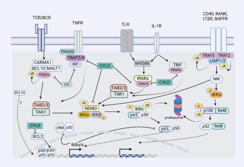
Background: The nuclear factor kappa B (NF-κB) is a critical pathway that regulates various cellular functions, including immune response, proliferation, growth, and apoptosis. Furthermore, this pathway is tightly regulated to ensure stability in the presence of immunogenic triggers or genotoxic stimuli. The lack of control of the NF-κB pathway can lead to the initiation of different diseases, mainly autoimmune diseases and cancer, including Renal cell carcinoma (RCC). RCC is the most common type of kidney cancer and is characterized by complex genetic composition and elusive molecular mechanisms.

Aim of review: The current review summarizes the mechanism of NF-κB dysregulation in different subtypes of RCC and its impact on pathogenesis.

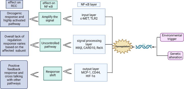
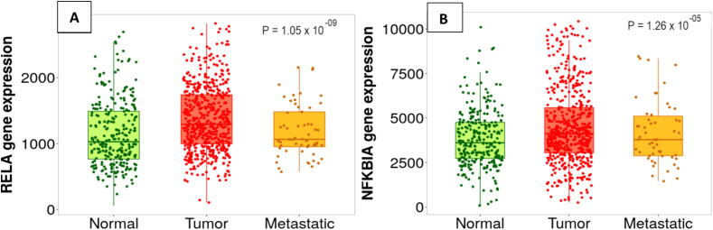
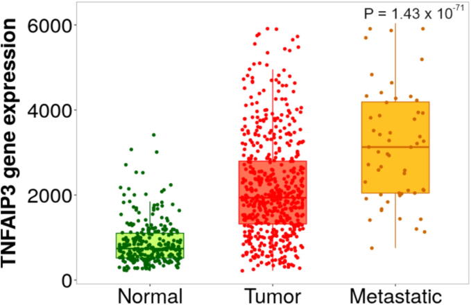
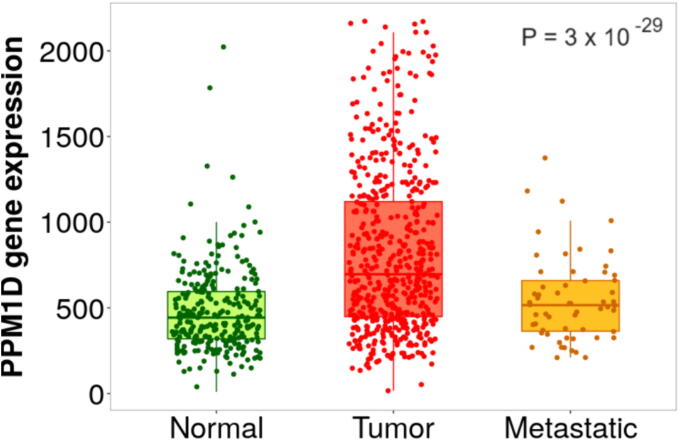
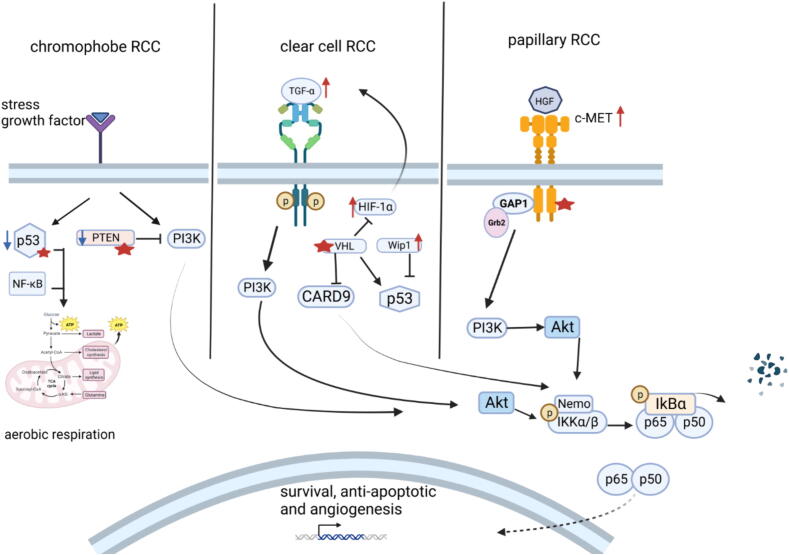
Key scientific concept of review: This review highlights the prominent role of NF-κB in RCC development and progression by driving the expression of multiple genes and interplaying with different pathways, including the phosphatidylinositol 3-kinase (PI3K)/protein kinase B (Akt) pathway. In silico analysis of RCC cohorts and molecular studies have revealed that multiple NF-κB members and target genes are dysregulated. The dysregulation includes receptors such as TLR2, signal-transmitting members including RelA, and target genes, for instance, HIF-1α. The lack of effective regulatory mechanisms results in a constitutively active NF-κB pathway, which promotes cancer growth, migration, and survival. In this review, we comprehensively summarize the role of dysregulated NF-κB-related genes in the most common subtypes of RCC, including clear cell RCC (ccRCC), chromophobe RCC (chRCC), and papillary RCC (PRCC).

Keywords: Genetic modification; Molecular mechanism; NF-κB dysregulation; Renal cell carcinoma.


Conflict of interest statement

Declaration of competing interest The authors declare that they have no known competing financial interests or personal relationships that could have appeared to influence the work reported in this paper.

Figures

None
Graphical abstract
Fig. 1
Fig. 1
Canonical and non-canonical NF-κB and the different mechanisms of regulations. The input layer of canonical NF-κB comprises different types of receptors, including BCR, TCR, TLR, IL-1R, and TNFR. These are activated in response to different triggers, resulting in canonical NF-κB pathway activation. Under basal conditions, IκBα masks the nuclear localization signal of p65 and p50, hence hindering NF-κB transcription factors from translocating to the nucleus. Stimulation of the receptors in the input layer transmits the signal to the processing layer through recruiting various adapters, resulting in β complex activation in the canonical NF-κB pathway. Subsequently, IKK phosphorylates IκBα to release p65-p50 and/or c-REL-p50 heterodimer that translocates to the nucleus and binds to the target gene promotor. Conversely, the non-canonical pathway is regulated by the TRAF-cIAP complex. Under basal conditions, NIK is instantly bound to TRAF3 and recruited to the E3 ubiquitin ligase complex consisting of TRAF2 and cIAP1/2. Thus, NIK will be ubiquitinated and degraded. Upon stimulation of the non-canonical NF-κB pathway by triggering lymphotoxin β, CD40, RANK, or BAFFR, NIK phosphorylates and activates IKKα that in turn phosphorylates p100, hence mediates its proteasomal processing. p52-RelB will translocate to the nucleus and stimulate gene transcription, producing the output response. NF-κB activity is regulated by several enzymes and proteins that keep NF-κB balanced. Mainly, IκBα/ε is induced in response to NF-κB activation to produce a negative feedback loop. In contrast, CYLD detaches the K63-ubiquitin chain and deactivates different subunits, including TRAF2, TRAF6, NEMO, and BCL3, therefore inactivating IKK kinase complex and inhibiting canonical NF-κB pathway activation. A20 regulates NF-κB through physically binding to TRAF2 to impede NF-κB activation, interfere with TRAF6 ubiquitination, and target RIP-1 for degradation. Also, A20 competes for the binding of ubiquitin chain on NEMO added by LUBAC, hence impeding IKKβ recognition by TAK-1deubiquitinating TRAF6 and RIP1 as well as interfering with NEMO ubiquitination. Moreover, BCL3 regulates NF-κB and affects its transcriptional activity through binding to p50 and p52 homodimers. This figure was generated using Biorender. BAFFR, B-cell activating factor receptor; BCR, B-cell receptor; cIAP1/2, cellular inhibitor of apoptosis 1 and 2; CYLD, cylindromatosis; IL-1R, interleukin-1 receptor; IKK, inhibitor of nuclear factor-κB kinase; IκB, inhibitor of nuclear factor-κB; LTβR, lymphotoxin β receptor; MyD88, myeloid differentiation primary response gene 88; NEMO, NF-κB essential modulator; TRIF, TIR domain-containing adaptor inducing interferon-beta; RIP1, receptor-interacting protein 1; NIK, NF-κB-inducing kinase; TAK1, TGF-β-activated protein kinase 1; TCR, T-cell receptor; TLR, toll-like receptor; TNFR, Tumor necrosis factor-α receptor; TRADD, TNF-R-associated death domain; TRAF2/5/6, TNF-R-associated factor 2, 5 and 6.
Fig. 2
Fig. 2
Canonical NF-κB-related genes are elevated in kidney tumor tissues. A TNM box plot of RELA gene expression in kidney normal, tumor, and metastatic samples. B TNM box plot of NFKBIA gene expression in kidney 277 normal, 556 tumors, and 58 metastatic samples (N = 891).
Fig. 3
Fig. 3
TNFAIP3 expression is correlated positively with the tumor stage. TNM box plot of TNFIAP3 gene in kidney 277 normal, 556 tumors, and 58 metastatic samples (N = 891).
Fig. 4
Fig. 4
PPM1D NF-κB-responsive gene is highly expressed in kidney tumor tissues. TNM box plot of PPM1D gene in 277 normal, 556 tumors, and 58 metastatic cancer kidney samples (N = 891).
Fig. 5
Fig. 5
The activation of the NF-κB pathway by various upstream receptors and triggers in different subtypes of RCC. NF-κB is activated in RCC different subtypes through different dysregulations that occur upstream, mainly in the PI3K/Akt pathway. In chromophobe RCC, mutations, and deletion of p53 and PTEN allow the overactivation of PI3K/Akt by growth factors and hormones that will, in turn, activate the NF-κB pathway and support aerobic oxidation mechanisms. Chromophobe RCC cancer cells exhibit increased expression of several Kreb’s cycle enzymes including citrate synthase and acotinase2. Also, NF-κB could enhance p53 suppression by inducing the expression of MDM2, providing a positive feedback response and shifting the response to survival and proliferation. In clear cell RCC, VHL mutation causes the accumulation and activation of HIF-1α and NF-κB, respectively. HIF-1α accumulation induces the expression of TGF-α that binds to the epidermal growth factor receptor and activates NF-κB in an Akt-mediated manner. Moreover, NF-κB provides a positive feedback loop by inducing the expression of multiple genes, including HIF-1α. p53 activity is suppressed in clear cell RCC through VHL loss. Additionally, Wip1 is another inhibitor of p53 that will suppress its function by dephosphorylating ser15 which will further potentiate NF-κB pathway. Besides, TGF-α activates PI3K providing an indirect NF-κB pathway activation. Papillary RCC is characterized by mutations in the c-MET receptor that will constantly activate its tyrosine kinase domain. Upon the dimerization of the receptor and phosphorylation and activation of the tyrosine kinase domain, GAP1 and Grb2 are recruited and bind to the multifactional docking site of c-MET, resulting in PI3K/Akt activation. Consequently, Akt phosphorylates IKK and activates the canonical NF-κB pathway. Accordingly, the p65-p50 heterodimer binds to multiple genes, which sustains different cancer hallmarks such as survival and migration. This figure was designed using Biorender. CARD9, caspase recruitment domain family member 9; c-MET, mesenchymal-epithelial transition factor; GAP1, general amino acid permease; Grb2, growth factor receptor-bound protein 2; HGF, hepatocyte growth factor, HIF-1α, hypoxia-inducible factor 1-α; IKK, inhibitor of nuclear factor-κB kinase; IκB, inhibitor of nuclear factor-κB; NEMO, NF-κB essential modulator; NF-κB, nuclear factor kappa light chain enhancer of activated B cells; PI3K, phosphatidylinositol 3-kinase; PTEN, phosphatase and tensin homolog; RCC; renal cell carcinoma TGF-α, Tissue growth factor-α; VHL, von Hippel_Lindau; Wip1, wild-type p53-induced phosphatase 1Different subsets of RCC exploit intricate pathways to sustain various cancer hallmarks. In fact, different genetic alteration patterns are observed within the same RCC subtype. Curiously, a biopsy from clear cell carcinoma patients revealed intratumor heterogeneity, where different regions in the same tumor mass exhibit unique mutational landscapes, and only 34 % of the mutations are shared between these regions . This evidence suggests that RCC comprises a heterogeneous group of cancer cells with distinct molecular profiles. However, overall, each subtype of RCC is characterized by some biomarkers or gene signature. While VHL mutation or deletion is detected in more than 70 % of ccRCC , another hereditary condition, Birt–Hogg–Dubé (BHD) syndrome, is associated with chRCC. Around 30 % of BHD patients develop renal tumors, and 34 % of the developed BHD-related renal tumors are chRCC . BHD syndrome is associated with FLCN gene mutation, which encodes folliculin. In detail, folliculin executes its tumor-suppressing action by regulating multiple energy metabolic pathways, including mTOR and MAPK. Loss of function of FLCN allows AMPK and mTOR pathways to be constitutively active, which aligns with the augmented mitochondrial biogenesis observed in chRCC . Lastly, heredity mutation in the MET gene results in Hereditary papillary renal carcinoma (HPRC), which significantly increases the risk of developing type 1 PRCC (which accounts for >80 % of PRCC) . In light of the cohort and translational studies of different subtypes of RCC, each subtype employs specific NF-κB genes based on the cancer hallmarks required. Moreover, several factors contribute to somatic mutations, including immune surveillance, cell-specific mutations, and environmental factors that patients may be exposed to, including ultraviolet (UV) light . Based on the above observations, ccRCC is associated with increased expression and dysregulation of several NF-κB genes, which are utilized to drive ccRCC development and progression. In contrast, chRCC and PRCC mainly drive NF-κB activation indirectly, hence specific genes of NF-κB pathway will be dysregulated.

Similar articles

Cited by

References

    1. Siegel R.L., Giaquinto A.N., Jemal A. Cancer statistics, 2024. CA Cancer J Clin. 2024;74(1):12–49. doi: 10.3322/caac.21820. - DOI - PubMed
    1. Hu M., Fan J.Y., Zhou X., Cao G.W., Tan X. Global incidence and mortality of renal cell carcinoma in 2020. Zhonghua Liu Xing Bing Xue Za Zhi. 2023;44(4):575–580. doi: 10.3760/cma.j.cn112338-20220624-00558. - DOI - PubMed
    1. Karin M. Nuclear factor-kappaB in cancer development and progression. Nature. 2006;441(7092):431–436. doi: 10.1038/nature04870. - DOI - PubMed
    1. Chen L., Deng H., Cui H., Fang J., Zuo Z., Deng J., et al. Inflammatory responses and inflammation-associated diseases in organs. Oncotarget. 2018;9(6):7204–7218. doi: 10.18632/oncotarget.23208. - DOI - PMC - PubMed
    1. Singh N., Baby D., Rajguru J.P., Patil P.B., Thakkannavar S.S., Pujari V.B. Inflammation and cancer. Ann Afr Med. 2019;18(3):121–126. doi: 10.4103/aam.aam_56_18. - DOI - PMC - PubMed

MeSH terms