. 2024 Aug 3;15(1):6570.
doi: 10.1038/s41467-024-50888-8.

Structural basis of the obligatory exchange mode of human neutral amino acid transporter ASCT2

Affiliations

Structural basis of the obligatory exchange mode of human neutral amino acid transporter ASCT2

Anna M Borowska et al. Nat Commun. .

Abstract

ASCT2 is an obligate exchanger of neutral amino acids, contributing to cellular amino acid homeostasis. ASCT2 belongs to the same family (SLC1) as Excitatory Amino Acid Transporters (EAATs) that concentrate glutamate in the cytosol. The mechanism that makes ASCT2 an exchanger rather than a concentrator remains enigmatic. Here, we employ cryo-electron microscopy and molecular dynamics simulations to elucidate the structural basis of the exchange mechanism of ASCT2. We establish that ASCT2 binds three Na+ ions per transported substrate and visits a state that likely acts as checkpoint in preventing Na+ ion leakage, both features shared with EAATs. However, in contrast to EAATs, ASCT2 retains one Na+ ion even under Na+-depleted conditions. We demonstrate that ASCT2 cannot undergo the structural transition in TM7 that is essential for the concentrative transport cycle of EAATs. This structural rigidity and the high-affinity Na+ binding site effectively confine ASCT2 to an exchange mode.


Conflict of interest statement

The authors declare no competing interests.

Figures

Fig. 1
Fig. 1. Cryo-EM structure of ASCT2 in lipid nanodiscs in the presence of Na+ and glutamine.
a Impact of Na+ ions on glutamine transport mediated by ASCT2 measured in a radioactive uptake assay at indicated salt conditions. Data show the exchange of glutamine in the presence of internal and external Na+ ions (green circles); exchange of glutamine with internal K+ ions and external Na+ ions (yellow triangle); exchange of glutamine with internal Na+ ions and external K+ ions (pink square); and exchange of glutamine in the presence of internal and external K+ ions (blue diamond). Data points and error bars represent mean values ± SEM from n = 2 biologically independent experiments, each done in two or three technical replicates. Source data are provided as a Source Data file. b Cryo-EM map of trimeric ASCT2 in lipid nanodiscs in the presence of Na+ ions and glutamine in an outward-facing state with closed HP2 gate (Gln-Na+-OFSHP2-cld) at 2.6 Å resolution, countered at 5σ. Each protomer is colored according to respective domains: scaffold domain in yellow, transport domain in blue, and HP2 region in salmon. The surrounding nanodisc is shown as a transparent unsharpened map at a lower countered level (3.7σ). The view is shown from the membrane side and the membrane boundaries are indicated as lines. c The cryo-EM structure of one ASCT2 protomer in the outward-occluded state (Gln-Na+-OFSHP2-cld). The scaffold domain is colored in yellow and the transport domain in blue. The closed HP2 gate is displayed in salmon, while glutamine and Na+ ions are in light cyan and pink, respectively. The membrane boundaries are indicated as lines. d Close-up view of the substrate binding pocket highlighting the interactions between glutamine (light cyan) and the transport domain with the coordinating displayed as sticks. eg A close-up view of the Na+ ions (pink) bound at Na1 (e), Na2 (f), and Na3 (g) sites with displayed coordinating residues.
Fig. 2
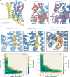
Fig. 2. Structural comparison of ASCT2 in the presence of Na+ ions and under Na+-depleted conditions.
a Superimposition of ASCT2 protomers imaged with the presence of glutamine and high Na+ ion concentration (Gln-Na+-OFSHP2cld, transport domain in light blue and closed HP2 in salmon) and in the absence of substrate under the Na+-depleted conditions (Na+-depleted OFSHP2op, transport domain in dark blue and open HP2 in purple). The scaffold domain in both structures is in yellow. b Comparison of interactions formed within the HP2a and HP2b in substrate-bound (left, transport domain in light blue and HP2 gate in salmon) and substrate-free (right, transport domain in dark blue and HP2 gate in purple) ASCT2. c Comparison of the conformation of the central region of TM7 in substrate-bound (left) and substrate-free (right) ASCT2. TM7 adopts the compact form (yellow) in substrate-bound ASCT2, forming the Na1 binding site (Na+ ion shown in pink). In the substrate-free ASCT2, TM7 is still in the compact form (yellow) that facilitates the binding of a Na+ ion at the Na1 binding site. Residues from the conserved NMDG motif are shown as sticks. d Superimposition of ASCT2 (dark blue) and GltTk (gray, PDB 5WDY) structures solved under similar conditions with 200 mM KCl. GltTk adopted the extended TM7 helix (orange), while ASCT2 still maintains the compact form of TM7 (yellow). Residues from the conserved NMDG motif are shown as sticks. e, f Free energy surface (FES) associated with the Na+ ion binding in ASCT2 (e) and GltTk (f) reweighted on the Z distance and the sodium number of contacts, showing a more tightly bound Na+ ion at the Na1 in ASCT2 than in concentrative transporters such as GltTk. The minimum energy path for the Na+ ion dissociation is depicted as a black dashed line.
Fig. 3
Fig. 3. The structural transition of TM7.
a The changes in torsional angle observed over the trajectory run in GltTk. Close-ups represent the starting conformation, in which TM7 is in the compact form (yellow) found in substrate-bound states (lower left), and the final conformation with an extended TM7 (orange; upper right). b The distribution of torsional angle observed in the simulation of GltTk as seen in (a). c, The changes in torsional angle observed over the trajectory run in ASCT2. Close-up represents the starting conformation, in which TM7 is in the compact form (yellow), maintained throughout the entire simulation time (lower right). d The distribution of torsional angle observed in the simulation of ASCT2 as seen in (c).
Fig. 4
Fig. 4. Conformational plasticity of ASCT2 protomers in lipid nanodiscs identified under substrate-bound and substrate-free conditions.
a Structures of substrate-bound ASCT2 at three distinct conformational states: Gln-Na+-OFSHP2cld (blue), Gln-Na+-iOFS-upHP2cld (light pink), and Gln-Na+-iOFS-downHP2cld (teal). b Structures of substrate-free ASCT2 at two distinct conformational states: Na+-depleted OFSHP2op (blue) and Na+-depleted iOFS-upHP2op (light pink). Each protomer is shown as a cartoon representation of the transport domain displayed in unique colors, and a surface colored in gray represents the scaffold domain. The scaffold domain acts as a reference point for the position of the transport domain in the membrane. The HP2 is displayed in salmon (closed conformation as seen in a) or purple (open conformation as seen in b).
Fig. 5
Fig. 5. Comparison of conformational states of ASCT2 in lipid nanodiscs.
a Conformational changes observed in the elevator transition from the Gln-Na+-OFS (blue) to the Gln-Na+-iOFS-up (light pink) states in substrate-bound ASCT2 with a closed HP2. b Conformational changes observed in the elevator transition from the Gln-Na+-iOFS-up (light pink) to the Gln-Na+-iOFS-down (teal) states in ASCT2 with a closed HP2. c Structural transition observed in substrate-free ASCT2 under Na+-depleted conditions from the Na+-depleted OFS (blue) state to the Na+-depleted iOFS-up (light pink), featuring an open HP2 gate. d Superimposition of two iOFS-up (light pink) states in substrate-bound and substrate-free ASCT2, displaying structural differences in the HP2 region (closed in salmon and open in purple). The structures in all panels were superimposed based on atoms of scaffold domains; however, the scaffold domains were hidden for a clear representation of conformational differences.
Fig. 6
Fig. 6. A scheme of the transport cycle and structural differences in concentrators and exchangers from the SLC1A family.
The upper part depicts outward-facing protomers displaying the structural rearrangements of HP2 and TM7 at various stages of the transport cycle, with the respective schematic representation of protomers below. Considering the reversible nature of secondary transporters and the structural equivalence of the binding sites in the outward- and inward-facing states, the structural transitions apply to both outward- and inward-facing conformations. The transport and scaffold domains are visualized in blue and yellow, respectively, with displayed HP2 gate (closed in salmon and open in purple) and TM7 (compact in dark yellow and extended in orange). The membrane boundaries are shown as gray shadows. The part of the transport cycle restricted in exchangers is indicated by a red box, and crossed arrows signal the transition to the conformation that exchangers do not sample. In contrast to concentrators, upon substrate release, ASCT2 retains a bound Na+ ion with a rigid TM7 in a compact state, prohibiting closure of the HP2 gate and a reorientation to the apo state, thereby effectively confining ASCT2 to an exchange mode.

References

    1. Bröer, S. & Bröer, A. Amino acid homeostasis and signalling in mammalian cells and organisms. Biochem. J.474, 1935–1963 (2017). 10.1042/BCJ20160822 - DOI - PMC - PubMed
    1. Chen, M. et al. Loss of RACK1 promotes glutamine addiction via activating AKT/mTOR/ASCT2 axis to facilitate tumor growth in gastric cancer. Cell Oncol. 10.1007/s13402-023-00854-1 (2023). - PubMed
    1. Ni, F. et al. Critical role of ASCT2-mediated amino acid metabolism in promoting leukaemia development and progression. Nat. Metab.1, 390–403 (2019). 10.1038/s42255-019-0039-6 - DOI - PMC - PubMed
    1. Lin, J. et al. SLC1A5 silencing inhibits esophageal cancer growth via cell cycle arrest and apoptosis. Cell. Physiol. Biochem.48, 397 (2018). 10.1159/000491769 - DOI - PubMed
    1. Toda, K. et al. Clinical role of ASCT2 (SLC1A5) in KRAS-mutated colorectal cancer. Int. J. Mol. Sci.18, 1632 (2017). - PMC - PubMed

MeSH terms

LinkOut - more resources