. 2024 Oct;38(10):2183-2195.
doi: 10.1038/s41375-024-02351-2. Epub 2024 Aug 3.

Rational combinatorial targeting by adapter CAR-T-cells (AdCAR-T) prevents antigen escape in acute myeloid leukemia

Affiliations

Rational combinatorial targeting by adapter CAR-T-cells (AdCAR-T) prevents antigen escape in acute myeloid leukemia

Daniel Atar et al. Leukemia. 2024 Oct.

Abstract

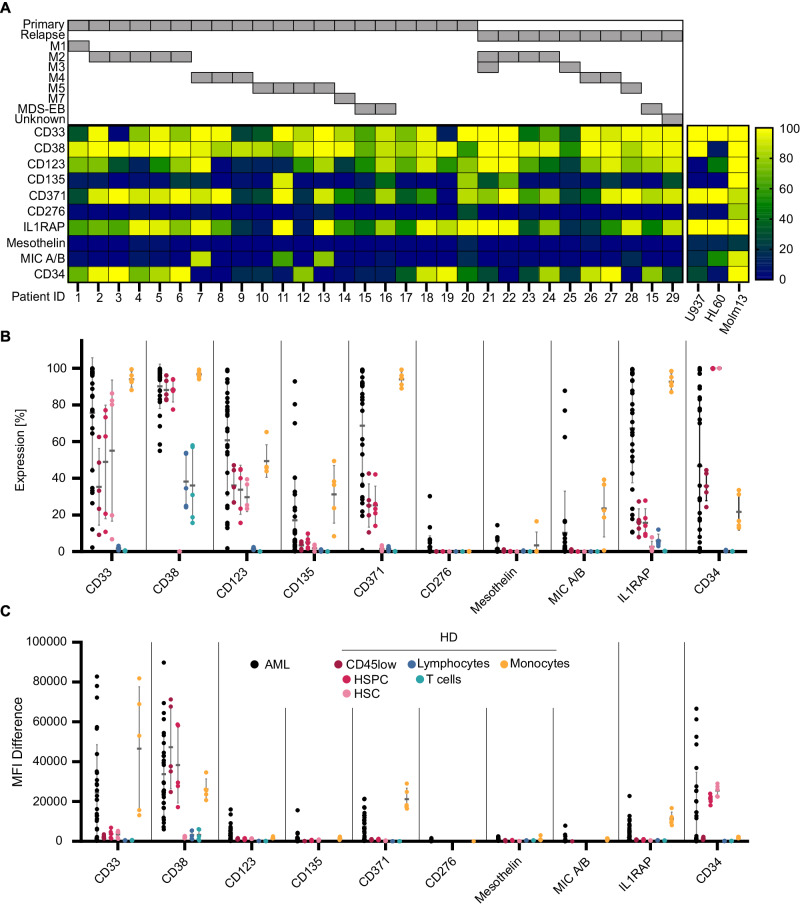
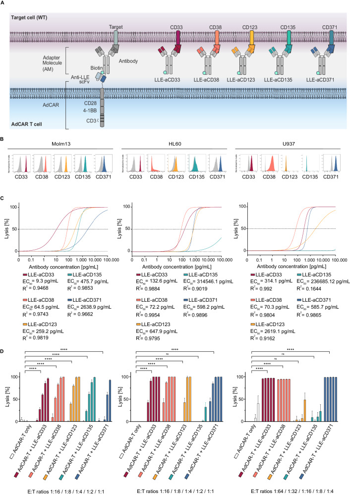
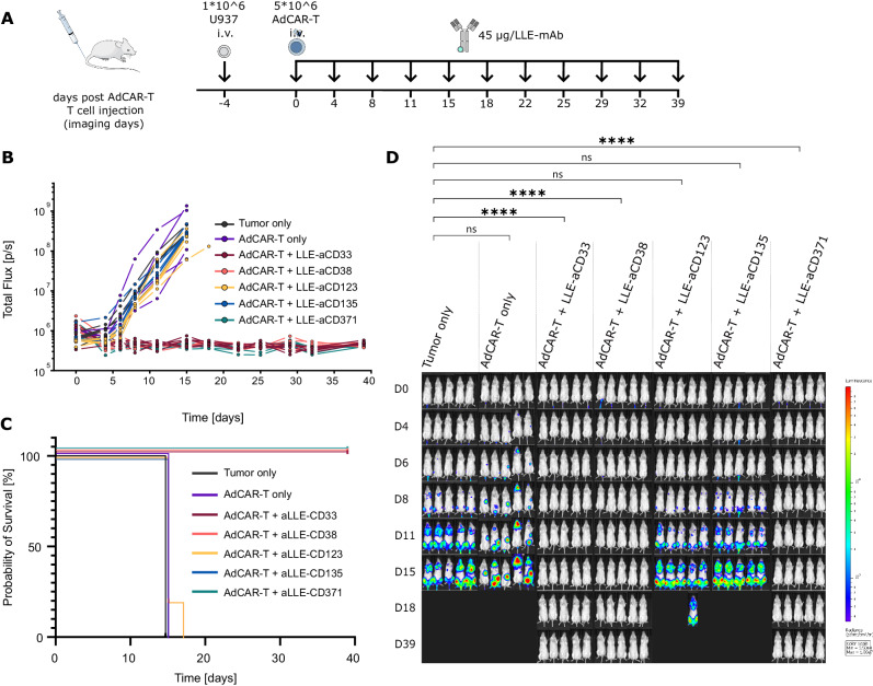
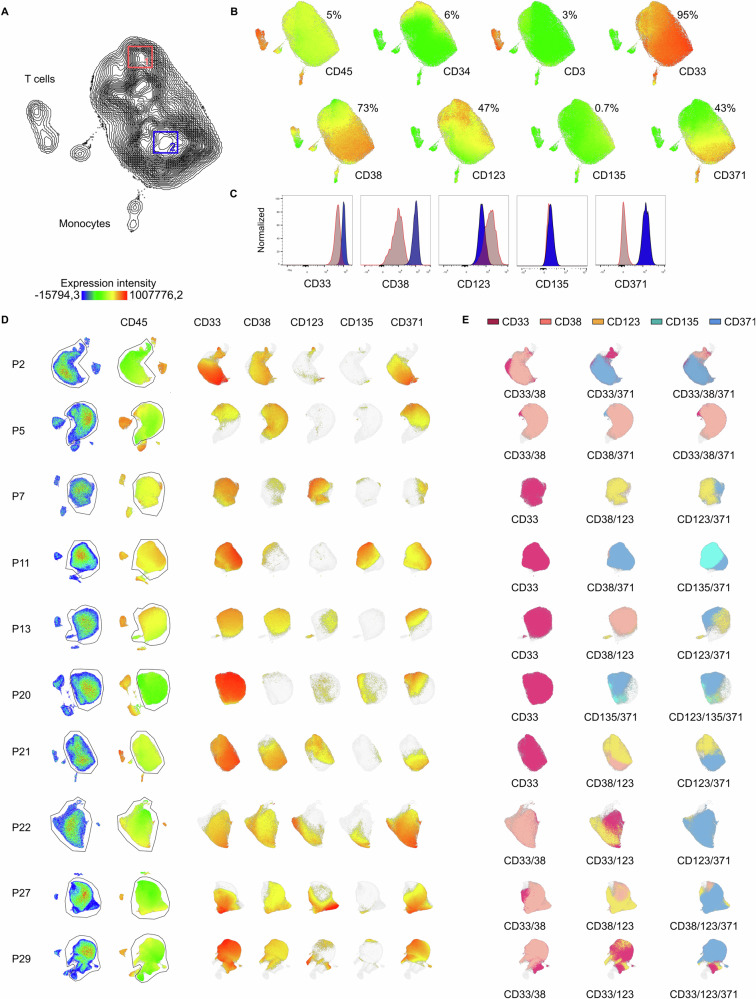
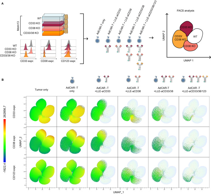
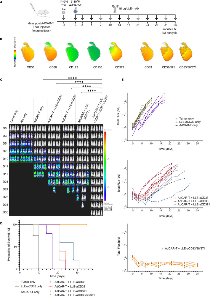
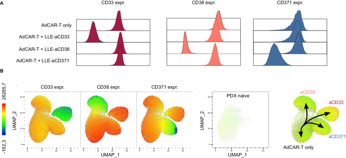
Targeting AML by chimeric antigen receptor T-cells (CAR-T) is challenging due to the promiscuous expression of AML-associated antigens in healthy hematopoiesis and high degree of inter- and intratumoral heterogeneity. Here, we present single-cell expression data of AML-associated antigens in 30 primary pediatric AML samples. We identified CD33, CD38, CD371, IL1RAP and CD123 as the most frequently expressed. Notably, high variability was observed not only across the different patient samples but also among leukemic cells of the same patient suggesting the necessity of multiplexed targeting approaches. To address this need, we utilized our modular Adapter CAR (AdCAR) platform, enabling precise qualitative and quantitative control over CAR-T-cell function. We show highly efficient and target-specific activity for newly generated adapter molecules (AMs) against CD33, CD38, CD123, CD135 and CD371, both in vitro and in vivo. We reveal that inherent intratumoral heterogeneity in antigen expression translates into antigen escape and therapy failure to monotargeted CAR-T therapy. Further, we demonstrate in PDX models that rational combinatorial targeting by AdCAR-T-cells can cure heterogenic disease. In conclusion, we elucidate the clinical relevance of heterogeneity in antigen expression in pediatric AML and present a novel concept for precision immunotherapy by combinatorial targeting utilizing the AdCAR platform.


Conflict of interest statement

The employing institution of DA, LR, ASM, SK, MM, SS, BK, MF, AC, KW, KS, ME, JS, PL, RH, and CMS (University Children’s Hospital Tuebingen) received research support from Miltenyi Biotec GmbH on the basis of a collaboration research agreement. CMS receives research funding from Miltenyi Biotec GmbH not related to the presented work. MS receives industry research support from Amgen, Bristol-Myers Squibb/Celgene, Gilead, Janssen, Miltenyi Biotec, Morphosys, Novartis, Roche, Seattle Genetics, and Takeda and serves as a consultant/advisor to AvenCell, CDR-Life, Ichnos Sciences, Incyte Biosciences, Janssen, Molecular Partners, and Takeda. She serves on the speakers’ bureau at Amgen, AstraZeneca, BMS/Celgene, Gilead, GSK, Janssen, Novartis, Pfizer, Roche, and Takeda. Furthermore, PL, RH, JM, PS and CMS are coinventors on a patent (WO2018078066A1) focusing on Adapter CAR technology. Other authors declare no relevant conflicts of interest.

Figures

Fig. 1
Fig. 1. Expression of target antigens in pediatric AML and healthy bone marrow.
A Heatmap visualizing the percentage of leukemic blasts, defined as CD45dim cells within the BM of pediatric AML patients (n = 30), expressing the target antigens CD33, CD38, CD123, CD135, CD371, CD276, IL1RAP, Mesothelin, or MIC A/B as well as CD34, analyzed by flow cytometry. Patients are grouped by primary disease vs. relapse as well as FAB classification/MDS-EB. Target expression of the used AML cell lines are visualized as heatmap next to patient data (right). Comparison of target antigen expression between AML blasts and major cell populations (defined as follows: HSC (CD45dim/CD34high/CD38low), HSPC (CD45dim/CD34high/CD38high), lymphocytes (CD45high/SSClow), T cells (CD45high/SSClow/CD3high), and monocytes (CD45high/SSCmedian)) in bone marrow from healthy donors (n = 5). In B the percentage of antigen-positive cells is provided; in C the mean fluorescence intensity difference (MFID). Each data point represents an individual patient or heathy donor, and horizontal lines represent the mean value ± standard deviation (SD).
Fig. 2
Fig. 2. Design and in vitro evaluation of novel AML-targeted AMs.
A Schematic illustration of the AdCAR-T system. AdCAR-T cells are directed to AML-associated target antigens via LLE-conjugated mAbs (LLEa-CD33, LLEa-CD38, LLEa-CD123, LLEa-CD135, and LLEa-CD371), referred to as AMs. B Histograms of target antigen expression (CD33, CD38, CD123, CD135 and CD371) on AML cell lines (Molm13, HL60 and U937) stained with the indicated AM and secondary anti-LLE mAb, analyzed by flow cytometry. C Cytotoxicity of AdCAR-T against the indicated luciferase-expressing AML cell lines as determined by LCA after 48 h at an E:T of 1:1 (Molm13, HL60) or 1:4 (U937), mediated by increasing concentrations, logarithmic titration steps from 0.1 pg/mL to 100 ng/mL, of the indicated AMs. EC50 values are provided for each AM. D Cytotoxicity of AdCAR-T cells against the indicated AML cell lines as determined by LCA after 48 h at fixed AM concentrations (10 ng/mL) and indicated E:T ratios. AdCAR-T cells in the absence of AM served as a negative control. Data shown in C and D represent the mean ± SD of (n = 6). Data shown in C were transformed by taking the logarithm of the x values and then fitted by nonlinear regression. Statistical analysis was performed using two-way ANOVA and Tukey’s multiple comparison test. ns, not significant. *p ≤ 0.0332 **p ≤ 0.021. ***p ≤ 0.002. ****p ≤ 0.0001. The full table of the statistical analysis is provided in Additional file 1.
Fig. 3
Fig. 3. In vivo validation of target antigen-specific activity of AdCAR-T cells.
A Schematic depiction of the in vivo experiment: NSG mice were engrafted with 1 × 106 U937luc/CD19t (CD33high, CD38high, CD123low, CD135low, CD371high) on day −4 via tail vein injection (i.v.). A total of 5 × 106 AdCAR-T cells were injected i.v. on day 0. Forty-five micrograms of the indicated AM (LLE-aCD33, LLE-aCD38, LLE-aCD123, LLE-aCD135 or LLE-aCD371) was injected subcutaneously (s.c.) twice a week starting on day 0. Untreated mice (tumor only) served as a negative control (n = 5 per group). Tumor load was monitored by BLI. Mice were sacrificed when they reached the endpoint criteria. B Total flux [photons/second] of in vivo bioluminescence blotted over time for individual animals. C Kaplan‒Meier curves for reaching endpoint criteria. D BLI images at the indicated time points (exposure time 10 sec.). Statistical analysis was performed using two-way ANOVA and Tukey’s multiple comparison test. ns, not significant. *p ≤ 0.0332 **p ≤ 0.021. ***p ≤ 0.002. ****p ≤ 0.0001. The full table of the statistical analysis is provided in Additional file 1.
Fig. 4
Fig. 4. Intratumoral heterogeneity in target antigen expression in pediatric AML.
A UMAP based on the expression of CD45, CD3, CD34, CD33, CD38, CD123, CD135 and CD371 as well as FSC and SSC signals, as determined by flow cytometry, of an exemplary pediatric AML bone marrow sample (P21). B Color-coded intensity of CD45, CD3, CD34, CD33, CD38, CD123, CD135 and CD371 expression, with each dot representing one cell. C To highlight intratumoral heterogeneity in target antigen expression, expression of CD33, CD38, CD123, CD135 and CD371, plotted as histograms, in two different areas of the AML blast population, gated and labeled as 1 (LSC-like) and 2 (bulk). D UMAPs of AML bone marrow samples (P2, P5, P7, P11, P13, P20, P21, P22, P27, and P29); the left row shows bulk bone marrow as a pseudo color dot plot, and the second row shows CD45 expression and AML blast gates, followed by the expression of target antigens CD33, CD38, CD123, CD135 and CD371. E Suggestions of possible target combinations to cover most leukemic blasts.
Fig. 5
Fig. 5. In vitro evaluation of multiplex targeting by AdCAR-T.
A Schematic depiction of the flow cytometry-based cytotoxicity assay: Molm13 wild type (WT), Molm13 CD33KO, Molm13 CD38KO, and Molm13 CD33/CD38 KO were mixed at a 1:1:1:1 ratio. The expression of CD33, CD38, and CD123 in individual cell populations is shown on the lower left. AdCAR-T cells were added at an E:T ratio of 1:1. LLE-aCD33, LLE-aCD38, or LLE-aCD123 as well as combinations thereof were added to reach a final concentration of 10 ng/mL. B UMAP representing batched surviving target cells after 48 h of incubation of all conditions (n = 3 each condition), based on the expression of CD33, CD38 and CD123, as determined by flow cytometry. From left to right, viable target cells for the indicated conditions plotted in color. From top to bottom, color-coded expression of CD33, CD38, and CD123.
Fig. 6
Fig. 6. In vivo validation of multiplex targeting by AdCAR-T cells in a PDX model.
A Schematic depiction of the in vivo experiment: NSG mice were engrafted with 1 × 106 PDX luc/CD19t (P21) cells on day −3 via tail vein injection (i.v.). A total of 5 × 106 AdCAR-T cells were injected i.v. on day 0. A total of 45 µg of the indicated AM (LLE-aCD33, LLE-aCD38, or LLE-aCD371) or a combination thereof was injected subcutaneously (s.c.) twice a week starting on day 0. Untreated mice (tumor only), mice injected with PBS instead of AM (AdCAR-T only), and mice injected with LLE-aCD33, LLE-aCD38 and LLE-aCD371 but not AdCAR-T (AM only) served as negative controls (n = 5 per group, n = 4 in tumor only). Tumor load was monitored by BLI. Mice were sacrificed when they reached the endpoint criteria. B UMAP based on the expression of CD45, CD3, CD34, CD33, CD38, CD123, CD135 and CD371 as well as FSC and SSC signals, as determined by flow cytometry, of PDX cells prior to injection, expression of CD33, CD38, CD123, CD135 and CD371 on AML blasts (left) and suggestions for combinatorial targeting (right). C BLI images at the indicated time points (exposure time 10 s). D Total flux [photons/second] of in vivo bioluminescence blotted over time for individual animals. E Kaplan‒Meier curves for reaching endpoint criteria. Statistical analysis was performed using two-way ANOVA and Tukey’s multiple comparison test. ns, not significant. *p ≤ 0.0332 **p ≤ 0.021. ***p ≤ 0.002. ****p ≤ 0.0001. The full table of the statistical analysis is provided in Additional file 1.
Fig. 7
Fig. 7. Analysis of resistance mechanisms to mono-targeting.
A Representative flow cytometry analyses of bone marrow from relapsed mice. Expression of CD33, CD38 and CD371 on viable AML blasts for the indicated groups plotted as histograms. B UMAP, representing batched viable AML blasts in bone marrow from tumor-only mice, AdCAR-T-only mice and mice treated with AdCAR-T plus LLE-aCD33, LLE-aCD38, or LLE-aCD123 (n = 3 each condition), based on the expression of CD33, CD38 and CD123, as determined by flow cytometry. From left to right, expression of CD33, CD38, and CD371 color-coded, spatial localization of treatment naïve PDX cells ( = tumor only) and pseudotrajectories enforced by mono-targeting.

References

    1. Maude SL, Laetsch TW, Buechner J, Rives S, Boyer M, Bittencourt H, et al. Tisagenlecleucel in children and young adults with B-cell lymphoblastic leukemia. N. Engl J Med. 2018;378:439–48. - PMC - PubMed
    1. Cappell KM, Sherry RM, Yang JC, Goff SL, Vanasse DA, McIntyre L, et al. Long-term follow-up of anti-CD19 chimeric antigen receptor T-cell therapy. J Clin Oncol. 2020;38:3805–15. - PMC - PubMed
    1. Brudno JN, Maric I, Hartman SD, Rose JJ, Wang M, Lam N, et al. T cells genetically modified to express an anti-B-cell maturation antigen chimeric antigen receptor cause remissions of poor-prognosis relapsed multiple myeloma. J Clin Oncol. 2018;36:2267–80. - PMC - PubMed
    1. Gill S, Brudno JN. CAR T-cell therapy in hematologic malignancies: clinical role, toxicity, and unanswered questions. Am Soc Clin Oncol Educ Book. 2021;41:1–20. - PubMed
    1. Shlush LI, Mitchell A, Heisler L, Abelson S, Ng SWK, Trotman-Grant A, et al. Tracing the origins of relapse in acute myeloid leukaemia to stem cells. Nature. 2017;547:104–8. - PubMed

MeSH terms

Substances