. 2024 Aug 2;14(1):17916.
doi: 10.1038/s41598-024-68970-y.

The impact of CLDN18.2 expression on effector cells mediating antibody-dependent cellular cytotoxicity in gastric cancer

Affiliations

The impact of CLDN18.2 expression on effector cells mediating antibody-dependent cellular cytotoxicity in gastric cancer

Akira Matsuishi et al. Sci Rep. .

Abstract

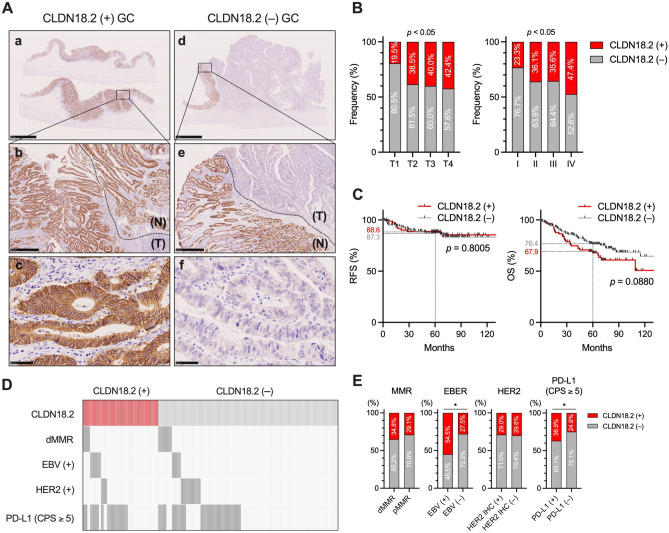
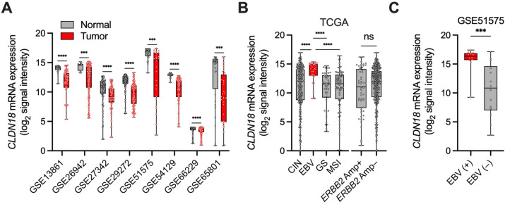
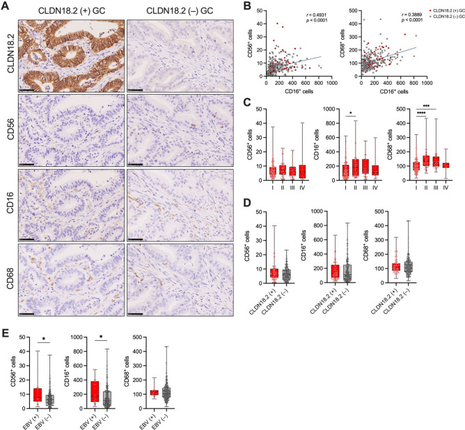
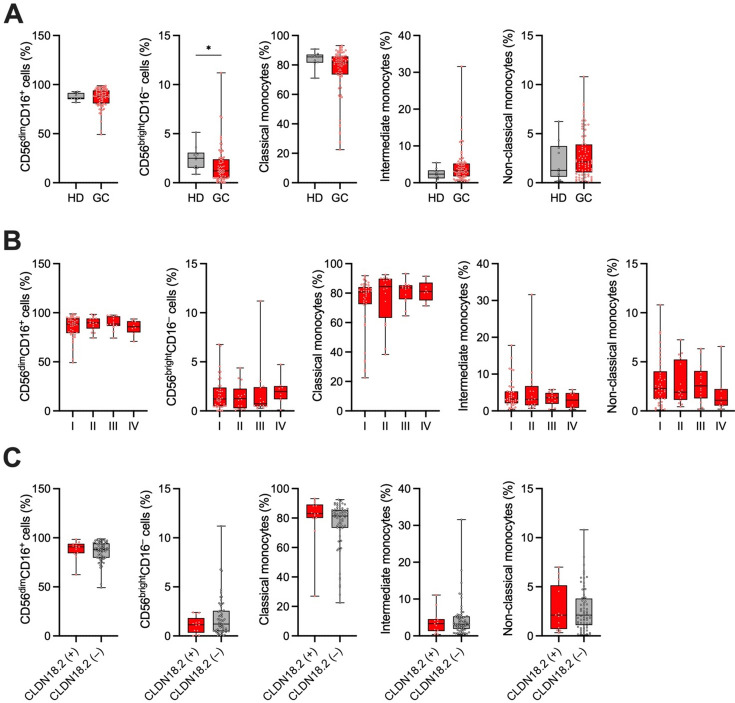
Activating antibody-dependent cellular cytotoxicity (ADCC) by targeting claudin-18 isoform 2 (CLDN18.2) using zolbetuximab, a monoclonal antibody against CLDN18.2, has been considered a promising novel therapeutic strategy for gastric cancer (GC). However, the impact of CLDN18.2 expression on natural killer (NK) cells and monocytes/macrophages-crucial effector cells of ADCC-in GC has not been fully investigated. In the present study, we assessed the impact of CLDN18.2 expression on clinical outcomes, molecular features, and the frequencies of tumor-infiltrating NK cells and macrophages, as well as peripheral blood NK cells and monocytes, in GC by analyzing our own GC cohorts. The expression of CLDN18.2 did not significantly impact clinical outcomes of GC patients, while it was significantly and positively associated with Epstein-Barr virus (EBV) status and PD-L1 expression. The frequencies of tumor-infiltrating NK cells and macrophages, as well as peripheral blood NK cells and monocytes, were comparable between CLDN18.2-positive and CLDN18.2-negative GCs. Importantly, both CLDN18.2 expression and the number of tumor-infiltrating NK cells were significantly higher in EBV-associated GC compared to other molecular subtypes. Our findings support the effectiveness of zolbetuximab in CLDN18.2-positive GC, and offer a novel insight into the treatment of this cancer type, highlighting its potential effectiveness for CLDN18.2-positive/EBV-associated GC.

Keywords: CLDN18.2; Gastric cancer; Monocytes/macrophages; NK cells.


Conflict of interest statement

The authors declare no competing interests.

Figures

Figure 1
Figure 1
Association of CLDN18.2 expression with clinicopathological features and clinical outcomes in GC (FMU cohort 1). (A) Representative IHC images for CLDN18.2 in CLDN18.2 ( +) and CLDN18.2 (–) GCs in FMU cohort 1 (n = 284). Scale bars: 5 mm for a and d, 500 μm for b and e, and 50 μm for c and f. N denotes the adjacent normal region, while T denotes the tumor region. (B) Associations between CLDN18.2 expression detected by IHC and the depth of tumor invasion (T1, n = 149; T2, n = 39; T3, n = 30; T4, n = 66) or pathological TNM stage (I, n = 159; II, n = 61; III, n = 45; IV, n = 19) in patients with GC. (C) Kaplan–Meier curves for RFS and OS according to CLDN18.2 expression detected by IHC in patients with GC [CLDN18.2 ( +), n = 84; CLDN18.2 (–), n = 200]. (D) A panel illustrating the association between CLDN18.2 expression and molecular characteristics detected by IHC or in situ hybridization (for EBV status) in GC (n = 284). Dark grey indicates positive cases, and light grey indicates negative cases for dMMR, EBV, HER2, and PD-L1 (CPS ≥ 5). dMMR stands for deficient mismatch repair, EBV for Epstein-Barr virus, HER2 for human epidermal growth factor receptor 2, PD-L1 for programmed cell death ligand 1, and CPS for a combined positive score. (E) Associations between CLDN18.2 expression and molecular characteristics detected by IHC or in situ hybridization (for EBV status) in GC [dMMR, n = 23 vs pMMR, n = 261; EBV ( +), n = 22 vs EBV (–), n = 262; HER2 IHC ( +), n = 31 vs HER2 IHC (–), n = 253; PD-L1 ( +), n = 111 vs PD-L1 (–), n = 173]. Statistical significance was determined by Fisher’s exact test or the Chi-square test (B,E) and the log-rank test (C). *p < 0.05.
Figure 2
Figure 2
Association of CLDN18 mRNA expression with molecular subtypes in GC (GC cohorts from TCGA and GEO). (A) mRNA expression of CLDN18 in GC samples and paired normal tissues across eight cohorts from GEO (GSE13861: Normal, n = 19 vs Tumor, n = 65; GSE26942: Normal, n = 12 vs Tumor, n = 202; GSE27342: Normal, n = 80 vs Tumor, n = 80; GSE29272: Normal, n = 134 vs Tumor, n = 134; GSE51575: Normal, n = 26 vs Tumor, n = 26; GSE54129: Normal, n = 21 vs Tumor, n = 111; GSE66229: Normal, n = 100 vs Tumor, n = 300; GSE65801: Normal, n = 32 vs Tumor, n = 32). (B) mRNA expression of CLDN18 in each molecular subtype of GC in TCGA cohort (CIN, n = 221; EBV, n = 30; GS, n = 50; MSI, n = 73; ERBB2 Amp + , n = 55; ERBB2 Amp–, n = 352). (C) mRNA expression of CLDN18 between EBV ( +) (n = 12) and EBV (–) (n = 14) GCs in GSE51575. Statistical significance was determined by the Mann–Whitney U test (AC) and the Kruskal–Wallis test with Dunn’s multiple comparisons test (B). ***p < 0.001, ****p < 0.0001.
Figure 3
Figure 3
Association of CLDN18.2 expression with the infiltration of NK cells and macrophages in GC (FMU cohort 1). (A) Representative IHC images for CLDN18.2, CD56, CD16, and CD68 in CLDN18.2 ( +) and CLDN18.2 (–) GCs in FMU cohort 1 (n = 284). Scale bars: 50 μm. (B) Correlations between the number of tumor-infiltrating CD16+ cells and CD56+ cells or CD68+ cells detected by IHC in GCs (n = 284). (C) Comparison of the number of CD56+ cells, CD16+ cells, and CD68+ cells detected by IHC across all pathological TNM stages (I, n = 159; II, n = 61; III, n = 45; IV, n = 19) in GC. (D) Associations between CLDN18.2 expression and infiltration of CD56+ cells, CD16+ cells, or CD68+ cells detected by IHC in GC [CLDN18.2 ( +), n = 84; CLDN18.2 (–), n = 200]. (E) Associations between EBV status and infiltration of CD56+ cells, CD16+ cells, or CD68+ cells detected by in situ hybridization (for EBV) or IHC in GC [EBV ( +), n = 22; EBV (–), n = 262]. Statistical significance was determined by the Spearman rank-correlation coefficient (B), the Kruskal–Wallis test with Dunn’s multiple comparisons test (C), and the Mann–Whitney U test (D,E). *p < 0.05, ***p < 0.001, ****p < 0.0001.
Figure 4
Figure 4
Association of CLDN18.2 expression with the frequencies of peripheral blood NK cells and monocytes in GC (FMU cohort 3). (A) Comparison of the frequencies of CD56dimCD16+ NK cells, CD56brightCD16 NK cells, classical monocytes, intermediate monocytes, and nonclassical monocytes in PBMCs detected by flow cytometry between healthy donors (HD) (n = 10) and GC patients (GC) (n = 79). (B) Comparison of the frequencies of the CD56dimCD16+ NK cells, CD56brightCD16 NK cells, classical monocytes, intermediate monocytes, and nonclassical monocytes in PBMCs detected by flow cytometry across all pathological TNM stages (I, n = 41; II, n = 16; III, n = 14; IV, n = 8) in patients with GC. (C) Associations between CLDN18.2 expression and the frequencies of CD56dimCD16+ NK cells, CD56brightCD16 NK cells, classical monocytes, intermediate monocytes, and nonclassical monocytes in PBMCs in patients with GC [CLDN18.2 ( +), n = 13; CLDN18.2 (–), n = 64]. The frequency of each subset of NK cells and monocytes was determined by flow cytometry, while CLDN18.2 positivity was assessed by IHC using surgically resected GC specimens. Statistical significance was determined by the the Mann–Whitney U test (A,C) and the Kruskal–Wallis test with Dunn’s multiple comparisons test (B). *p < 0.05.

References

    1. Smyth, E. C., Nilsson, M., Grabsch, H. I., van Grieken, N. C. & Lordick, F. Gastric cancer. Lancet396, 635–648. 10.1016/s0140-6736(20)31288-5 (2020). 10.1016/s0140-6736(20)31288-5 - DOI - PubMed
    1. Siegel, R. L., Miller, K. D., Fuchs, H. E. & Jemal, A. Cancer statistics, 2022. CA Cancer J. Clin.72, 7–33. 10.3322/caac.21708 (2022). 10.3322/caac.21708 - DOI - PubMed
    1. Sahin, U. et al. Claudin-18 splice variant 2 is a pan-cancer target suitable for therapeutic antibody development. Clin. Cancer Res.14, 7624–7634. 10.1158/1078-0432.Ccr-08-1547 (2008). 10.1158/1078-0432.Ccr-08-1547 - DOI - PubMed
    1. Sahin, U. et al. FAST: A randomised phase II study of zolbetuximab (IMAB362) plus EOX versus EOX alone for first-line treatment of advanced CLDN18.2-positive gastric and gastro-oesophageal adenocarcinoma. Ann. Oncol.32, 609–619. 10.1016/j.annonc.2021.02.005 (2021). - PubMed
    1. Lordick, F. et al. Patient-reported outcomes from the phase II FAST trial of zolbetuximab plus EOX compared to EOX alone as first-line treatment of patients with metastatic CLDN18.2+ gastroesophageal adenocarcinoma. Gastric Cancer24, 721–730. 10.1007/s10120-020-01153-6 (2021). - PMC - PubMed