Review
. 2024 Aug 2;21(1):54.
doi: 10.1186/s12979-024-00455-y.

The immunology of B-1 cells: from development to aging

Affiliations
Review

The immunology of B-1 cells: from development to aging

Matheus Silvério Mattos et al. Immun Ageing. .

Abstract

B-1 cells have intricate biology, with distinct function, phenotype and developmental origin from conventional B cells. They generate a B cell receptor with conserved germline characteristics and biased V(D)J recombination, allowing this innate-like lymphocyte to spontaneously produce self-reactive natural antibodies (NAbs) and become activated by immune stimuli in a T cell-independent manner. NAbs were suggested as "rheostats" for the chronic diseases in advanced age. In fact, age-dependent loss of function of NAbs has been associated with clinically-relevant diseases in the elderly, such as atherosclerosis and neurodegenerative disorders. Here, we analyzed comprehensively the ontogeny, phenotypic characteristics, functional properties and emerging roles of B-1 cells and NAbs in health and disease. Additionally, after navigating through the complexities of B-1 cell biology from development to aging, therapeutic opportunities in the field are discussed.

Keywords: Aging; Autoantibodies; B cell development; B-1 cell; Immunology; Natural antibody.


Conflict of interest statement

The authors declare no competing interests.

Figures

Fig. 1
Fig. 1
Key molecular events in B-1 cell development. B cell lymphopoiesis takes place in three waves. The first wave starts at E9.0 at the yolk sac. This wave is HSCs-independent and generates only B-1 cells. The second wave takes place in the FL, it is HSCs-dependent and can generate mainly B-1 but also B-2 cells while the third wave happens in the adult BM and mainly generates B-2 cells. (A) Lin28b is responsible to downregulate the micro–RNA Let-7 which normally prevents the activation of the transcriptional factor Arid3a. Activated Arid3a can migrate to the nucleus and bias the choice of VH during VDJ recombination. (B) Additionally, Arid3a can be palmitoylated in the cytoplasm and associate with BCR-containing lipid rafts, increasing the threshold required for BCR activation. (C) Bhlhe41 regulates the expression of the α-chain of the IL-5 receptor. (D) IL-5R signaling is required for B-1 cell proliferation and self-renewal. (E) PU.1 controls the expression of key genes in B-1 cell development such as IL-7Ra, Pax5 and Flt3. (F) CTLA4 is essential for the stability of the BCR and to control B-1 cell proliferation. (G) Absence of IL-7 signaling prevents pSTAT5-mediated inhibition of IgK recombination in FL
Fig. 2
Fig. 2
B-1 cell migration and tissue distribution. Progenitor B-1 cells (Lin CD93+ CD19+ B220low) migrate from FL to the neonate spleen where they can be found as transitional B-1 cells (IgM+ CD19+ CD23± CD93+) until the second week of life. At this moment, transitional B-2 cells starts to arrive in the spleen and the transitional B-1 cells migrates to the body cavities where they arrive trough the omentum and can be found as mature B-1 cells (IgMhigh IgDlow CD19high B220low CD23 CD43+ CD11b+ CD5+ or CD5). Upon activation, B-1 cells downregulate CD11b and CD9 and migrate to the spleen where they upregulate CD6 which is required for B-1 cell proliferation in this compartment. At spleen and in a less extent in the BM, B-1 cells spontaneously differentiate into plasma cells and secret natural IgM
Fig. 3
Fig. 3
Homeostatic B-1 cell functions. B-1 cells secrete NAbs that act as first-line defense against invading pathogens and tissue damage by promoting: A complement cascade activation; B pathogen neutralization; C NAbs-dependent phagocytosis of invading pathogens; D opsonization and NAbs-dependent phagocytosis of dead cells; E modulation the immune system by secreting cytokines. PC: phosphorylcholine
Fig. 4
Fig. 4
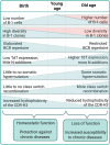
Age-related alterations in B-1 cells and NAbs. The number of B-1 cells in body cavities increases after birth until it peaks in the young adult and is kept until old age. However, tissue specific signals select B-1 cells expressing characteristic BCRs which culminate in the reduction in the diversity of B-1 cells. Although the B-1 cell population is maintained via self-renewal, B-1 cells can be generated de novo by adult BM-derived HSC, which now express TdT. This result in BCRs with more N-additions and more mutations. This, together with reduced hydrophobicity in the CDR-H3 loop, decreases the functionality of NAbs in the elderly, increasing the susceptibility to chronic diseases. The green shades indicate the general age-related alterations. The red shades indicate the age-related alterations in female mice

Similar articles

Cited by

References

    1. Parker DC. T cell-dependent B cell activation. Annu Rev Immunol. 1993;11:331–60. 10.1146/annurev.iy.11.040193.001555. 10.1146/annurev.iy.11.040193.001555 - DOI - PubMed
    1. Kantor AB, et al. An unbiased analysis of V(H)-D-J(H) sequences from B-1a, B-1b, and conventional B cells. J Immunol. 1997;158:1175–86. 10.4049/jimmunol.158.3.1175 - DOI - PubMed
    1. Lanier LL, et al. Expression of Lyt-1 antigen on certain murine B cell lymphomas. J Exp Med. 1981;153:998–1003. 10.1084/jem.153.4.998. 10.1084/jem.153.4.998 - DOI - PMC - PubMed
    1. Hayakawa K, et al. The “Ly-1 B” cell subpopulation in normal immunodefective, and autoimmune mice. J Exp Med. 1983;157:202–18. 10.1084/jem.157.1.202. 10.1084/jem.157.1.202 - DOI - PMC - PubMed
    1. Ghosn EE, et al. CD11b expression distinguishes sequential stages of peritoneal B-1 development. Proc Natl Acad Sci U S A. 2008;105:5195–200. 10.1073/pnas.0712350105. 10.1073/pnas.0712350105 - DOI - PMC - PubMed

LinkOut - more resources