. 2024 Aug 2;21(1):189.
doi: 10.1186/s12974-024-03176-7.

Toxoplasma infection induces an aged neutrophil population in the CNS that is associated with neuronal protection

Affiliations

Toxoplasma infection induces an aged neutrophil population in the CNS that is associated with neuronal protection

Kristina V Bergersen et al. J Neuroinflammation. .

Abstract

Background: Infection with the protozoan parasite Toxoplasma gondii leads to the formation of lifelong cysts in neurons that can have devastating consequences in the immunocompromised. In the immunocompetent individual, anti-parasitic effector mechanisms and a balanced immune response characterized by pro- and anti-inflammatory cytokine production establishes an asymptomatic infection that rarely leads to neurological symptoms. Several mechanisms are known to play a role in this successful immune response in the brain including T cell production of IFNγ and IL-10 and the involvement of CNS resident cells. This limitation of clinical neuropathology during chronic infection suggests a balance between immune response and neuroprotective mechanisms that collectively prevent clinical manifestations of disease. However, how these two vital mechanisms of protection interact during chronic Toxoplasma infection remains poorly understood.

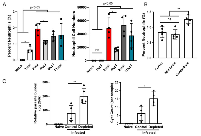
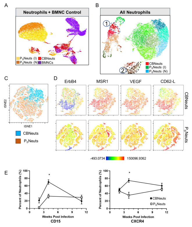
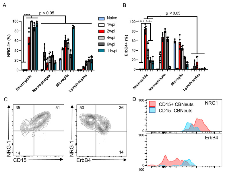
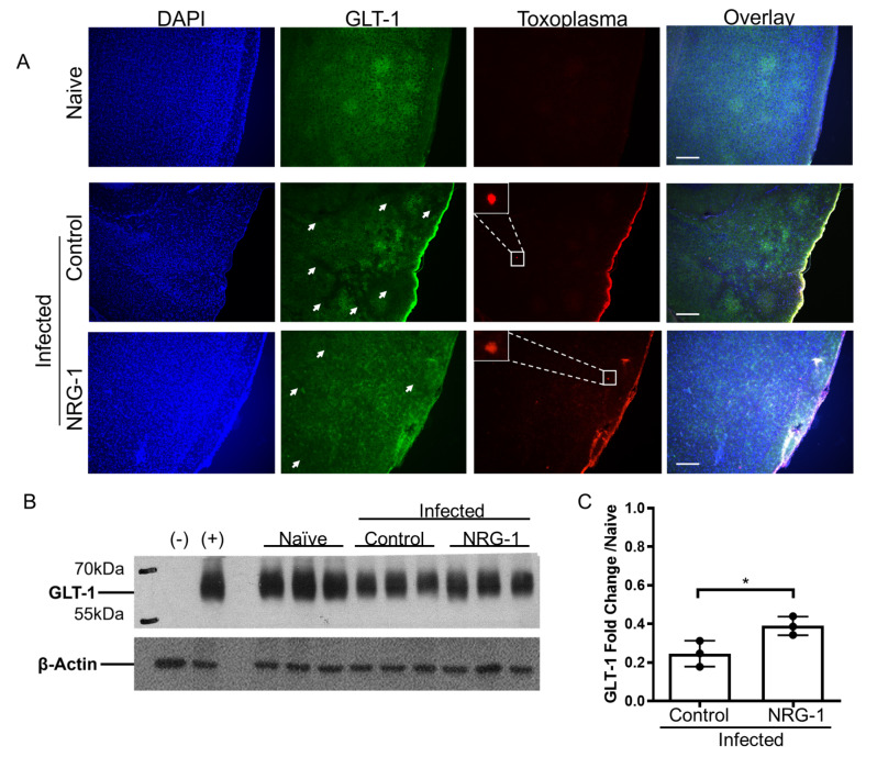
Main text: This study demonstrates a previously undescribed connection between innate neutrophils found chronically in the brain, termed "chronic brain neutrophils" (CBNeuts), and neuroprotective mechanisms during Toxoplasma infection. Lack of CBNeuts during chronic infection, accomplished via systemic neutrophil depletion, led to enhanced infection and deleterious effects on neuronal regeneration and repair mechanisms in the brain. Phenotypic and transcriptomic analysis of CBNeuts identified them as distinct from peripheral neutrophils and revealed two main subsets of CBNeuts that display heterogeneity towards both classical effector and neuroprotective functions in an age-dependent manner. Further phenotypic profiling defined expression of the neuroprotective molecules NRG-1 andErbB4 by these cells, and the importance of this signaling pathway during chronic infection was demonstrated via NRG-1 treatment studies.

Conclusions: In conclusion, this work identifies CBNeuts as a heterogenous population geared towards both classical immune responses and neuroprotection during chronic Toxoplasma infection and provides the foundation for future mechanistic studies of these cells.

Keywords: Toxoplasma gondii; Brain; Chronic infection; Immune response; Neuroprotection; Neutrophils.


Conflict of interest statement

BF holds patents related to the work being reported without direct corporate involvement at the time. The remaining authors declare that the research was conducted in the absence of any commercial or financial relationships that could be construed as a potential conflict of interest.

Figures

Fig. 1
Fig. 1
Neutrophil persistence in the brain during chronic infection is vital for control of parasite replication. C57BL/6J mice were infected intraperitoneally with 10 T. gondii cysts (n = 4 per time point) or injected with PBS as a control (n = 3) and analyzed for immune cells in the brain at both acute (1wpi) and chronic (2, 4, 6, and 11wpi) time points via flow cytometry. For location studies, mice (n = 5) were sacrificed at 4wpi, brains were dissected into 3 distinct regions, and BMNCs were analyzed via flow cytometry to determine neutrophil location in the brain. For neutrophil depletion, a cohort of infected mice received neutralizing Ly6G mAb treatment at 4wpi for 2 weeks to deplete neutrophils. (A) Frequencies (left) and numbers (right) of CD11b + Ly6G + neutrophils in brain at each time point. (B) Quantification of neutrophil percentages in each defined brain region. (C) Quantified parasite burden from whole brain DNA via RT-PCR using T. gondii B1 gene (left) and cyst burden quantified via direct counting from H&E-stained slides (right). * = P < 0.05, ** = P < 0.01, “p-val < 0.05” indicates varying degrees of significance between indicated time points; significance determined via One-way ANOVA (A-B) or unpaired student t-test (C), and error bars indicate SD
Fig. 2
Fig. 2
Transcriptomic and protein analysis of CBNeuts reveals a distinct tissue-specific profile. For transcriptomic profiling, CBNeuts (from n = 7 brains) and PSNeuts (from n = 3 spleens) were sorted from infected mice at 4wpi via flow cytometry and prepped for scRNAseq along with chronically infected BMNCs (n = 3 mice) and naïve spleen neutrophil (n = 3) controls. Phenotypic profiles from CBNeuts and PSNeuts were also evaluated via flow cytometry at 4wpi. For both, neutrophils were identified based on expression of CD11b and Ly6G. (A) UMAP plot of all aggregated scRNAseq samples: PSNeuts from infected mouse (I, yellow), PSNeuts from naïve mouse (N, orange), CBNeuts (red), and BMNCs (purple). (B) UMAP plot of aggregated sorted CBNeuts, PSNeuts from infected mouse spleen (I), and PSNeuts from naïve spleen (N). (C) tSNE plot of concatenated CBNeuts and PSNeuts at 4wpi. (D) tSNE plots of selected alternative molecules from concatenated CBNeuts (top) and PSNeuts (bottom). tSNE plot scale shows populations with low expression (blue) to high expression (red) of molecules. (E) Flow cytometry quantification of CD15 (left) and CXCR4 (right) expression by CBNeuts and PSNeuts at multiple chronic infection time points (2, 4, and 11wpi). * = p < 0.05; significance determined via unpaired student t-test, and error bars indicate SD
Fig. 3
Fig. 3
CBNeuts display age-dependent heterogeneity at the transcriptomic level. CBNeuts (from n = 7 brains) were sorted from infected mice at 4wpi via flow cytometry and prepped for scRNAseq. A) UMAP plot of CBNeuts (red = neutrophil populations) identified as: (1) “Aged Extravasated” (AE) (Ly6Gint, Fut4+, CXCR4+) or (2) “Recently Recruited” (RR) (Ly6Ghi, Fut4int, CXCR4-) subsets. Subsets also identified in aggregated dataset (Fig. 2B). Pink subset identified as red blood cells. B-C) Heat maps of (B) neuroprotective genes (top panel) and genes relating to classical (NETosis) vs. alternative (angiogenesis) functions (bottom panel). (C) GO enrichment terms from top 50 most significantly upregulated genes in AE and RR CBNeuts
Fig. 4
Fig. 4
Neuronal regeneration attempts during chronic infection are inhibited in the absence of neutrophils. C57BL/6J mice were infected intraperitoneally with 10 T. gondii cysts or injected with PBS as a control (n = 5 per group), and a cohort of infected mice received neutralizing Ly6G mAb treatment at 4wpi for 2 weeks to deplete neutrophils. (A) Immunofluorescence images of infected control and neutrophil-depleted brains, 40x images. Blue = DAPI, Green = SLPI, Red = T. gondii, Scale bar = 25 μm. White arrows indicate SLPI + cells. (B) Magnified images of selected regions (dashed white boxes) in (A) to show SLPI stained neuronal axons. (C) Blinded quantification of SPLI + cells. *=P < 0.05; significance determined via unpaired student t-test, and error bars indicate SD
Fig. 5
Fig. 5
CBNeuts express neuroprotective molecules NRG-1 and ErbB4. C57BL/6J mice were infected intraperitoneally with 10 T. gondii cysts (n = 4 per time point) or injected with PBS as a control (n = 3) and analyzed for immune cells in the brain and expression of neuroprotective molecules at both acute (1wpi) and chronic (2, 4, 6, and 11wpi) time points via flow cytometry. For NRG-1 treatment, a cohort of 2 week-infected mice were treated with 5 µg/kg/day of NRG-1 daily for 7 days. (A-B) Time course quantification of expression frequencies (shown by percent of positive cells) of NRG1 (A)and ErbB4 (B) by brain mononuclear cells (BMNCs). (C) Representative flow plots of NRG-1 and ErbB4 expression by CBNeuts at 4wpi. Numbers on flow plots represent the average percentage of expression (n = 4). (D) Expression overlap of neuroprotective molecules distinguished by CD15+ (pink) and CD15- (blue) CBNeuts. For graphs, ****=P < 0.0001 (remaining significance indicated as p < 0.05); significance determined via One-way ANOVA. For all graphs, error bars indicate SD
Fig. 6
Fig. 6
NRG-1 treatment rescues infection-induced GLT-1 levels associated with neurotoxicity. C57BL/6J mice were infected intraperitoneally with 10 T. gondii cysts (n = 4 per time point) or injected with PBS as a control (n = 3). For NRG-1 treatment, a cohort of 2 week-infected mice were treated with 5 µg/kg/day of NRG-1 daily for 7 days. (A) Immunofluorescence images of naïve, infected control, and infected NRG-1-treated mouse brain slices. DAPI (blue), GLT-1 (green), and Toxoplasma (red); white arrows indicate areas of decreased GLT-1 expression. Images taken at 10x magnification, Scale bar = 100 μm. (B) Western blot of GLT-1 from brain homogenate of naïve, infected control, and infected NRG-1-treated mice. Half brains were harvested and protein was extracted, normalized, and run on a Western blot. (C) Quantification of Western blot results using GLT-1:β-Actin ratios. * = P < 0.05; significance determined via unpaired student’s t-test., For all graphs, error bars indicate SD

References

    1. Montoya JG, Liesenfeld O, Toxoplasmosis. Lancet. 2004;363(9425):1965–76. - PubMed
    1. Wang Z-D, Liu H-H, Ma Z-X, Ma H-Y, Li Z-Y, Yang Z-B, et al. Toxoplasma Gondii Infection in Immunocompromised patients: a systematic review and Meta-analysis. Front Microbiol. 2017;8:389. - PMC - PubMed
    1. McGovern KE, Wilson EH. Role of chemokines and trafficking of Immune cells in parasitic infections. Curr Immunol Rev. 2013;9(3):157–68. - PMC - PubMed
    1. Sasai M, Yamamoto M. Innate, adaptive, and cell-autonomous immunity against Toxoplasma Gondii infection. Exp Mol Med. 2019;51(12):1–10. - PMC - PubMed
    1. Biswas A, French T, Düsedau HP, Mueller N, Riek-Burchardt M, Dudeck A, et al. Behavior of Neutrophil Granulocytes during Toxoplasma gondii infection in the Central Nervous System. Front Cell Infect Microbiol. 2017;7:259. - PMC - PubMed