. 2024 Sep 4;32(9):3101-3113.
doi: 10.1016/j.ymthe.2024.07.027. Epub 2024 Aug 5.

TIPE2 gene transfer ameliorates aging-associated osteoarthritis in a progeria mouse model by reducing inflammation and cellular senescence

Affiliations

TIPE2 gene transfer ameliorates aging-associated osteoarthritis in a progeria mouse model by reducing inflammation and cellular senescence

Ping Guo et al. Mol Ther. .

Abstract

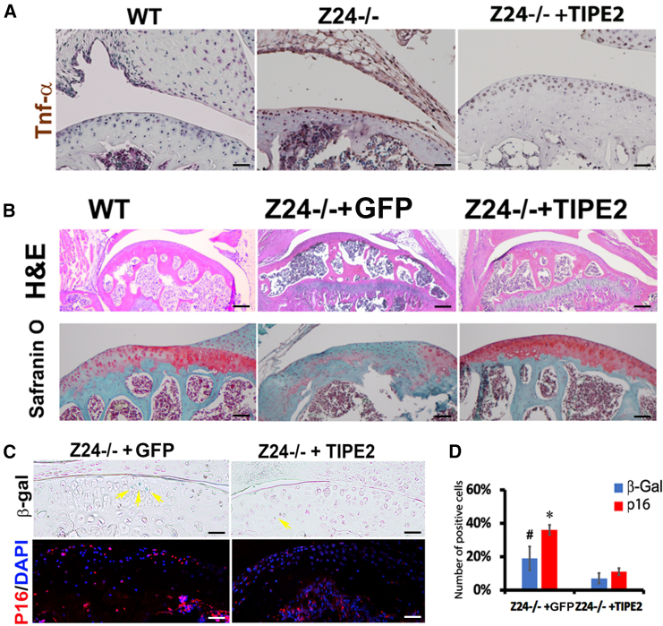
Osteoarthritis (OA) pain is often associated with the expression of tumor necrosis factor alpha (TNF-α), suggesting that TNF-α is one of the main contributing factors that cause inflammation, pain, and OA pathology. Thus, inhibition of TNF-α could potentially improve OA symptoms and slow disease progression. Anti-TNF-α treatments with antibodies, however, require multiple treatments and cannot entirely block TNF-α. TNF-α-induced protein 8-like 2 (TIPE2) was found to regulate the immune system's homeostasis and inflammation through different mechanisms from anti-TNF-α therapies. With a single treatment of adeno-associated virus (AAV)-TIPE2 gene delivery in the accelerated aging Zmpste24-/- (Z24-/-) mouse model, we found differences in Safranin O staining intensity within the articular cartilage (AC) region of the knee between TIPE2-treated mice and control mice. The glycosaminoglycan content (orange-red) was degraded in the Z24-/- cartilage while shown to be restored in the TIPE2-treated Z24-/- cartilage. We also observed that chondrocytes in Z24-/- mice exhibited a variety of senescent-associated phenotypes. Treatment with TIPE2 decreased TNF-α-positive cells, β-galactosidase (β-gal) activity, and p16 expression seen in Z24-/- mice. Our study demonstrated that AAV-TIPE2 gene delivery effectively blocked TNF-α-induced inflammation and senescence, resulting in the prevention or delay of knee OA in our accelerated aging Z24-/- mouse model.

Keywords: AAV; OA; TIPE2; adeno-associated virus; gene delivery; osteoarthritis; senescent cells; tumor necrosis factor α; tumor necrosis factor α-induced protein 8-like 2.


Conflict of interest statement

Declaration of interests The authors declare no competing interests.

Figures

None
Graphical abstract
Figure 1
Figure 1
Z24−/− mice grow slower than the littermates and develop OA spontaneously Photograph of 12-week-old Z24−/− and their littermates (A). Body weight retarded growth compared to their littermates (B). Knee joint was stained with Safranin O, which shows the process of OA development in Z24−/− mice (C). Representative images showing medial tibial plateau cartilage from WT (C-a) to severe in Z24−/− mice (C-f). OARSI score for quantification of osteoarthritic changes (bar graph) (D). ∗p < 0.05 (n = 18). Scale bar: 50 μm.
Figure 2
Figure 2
Downregulation of collagen II and increasing inflammatory factors in isolated chondrocytes from 12-week-old Z24−/− mice (A) Representative western blot of PPARγ, MMP-13, ADAMTS-5, HtrA1, iNOS, and collagen II in chondrocytes determined by western blot analysis normalized to GAPDH (n = 9). (B) Quantification of the protein expression relative to GAPDH. (C) IL-6, IL-1β, TNF-α, and COX-2 levels in chondrocytes measured by ELISA (n = 9). ∗∗∗∗p < 0.001, ∗∗p < 0.01, and ∗p < 0.05.
Figure 3
Figure 3
AAV6-TIPE2 vector construction and delivery to the cartilage in the Z24−/− mice knee (A) Schematic construction for AAV vector. (B) Protein expression of TIPE2. (C) GFP was visualized in mouse cartilage in transduced chondrocytes of the knee articular cartilage, and H&E shows morphology of cartilage. (D) Number of GFP-positive chondrocytes isolated from three Z24−/− mice. Scale bar: 50 μm. Ms1, Ms2, and Ms3 mean mouse numbers 1, 2, and 3.
Figure 4
Figure 4
AAV-TIPE2 gene delivery attenuates the OA pathology and decreases TNF-α expression and the number of senescent cells (A) TNF-α was high expression in the cartilage of 12 weeks of Z24−/− knee (middle panel) and decreased after TIPE2 treatment (right panel). (B) Representative histology of Z24−/− knee cartilage 8 weeks post-TIPE2 treatment and H&E and Safranin O staining. (C) β-gal- and p16-positive cells decreased in the TIPE2-treated knee articular cartilage. (D) Quantification of β-gal- and p16-positive cells. #p < 0.05 and ∗p < 0.01. Scale bar: 50 μm.
Figure 5
Figure 5
TIPE2 gene transduction decreases senescent cell markers in chondrogenic cells (A) TIPE2 gene transduction of ATDC5 cells does not affect the chondrogenic morphology. (B) Representative western blot image of TIPE2 gene transduced cells and quantification showed decrease in senescent p16 gene expression in the presence of TNF-α. (C) The number of β-gal-positive cells are decreased in AAV-TIPE2 transduced cells (n = 5). Bar graphs are the quantification of gene expression in (B) (∗∗p < 0.01 and ∗∗∗p < 0.005) and percentage of β-gal-positive cells in (C) (∗p < 0.01). Scale bar: 50 μm.
Figure 6
Figure 6
TIPE2 gene delivery altered OA marker expression Upregulation of collagen II and decreasing inflammatory factors in isolated AAV-TIPE2 transduced chondrocytes. (A) Representative western blot of iNOS, MMP-13, PPARγ HtrA1, collagen II, and ADAMTS-5 in the isolated chondrocytes determined by western blot analysis normalized to GAPDH (n = 9). TIPE2 expression was decreased in Z24−/− chondrocytes compared to normal controls. (B) Quantification of the protein expression (∗p < 0.05, ∗∗p < 0.01, ∗∗∗p < 0.005, and ∗∗∗∗p < 0.001). (C) IL-1β, IL-6, TNF-α, and COX-2 levels in chondrocytes measured by ELISA (n = 9). ∗∗∗∗p < 0.001.
Figure 7
Figure 7
TIPE2 gene delivery in OA chondrocytes activates multiple signaling pathways (A) Upregulation of p65 and decreasing Rac1 gene expression in isolated AAV-TIPE2 transduced chondrocytes (n = 9). (B) Bar graphs are the quantification of the protein expression in (A) (∗∗p < 0.01 and ∗∗∗∗p < 0.001). (C and D) Downregulation of TGF-β1 and upregulation of TGF-β3, Timp3, and Sirt1 in the isolated chondrocytes determined by western blot analysis normalized to GAPDH (n = 9) and quantified (∗∗∗∗p < 0.001). (E) miR21 and miR155 were downregulated in the isolated chondrocytes by TIPE2 treatment (n = 3, ∗∗∗∗p < 0.001).

References

    1. Yao Q., Wu X., Tao C., Gong W., Chen M., Qu M., Zhong Y., He T., Chen S., Xiao G. Osteoarthritis: pathogenic signaling pathways and therapeutic targets. Signal Transduct. Target. Ther. 2023;8:56. doi: 10.1038/s41392-023-01330-w. - DOI - PMC - PubMed
    1. Sofat N., Ejindu V., Kiely P. What makes osteoarthritis painful? The evidence for local and central pain processing. Rheumatology (Oxford) 2011;50:2157–2165. doi: 10.1093/rheumatology/ker283. - DOI - PubMed
    1. Sanchez-Lopez E., Coras R., Torres A., Lane N.E., Guma M. Synovial inflammation in osteoarthritis progression. Nat. Rev. Rheumatol. 2022;18:258–275. doi: 10.1038/s41584-022-00749-9. - DOI - PMC - PubMed
    1. Li H., Xie S., Qi Y., Li H., Zhang R., Lian Y. TNF-alpha increases the expression of inflammatory factors in synovial fibroblasts by inhibiting the PI3K/AKT pathway in a rat model of monosodium iodoacetate-induced osteoarthritis. Exp. Ther. Med. 2018;16:4737–4744. doi: 10.3892/etm.2018.6770. - DOI - PMC - PubMed
    1. Cho Y., Jeong S., Kim H., Kang D., Lee J., Kang S.B., Kim J.H. Disease-modifying therapeutic strategies in osteoarthritis: current status and future directions. Exp. Mol. Med. 2021;53:1689–1696. doi: 10.1038/s12276-021-00710-y. - DOI - PMC - PubMed

MeSH terms

Substances

LinkOut - more resources