

Phages produce persisters
- PMID: 39096350
- PMCID: PMC11297538
- DOI: 10.1111/1751-7915.14543
Phages produce persisters
Abstract
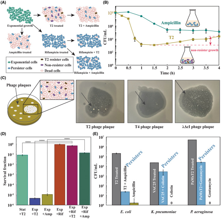
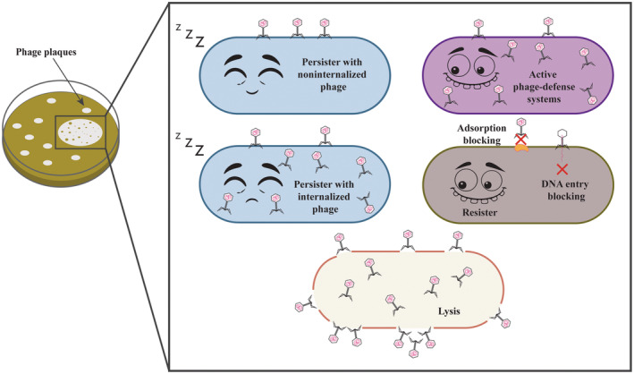
Arguably, the greatest threat to bacteria is phages. It is often assumed that those bacteria that escape phage infection have mutated or utilized phage-defence systems; however, another possibility is that a subpopulation forms the dormant persister state in a manner similar to that demonstrated for bacterial cells undergoing nutritive, oxidative, and antibiotic stress. Persister cells do not undergo mutation and survive lethal conditions by ceasing growth transiently. Slower growth and dormancy play a key physiological role as they allow host phage defence systems more time to clear the phage infection. Here, we investigated how bacteria survive lytic phage infection by isolating surviving cells from the plaques of T2, T4, and lambda (cI mutant) virulent phages and sequencing their genomes. We found that bacteria in plaques can escape phage attack both by mutation (i.e. become resistant) and without mutation (i.e. become persistent). Specifically, whereas T4-resistant and lambda-resistant bacteria with over a 100,000-fold less sensitivity were isolated from plaques with obvious genetic mutations (e.g. causing mucoidy), cells were also found after T2 infection that undergo no significant mutation, retain wild-type phage sensitivity, and survive lethal doses of antibiotics. Corroborating this, adding T2 phage to persister cells resulted in 137,000-fold more survival compared to that of addition to exponentially growing cells. Furthermore, our results seem general in that phage treatments with Klebsiella pneumonia and Pseudomonas aeruginosa also generated persister cells. Hence, along with resistant strains, bacteria also form persister cells during phage infection.
© 2024 The Author(s). Microbial Biotechnology published by John Wiley & Sons Ltd.
Conflict of interest statement
The authors declare no conflicts of interest.
Figures
References
-
- Abedon, S.T. (2018) Detection of bacteriophages: phage plaques. In: Harper, D.R. , Abedon, S.T. , Burrowes, B.H. & McConville, M.L. (Eds.) Bacteriophages: biology, technology, therapy. Cham: Springer International Publishing, pp. 1–32.
-
- Cruz‐Muñiz, M.Y. , López‐Jacome, L.E. , Hernández‐Durán, M. , Franco‐Cendejas, R. , Licona‐Limón, P. , Ramos‐Balderas, J.L. et al. (2017) Repurposing the anticancer drug mitomycin C for the treatment of persistent Acinetobacter baumannii infections. International Journal of Antimicrobial Agents, 49, 88–92. - PubMed
Publication types
MeSH terms
Associated data
- Actions
Grants and funding
LinkOut - more resources
Full Text Sources
