. 2024 Nov;27(4):691-699.
doi: 10.1007/s10456-024-09940-w. Epub 2024 Aug 3.

Mitochondrial control of hypoxia-induced pathological retinal angiogenesis

Affiliations

Mitochondrial control of hypoxia-induced pathological retinal angiogenesis

Hitomi Yagi et al. Angiogenesis. 2024 Nov.

Erratum in

Abstract

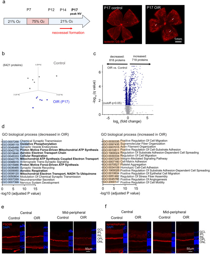
Objective: Pathological retinal neovascularization is vision-threatening. In mouse oxygen-induced retinopathy (OIR) we sought to define mitochondrial respiration changes longitudinally during hyperoxia-induced vessel loss and hypoxia-induced neovascularization, and to test interventions addressing those changes to prevent neovascularization.

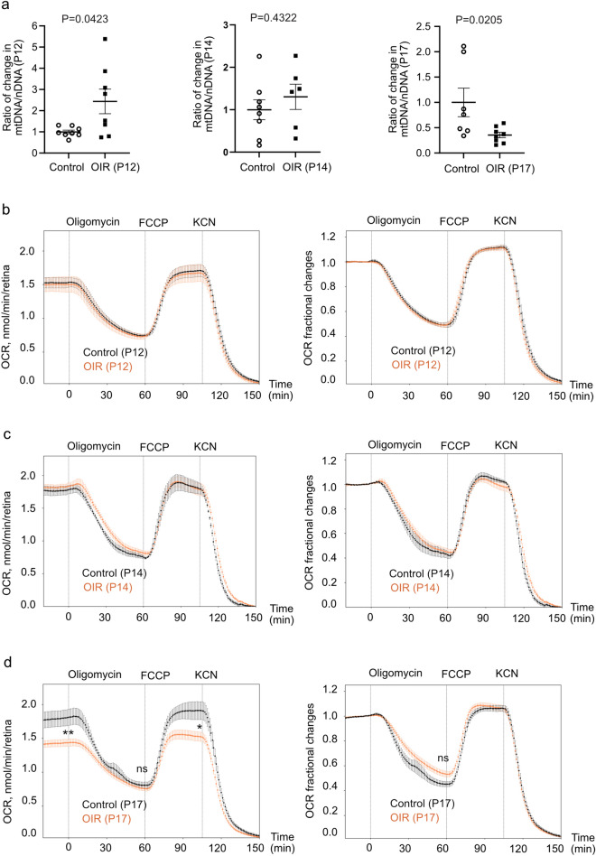
Methods: OIR was induced in C57BL/6J mice and retinal vasculature was examined at maximum neovessel formation. We assessed total proteome changes and the ratio of mitochondrial to nuclear DNA copy numbers (mtDNA/nDNA) of OIR vs. control retinas, and mitochondrial oxygen consumption rates (OCR) in ex vivo OIR vs. control retinas (BaroFuse). Pyruvate vs. vehicle control was supplemented to OIR mice either prior to or during neovessel formation.

Results: In OIR vs. control retinas, global proteomics showed decreased retinal mitochondrial respiration at peak neovascularization. OCR and mtDNA/nDNA were also decreased at peak neovascularization suggesting impaired mitochondrial respiration. In vivo pyruvate administration during but not prior to neovessel formation (in line with mitochondrial activity time course) suppressed NV.

Conclusions: Mitochondrial energetics were suppressed during retinal NV in OIR. Appropriately timed supplementation of pyruvate may be a novel approach in neovascular retinal diseases.

Keywords: Hypoxia; Mitochondrial respiration; Neovascularization; Oxygen-induced retinopathy; Retinal angiogenesis; Retinopathy of prematurity.


Conflict of interest statement

Declarations Competing interests IRS and MG have financial ties to EnTox Sciences (Mercer Island, WA), manufacturer of BaroFuse. All other authors declare no conflicts of interest.

Figures

Fig. 1
Fig. 1
Global proteomic analysis of OIR vs. control mouse retinas. a Schematic of the mouse OIR model (left). Neovessels form from postnatal day (P)14 and peak at P17. Representative images of retinal whole mounts (right) at P17 with isolectin GS-IB4 vessel staining (red). OIR retinas exhibited central vaso-obliteration (VO) and mid-peripheral neovascularization (NV, white arrows). Scale bar 1 mm. b Unfiltered principal component analysis of 6421 proteins depicting differences between retinal protein profiles of P17 OIR (blue) vs. control mice (grey). Each dot represents one retina (n = 6 retinas/group). c Volcano plot of differentially abundant proteins between P17 OIR vs. control retinas. Each data point represents a unique protein based on log2 (fold change) on the x-axis and -log10 (q value) on the y-axis. Cutoff corresponds to a false discovery rate-adjusted P value (q value) < 0.05. d Top-ranked Gene Ontology (GO) terms of biological processes with decreased (left) or increased (right) protein abundance (q < 0.05) in P17 OIR vs. control retinas. Pathways were sorted by -log10 (adjusted P value). Pathways for mitochondrial activity are highlighted in bold. e, f Immunostaining for retinal synapses in P17 OIR vs. control retinas: PSD95 (postsynaptic, left, red) and synaptophysin (presynaptic, right, red). Cell nuclei were labeled with 4′,6-diamidine-2′-phenylindole dihydrochloride (DAPI, blue). Images were taken in central and mid-peripheral retinal areas. GCL Ganglion cell layer, IPL inner plexiform layer, INL inner nuclear layer, OPL outer plexiform layer, ONL outer nuclear layer. Scale bar 50 µm
Fig. 2
Fig. 2
Impaired mitochondrial respiration in OIR vs. control retinas. a Altered mtDNA/nDNA in P12, P14, and P17 OIR vs. control retinas. Ratio of change was calculated and compared with control retinas. Unpaired t-test or Mann-Whitney test (n = 6-8 mice/group). bd Oxygen consumption rate (OCR, left) and factional changes of OCR with baseline set to 1 (right) in P12 (n = 6 retinas/group), P14 (n = 3/group), and P17 (OIR, n = 16; control, n = 5) OIR and control retinas. Oligomycin A (ATP synthase inhibitor), carbonyl cyanide 4-(trifluoromethoxy)phenylhydrazone (FCCP, uncoupler of mitochondrial respiration), and potassium cyanide (KCN, complex IV inhibitor) were sequentially applied. Decreased OCR in P17 OIR vs. control retinas at baseline and at FCCP-induced maximal respiratory capacity confirmed by unpaired t-test. P < 0.01 (**), P < 0.05 (*), ns not significant
Fig. 3
Fig. 3
Pyruvate protected against hypoxia-induced pathological retinal angiogenesis. a Replot of pyruvic acid in OIR vs. control retinas at P17 from our prior publication. Unpaired t-test (n = 3-4 samples/group with 6 retinas/sample). (Tomita et al., Diabetologia. 2021 Jan;64(1):70–82). b Pyruvate sustained mitochondrial respiration under glucose deprivation conditions in OIR retinas. BaroFuse analysis of OCR in P17 C57BL/6J OIR retinas perfused with Krebs-Ringer Solution supplemented with 1 mM glucose only (control) vs. 1 mM glucose and 10 mM pyruvate. BAY-876 (glucose transporter inhibitor, 20 µM) was injected into both media to block glucose entry into the cells. Unpaired t-test was applied to detect differences in OCR 30, 60, and 90 min after BAY-876 injection between retinas treated with pyruvate vs. control (n = 4-5 retinas/group, 2 independent experiments). **P < 0.01, ***P < 0.001. c Schematics of pyruvate supplementation in OIR mice. Mouse pups were intraperitoneally injected with 50 µg/g pyruvate or vehicle daily either prior to or during neovessel formation. d, e At P17, neovascular (NV) and vaso-obliterated (VO) area were examined after 50 µg/g pyruvate or vehicle treatment from P12-P14 d or P14-P16 e. Scale bar 1 mm. Ratio of change was calculated and compared with vehicle group. Unpaired t-test (n = 8-14 retinas/group). f, g Quantification of NV, VO in OIR pups after 10 µg/g f or 70 µg/g g pyruvate vs. vehicle treatment from P14-P16. Unpaired t-test (n = 12-13 retinas/group)

References

    1. Hartnett ME, Lane RH (2013) Effects of oxygen on the development and severity of retinopathy of prematurity. J AAPOS 17(3):229–234. 10.1016/j.jaapos.2012.12.155 - PMC - PubMed
    1. Mandala VK, Urakurva AK, Gangadhari S, Kotha R Sr (2023) The effects of early enteral and parental nutrition on retinopathy of prematurity: a systematic review. Cureus 15(11):e49029. 10.7759/cureus.49029 - PMC - PubMed
    1. Fu Z, Nilsson AK, Hellstrom A, Smith LEH (2022) Retinopathy of prematurity: metabolic risk factors. Elife. 10.7554/eLife.80550 - PMC - PubMed
    1. Sato T, Wada K, Arahori H, Kuno N, Imoto K, Iwahashi-Shima C, Kusaka S (2012) Serum concentrations of bevacizumab (avastin) and vascular endothelial growth factor in infants with retinopathy of prematurity. Am J Ophthalmol 153(2):327-333.e321. 10.1016/j.ajo.2011.07.005 - PubMed
    1. Zehden JA, Mortensen XM, Reddy A, Zhang AY (2022) Systemic and ocular adverse events with intravitreal anti-VEGF therapy used in the treatment of diabetic retinopathy: a review. Curr Diab Rep 22(10):525–536. 10.1007/s11892-022-01491-y - PubMed

MeSH terms

LinkOut - more resources