Review
. 2024 Aug 3;19(1):121.
doi: 10.1186/s11671-024-04072-y.

Biogenic nanoparticles: pioneering a new era in breast cancer therapeutics-a comprehensive review

Affiliations
Review

Biogenic nanoparticles: pioneering a new era in breast cancer therapeutics-a comprehensive review

Shahnawaz Ahmad Bhat et al. Discov Nano. .

Abstract

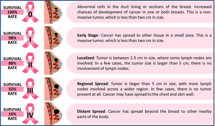
Breast cancer, a widespread malignancy affecting women globally, often arises from mutations in estrogen/progesterone receptors. Conventional treatments like surgery, radiotherapy, and chemotherapy face limitations such as low efficacy and adverse effects. However, nanotechnology offers promise with its unique attributes like targeted delivery and controlled drug release. Yet, challenges like poor size distribution and environmental concerns exist. Biogenic nanotechnology, using natural materials or living cells, is gaining traction for its safety and efficacy in cancer treatment. Biogenic nanoparticles synthesized from plant extracts offer a sustainable and eco-friendly approach, demonstrating significant toxicity against breast cancer cells while sparing healthy ones. They surpass traditional drugs, providing benefits like biocompatibility and targeted delivery. Thus, this current review summarizes the available knowledge on breast cancer (its types, stages, histopathology, symptoms, etiology and epidemiology) with the importance of using biogenic nanomaterials as a new and improved therapy. The novelty of this work lies in its comprehensive examination of the challenges and strategies for advancing the industrial utilization of biogenic metal and metal oxide NPs. Additionally; it underscores the potential of plant-mediated synthesis of biogenic NPs as effective therapies for breast cancer, detailing their mechanisms of action, advantages, and areas for further research.

Keywords: Biogenic nanoparticles; Breast cancer; Conventional treatment; Medicinal plants; Nanomaterials.


Conflict of interest statement

The authors declare no competing interests.

Figures

Fig. 1
Fig. 1
Graphical representation of age-standardized mortality rate among all ages due to different types of cancer in 2020 globally [8]
Fig. 2
Fig. 2
Graphical representation illustrating age standardized BC incidence and mortality statistics worldwide categorized by region (Data source: Ref. [25])
Fig. 3
Fig. 3
Different stages of breast cancer
Fig. 4
Fig. 4
Breast cancer imaging. A postmenopausal woman 53 years of age with no family history and no clinical findings underwent routine breast screening with mammography, which detected a lesion in the right breast (panel a, cranio-caudal view (left) and mediolateral oblique view (right)). The images were also acquired with digital breast tomosynthesis, which showed a small spiculated lesion in the lower inner quadrant (panel b, mediolateral oblique view). The lesion was investigated with ultrasonography (panel c), and biopsy confirmed an invasive ductal carcinoma on histology. MRI showed the enhancing spiculated mass (panel d). The tumour is indicated within the dashed lines in each panel. [89]
Fig. 5
Fig. 5
Benign breast lumps on standard of care ultrasound and volume sweep imaging (VSI). Benign anechoic cyst with posterior acoustic enhancement seen on standard of care ultrasound (A) and VSI (B). Hypoechoic well-circumscribed oval fibroadenoma seen on standard of care ultrasound (C) and VSI (D). Isoechoic or hyperechoic lipoma (arrows) in the superficial breast tissue on standard of care ultrasound (E) and VSI (F) [92]
Fig. 6
Fig. 6
Breast cancer histological types and molecular alterations, The WHO classification distinguishes various invasive breast cancer subtypes that have particular molecular changes, some of which are shown here [98]. For instance, CDH1 mutations are present in 85% of instances of lobular carcinomas and their progenitors (lobular neoplasia), which results in the pathognomonic decrease of E-Cadherin expression [99], [100]. Additionally, they have copy number increases in ESR1, PIK3CA, PTEN, AKT1, ERBB2 and ERBB3 mutations, and PTEN. Secretory carcinomas have a particular translocation, t(12;15), which results in a gene fusion. NTRK3–ETV6 [101]. whereas t(6;9) and the fusion gene MYB-NFIB32 define adenoid cystic carcinoma [102]. Understanding these characteristics may aid in the creation of treatments that are specifically suited for certain histological subtypes [99]. Oestrogen receptor (ER), human epidermal growth factor receptor (HER2), and tall cell carcinoma with reverse polarity were identified by G. MacGrogan of the Institut Bergognié in France. (Remove the reference in the legend and cite the article from where the image has been taken)
Fig. 7
Fig. 7
Illustrates a comparison between passive and active targeting of drugs for breast cancer treatment [123]
Fig. 8
Fig. 8
Graphical demonstration of inorganic nanoparticles along with their conjugating materials and their potential applications
Fig. 9
Fig. 9
Outlines an overview of review articles focusing on the green synthesis of various metallic nanoparticles using different biological agents, along with their potential breast cancer activity
Fig. 10
Fig. 10
Illustration depicting plant-mediated approaches for nanoparticle fabrication
Fig. 11
Fig. 11
An overview of in vivo and in vitro assessment of grass-mediated biogenic synthesis of AgNPs. Symbol ‘g’ represent ‘Gram’, ‘%’ represents ‘Percentage’, ‘*’ signifies ‘p ≤ 0.05’ and ‘**’ signifies ‘P ≤ 0.001’. Reprinted with permission from Ref. [208]. Copyright 2021. Elsevier
Fig. 12
Fig. 12
Apoptosis induced by Au-NPs and AgNPs in MCF-7 cells. a Photomicrographs of MCF-7 cells stained with AO/EB after 24 h incubation with Au-NPs or AgNPs. Viable cells appear light green, early apoptotic cells appear in bright green fluorescence, late apoptosis cells appear in red to orange fluorescence and necrosis cells appear in red fluorescence. b Representative fluorescent micrographs of MCF-7 cells stained with Hoechst 33,258 fluorescent dye after 24 h incubation with either Au-NPs or AgNPs at ×200 Magnification. c Percentage distribution of apoptotic cells including viable, early apoptotic, late apoptotic and necrotic cells. d AgNPs induced inter-nucleosomal DNA fragmentation. MCF-7 cells were treated with different concentrations of AgNPs (0, 100 and 200 μl ml−1) for 24 h. Cells were harvested, and DNA was extracted. Fragmented DNA was extracted and analyzed via agarose gel electrophoresis. Representative gels from one of the three experiments. Reprinted with permission from Ref. [259]. Copyright 2011 RSC advances
Fig. 13
Fig. 13
The antiproliferative activity of CuO NPs against AMJ-13, MCF-7, and HBL-100 cancer cell lines. Triplicate samples of each cancer cell lines were prepared (concentration of 1 × 104 cells/mL) and treated with CuO NPs, which were subjected to 72 h incubation. Viability of cells was assessed via MTT assay by measuring the absorbance at a 492 nm wavelength. (Under Creative Commons CC BY License)
Fig. 14
Fig. 14
Fluorescence microscopy images illustrating the effects of CdS QDs on MCF-7 cell line. a Representative image of untreated MCF-7 cells. Images of MCF-7 cells treated with different concentrations of CdS QDs: b 15 μg/mL, c 30 μg/mL, and d 45 μg/mL. The cells were stained with AO/EtBr, where live cells appeared green, early and late apoptotic cells appeared yellow and reddish orange, respectively. Additional images of MCF-7 cells stained with DAPI: e untreated cells, f 15 μg/mL, g 30 μg/mL, and h 45 μg/mL of CdS QDs treated cells after 24 h. Apoptotic analysis using Annexin V/FITC staining: i untreated MCF-7 cells and CdS QDs treated MCF-7 cells with different concentrations: j 15 μg/mL, k 30 μg/mL, and l 45 μg/mL. Apoptotic cells are indicated by red color spots (arrow). These findings provide insights into the apoptotic effects of CdS QDs on MCF-7 cell line, as observed through fluorescence microscopy and Annexin V/FITC staining. Reprinted with permission from Ref. [294]. Copyright 2019 Elsevier

Similar articles

Cited by

References

    1. Siegel RL, Wagle NS, Cercek A, Smith RA, Jemal A. Colorectal cancer statistics, 2023. CA Cancer J Clin. 2023;73(3):233–54. 10.3322/CAAC.21772. 10.3322/CAAC.21772 - DOI - PubMed
    1. Olsen M, Lof P, Stiekema A, van den Broek D, Wilthagen EA, Bossuyt PM, Lok CAR. The diagnostic accuracy of human epididymis protein 4 (HE4) for discriminating between benign and malignant pelvic masses: a systematic review and meta-analysis. Acta Obstet Gynecol Scand. 2021;100(10):1788–99. 10.1111/AOGS.14224. 10.1111/AOGS.14224 - DOI - PubMed
    1. Sarkar S, Horn G, Moulton K, Oza A, Byler S, Kokolus S, Longacre M. Cancer development, progression, and therapy: an epigenetic overview. Int J Mol Sci. 2013;14(10):21087. 10.3390/IJMS141021087. 10.3390/IJMS141021087 - DOI - PMC - PubMed
    1. Cancer Tomorrow https://gco.iarc.fr/tomorrow/en. Accessed Mar 9, 2023.
    1. Bray F, Ferlay J, Soerjomataram I, Siegel RL, Torre LA, Jemal A. Global cancer statistics 2018: GLOBOCAN estimates of incidence and mortality worldwide for 36 cancers in 185 countries. CA Cancer J Clin. 2018;68(6):394–424. 10.3322/caac.21492. 10.3322/caac.21492 - DOI - PubMed

LinkOut - more resources