. 2024 Aug 20;5(8):101678.
doi: 10.1016/j.xcrm.2024.101678. Epub 2024 Aug 2.

β-resorcylic acid released by Limosilactobacillus reuteri protects against cisplatin-induced ovarian toxicity and infertility

Affiliations

β-resorcylic acid released by Limosilactobacillus reuteri protects against cisplatin-induced ovarian toxicity and infertility

Yinglin Feng et al. Cell Rep Med. .

Abstract

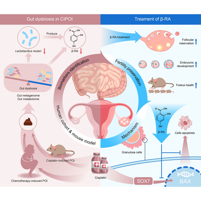
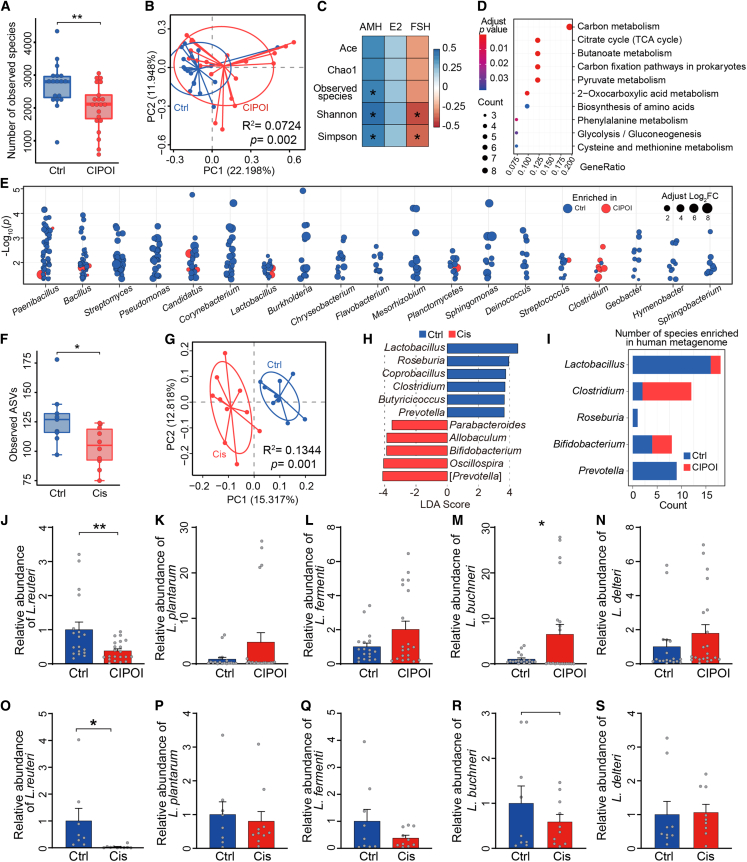
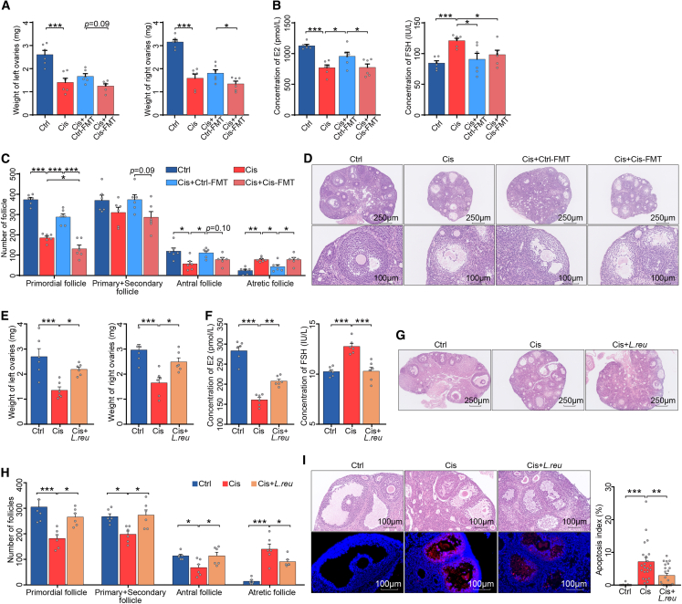
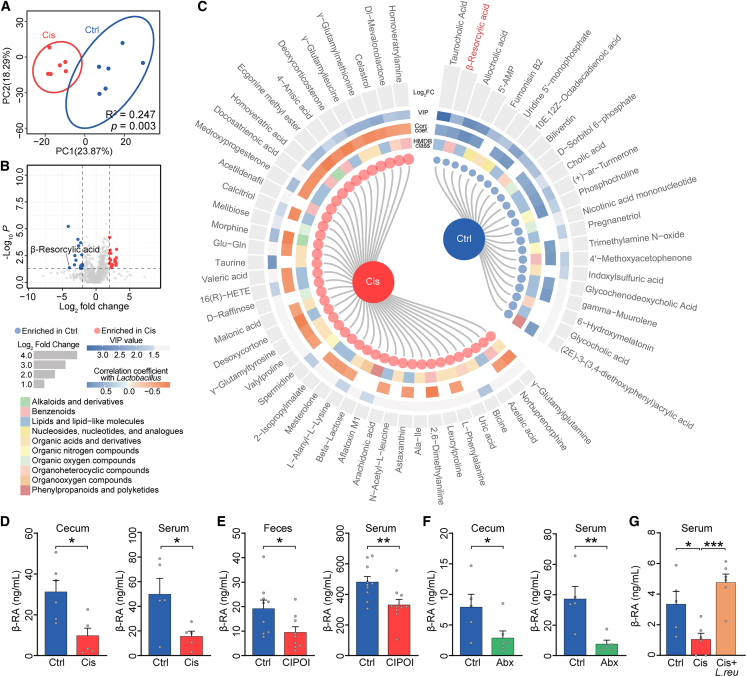
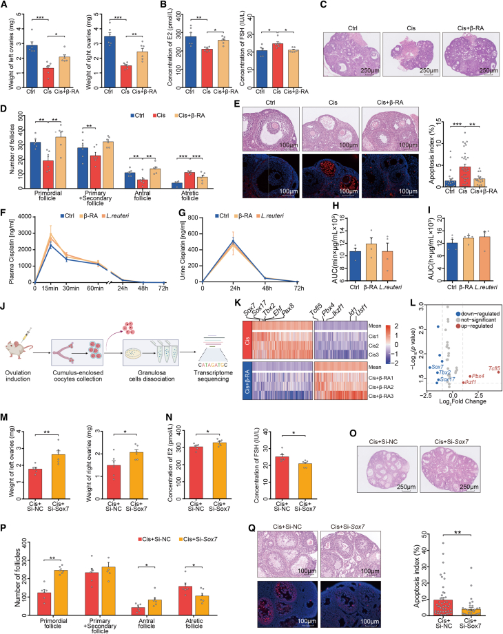
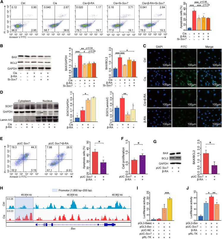
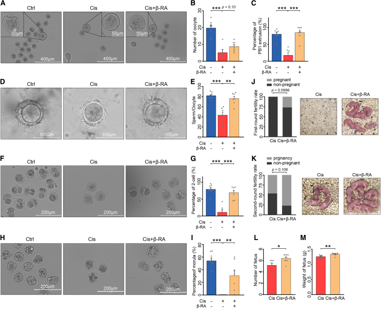
Chemotherapy-induced premature ovarian insufficiency (CIPOI) triggers gonadotoxicity in women undergoing cancer treatment, leading to loss of ovarian reserves and subfertility, with no effective therapies available. In our study, fecal microbiota transplantation in a cisplatin-induced POI mouse model reveals that a dysbiotic gut microbiome negatively impacts ovarian health in CIPOI. Multi-omics analyses show a significant decrease in Limosilactobacillus reuteri and its catabolite, β-resorcylic acid , in the CIPOI group in comparison to healthy controls. Supplementation with L. reuteri or β-RA mitigates cisplatin-induced hormonal disruptions, morphological damages, and reductions in follicular reserve. Most importantly, β-RA pre-treatment effectively preserves oocyte function, embryonic development, and fetus health, thereby protecting against chemotherapy-induced subfertility. Our results provide evidence that β-RA suppresses the nuclear accumulation of sex-determining region Y-box 7, which in turn reduces Bcl-2-associated X activation and inhibits granulosa cell apoptosis. These findings highlight the therapeutic potential of targeting the gut-ovary axis for fertility preservation in CIPOI.

Keywords: Limosilactobacillus reuteri; cisplatin-induced premature ovarian insufficiency; gut metabolome; gut microbiota; β-resorcylic acid.


Conflict of interest statement

Declaration of interests The authors declare no competing interests.

Figures

None
Graphical abstract
Figure 1
Figure 1
Dysbiosis of gut microbiota is associated with POI (A) Comparison of the number of observed species between CIPOI and control (Ctrl) groups. ∗∗p < 0.01 by Wilcoxon rank-sum test. (B) Principal coordinate analysis (PCoA) based on Bray-Curtis distances revealed that the Ctrl bacterial communities clustered separately from CIPOI bacterial communities. Each point represents a single sample, colored by group. Differences in beta diversity between CIPOI and Ctrl were tested by permutational multivariate analysis of variance (PERMANOVA). (C) Correlation between alpha-diversity indices and serum hormone level. Spearman’s rho statistic was adjusted using the Benjamini and Hochberg method. ∗p < 0.05. (D) Significant microbial pathways associated with the differentially expressed microbial genes using over-representation analysis (ORA). (E) Gut microbiota showed significantly different abundances between the CIPOI and Ctrl groups. Each point represents one species, and genera comprising over 10 species are displayed. The colors correspond to enrichment in CIPOI or Ctrl, and the circle sizes refer to the Log2 fold change. (F) Comparison of amplicon sequence variants (ASVs) between Cis and Ctrl mice. ∗p < 0.05 by Wilcoxon rank-sum test. (G) PCoA based on unweighted UniFrac distances revealed that the Ctrl bacterial communities clustered separately from the Cis bacterial communities. Each point represents a single sample, colored by group. Differences in beta diversity between Cis and Ctrl were tested by PERMANOVA. (H) Linear discriminant analysis effect size identified the most differentially abundant taxa between the two groups. Ctrl-enriched genera are indicated with a positive linear discriminant analysis (LDA) score, and genera enriched in Cis controls had a negative score. Only taxa meeting an LDA significant threshold of >3 are shown. (I) Concordance of genus variations between the intestinal microbiota of the human and mouse model. The counts indicate the number of differential species in the human metagenome data. (J–N) Relative abundance of Lactobacillus spp. of fecal samples from participants of the healthy group and the CIPOI group (N = 19 for Ctrl and N = 21 for CIPOI) (J) L. reuteri; (K) L. plantarum; (L) L. fermenti; (M) L. buchneri; (N) L. delteri. (O–S) Relative abundance of Lactobacillus spp. of fecal samples from mice of the control group and the Cis-POI group (n = 8 for Ctrl and n = 10 for Cis-POI). (O) L. reuteri; (P) L. plantarum; (R) L. fermenti; (S) L. buchneri; (L) L. delteri. Data in (J)–(S) presented as the mean ± SEM. ∗p < 0.05, ∗∗∗p < 0.001; two-tailed Student’s t test.
Figure 2
Figure 2
The gut microbiota regulated ovarian toxicity induced by cisplatin (A) Ovarian weight of Ctrl, Cis, Cis+Ctrl-FMT, and Cis+Cis-FMT mice (n = 6). (B) Serum levels of E2 and FSH in Ctrl, Cis, Cis+Ctrl-FMT, and Cis+Cis-FMT mice (n = 6). (C) Quantification of primordial, primary + secondary, antral, and atretic follicles (n = 6). (D) Representative images of ovaries and antral follicles in recipient mice by H&E. (E) Ovarian weight of Ctrl, Cis-POI, and Cis-POI+ L. reuteri mice (n = 6). (F) Serum levels of E2 and FSH in Ctrl, Cis-POI, and Cis-POI+ L. reuteri group (n = 6). (G) H&E staining of ovaries from mice in each group. (H) Quantitative analysis of the primordial, primary + secondary, antral, and atretic follicles (n = 6). (I) Representative images of H&E-stained histological sections from antral follicles and TUNEL-based quantification of the apoptotic index in the antral follicles (n = 17 for Ctrl, n = 21 for Cis-POI, and n = 23 for Cis-POI+L. reuteri). The data are all presented as the mean ± SEM. ∗p < 0.05, ∗∗p < 0.01, ∗∗∗p < 0.001; one-way ANOVA following the Benjamini and Hochberg’s method for multiple adjustments in (A, B, and C); one-way ANOVA following the Dunnett’s multiple comparisons test in (F, G, I, and J).
Figure 3
Figure 3
The metabolite of gut microbiota, β-RA, is downregulated in individuals with CIPOI (A) Principal-component analysis of metabolites in samples from Cis mice (red points) and Ctrl mice (blue points). Permutational multivariate analysis of variance with the Bray-Curtis distance metric was used to assess the significance of differences between the two groups. (B) Volcano plots of the metabolites. The data were compared using a two-tailed unpaired Student’s t test. (C) The differential metabolites between Cis and Ctrl mice. Metabolites with VIP score >1 and p value <0.05 were significant. Blue points and red points in the internal cycle represent the enrichment in Ctrl and Cis mice, respectively. The second, third, and fourth inter-cycle represent the class of metabolites according to the Human Metabolome Database (HMDB) database, the correlation coefficient with Lactobacillus, and VIP scores, respectively. The external cycle shows the Log2 fold of change of each metabolite. (D) β-RA levels in cecum content and serum from Ctrl and Cis-POI mice (n = 5). (E) β-RA levels in feces and serum from Ctrl and CIPOI women (n = 8). (F) β-RA levels in cecal content and serum of mice treated with or without antibiotics (Abx) (n = 5). (G) β-RA levels in serum from CIPOI mice treated with L. reuteri (109 colony-forming units [CFUs] per day), gavaged to mice from 3 days before cisplatin exposure, and throughout the period of CIPOI modeling (n = 5 for Ctrl, n = 6 for Cis-POI, and Cis-POI+L. reuteri). The data are all presented as the mean ± SEM. ∗p < 0.05, ∗∗p < 0.01, ∗∗∗p < 0.001; Spearman’s rho correlation in (C); two-tailed Student’s t test in (D–F); one-way ANOVA following the Dunnett’s multiple comparisons test in (G).
Figure 4
Figure 4
β-RA prevents cisplatin-induced ovarian damage by suppressing granulosa cell apoptosis (A) Ovarian weight of Ctrl, Cis-POI, and Cis-POI+β-RA mice (n = 6). (B) Serum levels of E2 and FSH of mice in each group (n = 6). (C) H&E staining of ovaries from Ctrl, Cis-POI, and Cis-POI+β-RA group. (D) Quantification of the primordial, primary + secondary, antral, and atretic follicles (n = 6). (E) Representative images of antral follicles in recipient mouse ovaries by H&E staining and TUNEL-based quantification of apoptotic index in antral follicles (23, 32, and 16 antral follicles from five independent sections of Cis and Cis+β-RA group, respectively). (F–I) Measurement of serum and urine cisplatin concentration in mice given L. reuteri and β-RA 3 consecutive days and followed by a single injection of 1.5 mg/kg cisplatin via an intraperitoneal route. (F) Concentration of cisplatin in plasma at 0, 15, 30, and 60 min and 24, 48, and 72 h after cisplatin exposure. (n = 4) (G) Concentration of cisplatin in urine at 0, 24, 48, and 72 h after cisplatin administration. (n = 4) (H) The area under the plasma concentration-time curve (AUC) from time zero to the time last measurable plasma concentration (n = 4). (I) The area under the urine concentration-time curve (AUC) from time zero to the time last measurable plasma concentration (n = 4). (J) Experimental design for ovarian granulosa cell isolation and transcriptome analysis. (K) Heatmap analysis of gene expression profiles of ovarian granulosa cells. The top five differential genes are shown (n = 3). (L) Volcano plot showing the top three deficient transcription factors. (M) Ovarian weight of Cis+Si-NC and Cis+Si- Sox7 mice (n = 6). (N) Serum levels of E2 and FSH of Cis-POI mice (n = 6). (O) H&E staining of ovaries from Cis-POI mice treated with or without Sox7-siRNA. (P) Quantitative analysis of primordial, primary + secondary, antral, and atretic follicles (n = 6). (Q) Representative images of H&E-stained histological sections of antral follicles and TUNEL-based quantification of the apoptotic index in antral follicles (44 and 34 antral follicles from five independent sections of Cis+Si-NC and Cis+Si-Sox7 group, respectively). The data are all presented as the mean ± SEM. ∗p < 0.05, ∗∗p < 0.01, ∗∗∗p < 0.001; one-way ANOVA following the Dunnett’s multiple comparisons test in (A, B, D, E, H, and I); two-tailed Student’s t test in (M, N, P, and Q).
Figure 5
Figure 5
Nuclear SOX7 induces granulosa cell apoptosis by binding to the Bax promoter (A) Flow cytometric analysis of the proportion of apoptotic KGN cells (n = 6). (B) Western blotting of BAX and BCL2 and their quantification (n = 3). (C) Representative immunofluorescence images of SOX7 expression in KGN cells. Green represents SOX7 while blue represents DAPI. (D) Western blotting was performed to quantify SOX7 expression in the nucleus and cytoplasm (n = 3). (E) The proportion of apoptotic KGN cells was determined using Annexin V-fluorescein isothiocyanate (FITC)/propidium iodide (PI) staining and flow cytometry (n = 4). (F) Cell Counting Kit-8 was used to measure the relative cell viability (n = 5). (G) Representative western blot analysis of BAX and BCL2 from the lysis solution of KGN cells (n = 3). (H) Chromatin from KGN cells treated with or without cisplatin was collected for immunoprecipitation with SOX7 antibody. The Integrative Genomics Viewer (IGV) track of the input-subtracted SOX7 chromatin immunoprecipitation sequencing (ChIP-seq) signal at the promoter of the Bax gene (200–1,800 bp) is displayed. (I) The effect of Sox7 overexpression on Bax transcriptional activity. KGN cells were co-transfected with pGL3-basic vector or pGL3-Bax, pUC-NC, or pUC-Sox7 along with the pRL-TK plasmid and cultured for 48 h. Luciferase activity was detected and normalized to Renilla (RL) activity (n = 4). (J) KGN cells were transfected with pGL3-Bax and pUC-Sox7 for 48 h. KGN cells were then treated with or without β-RA for 6 h. Relative luciferase activity in each group (n = 4). The data are all presented as the mean ± SEM. ∗p < 0.05, ∗∗p < 0.01, ∗∗∗p < 0.001; one-way ANOVA following the Dunnett’s multiple comparisons test in (A, B, D, I, and J); two-tailed Student’s t test in (E–G).
Figure 6
Figure 6
β-RA supplementation improved the fertilization and fertility deficits in cisplatin-treated mice (A) Representative photomicrographs of in vivo-matured oocytes collected from control, Cis-POI, and Cis-POI+β-RA mice. Scale bar: 400 μm and 50 μm. (B) Ovulated oocytes were counted in control (n = 8), Cis-POI (n = 6), and Cis-POI+β-RA (n = 7) mice. (C)The rate of the first polar body extrusion (PBE) was recorded in control (n = 8), Cis-POI (n = 6), and Cis-POI+β-RA (n = 7) mice oocytes. (D–M) Protective effect of β-RA on fertilization and fertility in Cis-POI mice. (D) Representative image of sperm binding to zona pellucida of control (n = 8), Cis-POI (n = 6), and Cis-POI+β-RA (n = 7) oocytes. Scale bar: 100 μm. (E) The sperm binding to the surface of the zona pellucida surrounding oocytes from the control (n = 8), Cis-POI (n = 6), and Cis-POI+β-RA (n = 7) groups was counted. (F and H) Representative image 2-cell (F) and morula (H) of control (n = 8), Cis-POI (n = 6), and Cis-POI+β-RA (n = 7) oocytes. Scale bar: 200 μm. (G) The percentage of 2-cell embryos was recorded in the control (n = 8), Cis-POI (n = 6), and Cis-POI+β-RA (n = 7) groups. (I) The percentage of morula was recorded in the control (n = 8), Cis-POI (n = 6), and Cis-POI+β-RA (n = 7) groups. (J) Percentage of pregnant mice in the first round between Cis-POI and Cis-POI+β-RA was compared using Fisher’s exact test. (K) Percentages of pregnant mice in the second round between Cis-POI and Cis-POI+β-RA were compared using Fisher’s exact test. (L and M) The fertility of Cis-POI (n = 6) and Cis-POI+β-RA (n = 8) mice was assessed by mating with male mice in the second round and recording the cumulative number (L) and weight of pups (M). The data are all presented as the mean ± SEM. ∗p < 0.05, ∗∗p < 0.01, ∗∗∗p < 0.001; one-way ANOVA following the Dunnett’s multiple comparisons test in (B, C, E, G, and I); Fisher’s exact test in (J and K); two-tailed Student’s t test in (L and M).

References

    1. van Dorp W., Haupt R., Anderson R.A., Mulder R.L., van den Heuvel-Eibrink M.M., van Dulmen-den Broeder E., Su H.I., Winther J.F., Hudson M.M., Levine J.M., Wallace W.H. Reproductive Function and Outcomes in Female Survivors of Childhood, Adolescent, and Young Adult Cancer: A Review. J. Clin. Oncol. 2018;36:2169–2180. doi: 10.1200/JCO.2017.76.3441. - DOI - PMC - PubMed
    1. Ugai T., Sasamoto N., Lee H.Y., Ando M., Song M., Tamimi R.M., Kawachi I., Campbell P.T., Giovannucci E.L., Weiderpass E., et al. Is early-onset cancer an emerging global epidemic? Current evidence and future implications. Nat. Rev. Clin. Oncol. 2022;19:656–673. doi: 10.1038/s41571-022-00672-8. - DOI - PMC - PubMed
    1. Gupta S., Harper A., Ruan Y., Barr R., Frazier A.L., Ferlay J., Steliarova-Foucher E., Fidler-Benaoudia M.M. International Trends in the Incidence of Cancer Among Adolescents and Young Adults. J. Natl. Cancer Inst. 2020;112:1105–1117. doi: 10.1093/jnci/djaa007. - DOI - PMC - PubMed
    1. Carson S.A., Kallen A.N. Diagnosis and Management of Infertility: A Review. JAMA. 2021;326:65–76. doi: 10.1001/jama.2021.4788. - DOI - PMC - PubMed
    1. De Vos M., Devroey P., Fauser B.C. Primary ovarian insufficiency. Lancet. 2010;376:911–921. doi: 10.1016/S0140-6736(10)60355-8. - DOI - PubMed

MeSH terms