Extracellular matrix regulation of cell spheroid invasion in a 3D bioprinted solid tumor-on-a-chip
- PMID: 39097123
- PMCID: PMC11390304
- DOI: 10.1016/j.actbio.2024.07.040
Extracellular matrix regulation of cell spheroid invasion in a 3D bioprinted solid tumor-on-a-chip
Abstract
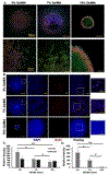
Tumor organoids and tumors-on-chips can be built by placing patient-derived cells within an engineered extracellular matrix (ECM) for personalized medicine. The engineered ECM influences the tumor response, and understanding the ECM-tumor relationship accelerates translating tumors-on-chips into drug discovery and development. In this work, we tuned the physical and structural characteristics of ECM in a 3D bioprinted soft-tissue sarcoma microtissue. We formed cell spheroids at a controlled size and encapsulated them into our gelatin methacryloyl (GelMA)-based bioink to make perfusable hydrogel-based microfluidic chips. We then demonstrated the scalability and customization flexibility of our hydrogel-based chip via engineering tools. A multiscale physical and structural data analysis suggested a relationship between cell invasion response and bioink characteristics. Tumor cell invasive behavior and focal adhesion properties were observed in response to varying polymer network densities of the GelMA-based bioink. Immunostaining assays and reverse transcription-quantitative polymerase chain reaction (RT-qPCR) helped assess the bioactivity of the microtissue and measure the cell invasion. The RT-qPCR data showed higher expressions of HIF-1α, CD44, and MMP2 genes in a lower polymer density, highlighting the correlation between bioink structural porosity, ECM stiffness, and tumor spheroid response. This work is the first step in modeling STS tumor invasiveness in hydrogel-based microfluidic chips. STATEMENT OF SIGNIFICANCE: We optimized an engineering protocol for making tumor spheroids at a controlled size, embedding spheroids into a gelatin-based matrix, and constructing a perfusable microfluidic device. A higher tumor invasion was observed in a low-stiffness matrix than a high-stiffness matrix. The physical characterizations revealed how the stiffness is controlled by the density of polymer chain networks and porosity. The biological assays revealed how the structural properties of the gelatin matrix and hypoxia in tumor progression impact cell invasion. This work can contribute to personalized medicine by making more effective, tailored cancer models.
Keywords: Bioprinting; Gelatin; Mechanobiology; Microstructure; Solid tumor spheroid.
Copyright © 2024 Acta Materialia Inc. Published by Elsevier Ltd. All rights reserved.
Conflict of interest statement
Declaration of competing interest The authors declare that they have no known competing financial interests or personal relationships that could have appeared to influence the work reported in this paper.
Figures


References
-
- Buchanan M, Chabot C, Lafleur J, Krzemien U, Lan C, Savichtcheva O, Boileau J-F, Ferrario C, Savage P, Park M, Use of PDXs and patient-derived cell lines to uncover unconventional drug therapies and combinations for the treatment of drug-resistant cancers, AACR, 2018.
-
- A.C. Society, What is a soft tissue sarcoma?, May 8, 2019. https://www.cancer.org/cancer/soft-tissue-sarcoma/about/soft-tissue-sarc.... (Accessed March 29 2020).
-
- Khan MM, Waqar S, Mahmood RA, Zahid M, Surgical management of soft tissue sarcoma, Journal of Rawalpindi Medical College (2018) 58–62.
Publication types
MeSH terms
Substances
Grants and funding
LinkOut - more resources
Full Text Sources
Research Materials
Miscellaneous

