. 2024 Aug;30(8):e14878.
doi: 10.1111/cns.14878.

Honokiol relieves hippocampal neuronal damage in Alzheimer's disease by activating the SIRT3-mediated mitochondrial autophagy

Affiliations

Honokiol relieves hippocampal neuronal damage in Alzheimer's disease by activating the SIRT3-mediated mitochondrial autophagy

Haitao Li et al. CNS Neurosci Ther. 2024 Aug.

Abstract

Background: This work elucidated the effect of honokiol (HKL) on hippocampal neuronal mitochondrial function in Alzheimer's disease (AD).

Methods: APP/PS1 mice were used as AD mice models and exposed to HKL and 3-TYP. Morris water maze experiment was performed to appraise cognitive performance of mice. Hippocampal Aβ+ plaque deposition and neuronal survival was evaluated by immunohistochemistry and Nissl staining. Hippocampal neurons were dissociated from C57BL/6 mouse embryos. Hippocampal neuronal AD model was constructed by Aβ oligomers induction and treated with HKL, CsA and 3-TYP. Neuronal viability and apoptosis were detected by cell counting kit-8 assay and TUNEL staining. mRFP-eGFP-LC3 assay, MitoSOX Red, dichlorodihydrofluorescein diacetate, and JC-1 staining were performed to monitor neuronal autophagosomes, mitochondrial reactive oxygen species (ROS), neuronal ROS, and mitochondrial membrane potential. Autophagy-related proteins were detected by Western blot.

Results: In AD mice, HKL improved cognitive function, relieved hippocampal Aβ1-42 plaque deposition, promoted hippocampal neuron survival, and activated hippocampal SIRT3 expression and mitochondrial autophagy. These effects of HKL on AD mice were abolished by 3-TYP treatment. In hippocampal neuronal AD model, HKL increased neuronal activity, attenuated neuronal apoptosis and Aβ aggregation, activated SIRT3 and mitochondrial autophagy, reduced mitochondrial and neuronal ROS, and elevated mitochondrial membrane potential. CsA treatment and 3-TYP treatment abrogated the protection of HKL on hippocampal neuronal AD model. The promotion of mitochondrial autophagy by HKL in hippocampal neuronal AD model was counteracted by 3-TYP.

Conclusions: HKL activates SIRT3-mediated mitochondrial autophagy to mitigate hippocampal neuronal damage in AD. HKL may be effective in treating AD.

Keywords: Alzheimer's disease; SIRT3; honokiol; mitochondrial autophagy; neuronal damage.


Conflict of interest statement

The authors have no conflict of interest to report.

Figures

FIGURE 1
FIGURE 1
HKL was effective in improving cognitive performance of AD mice, and reducing Aβ1–42 plaque deposition in hippocampus and cortex in AD mice. (A–D) Morris water maze experiment supported the effectiveness of HKL in improving cognitive performance of AD mice. **p < 0.01, and ***p < 0.001 versus the WT group. # p < 0.05, and ## p < 0.01 versus the HKL‐H group. (E) Congo‐red staining implied the suppression of HKL on plaque deposition in hippocampus and cortex of AD mice. (F–H) Immunohistochemistry revealed the blockage of HKL on Aβ1–42 plaque deposition in AD mice. **p < 0.01, ***p < 0.001.
FIGURE 2
FIGURE 2
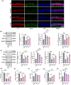
HKL activated hippocampal SIRT3 expression and hippocampal mitochondrial autophagy in AD mice. (A) Immunofluorescence staining indicated the promotion of HKL on hippocampal autophagy in AD mice. (B) The enhancement of HKL on hippocampal autophagy in AD mice was demonstrated by Western blot. (C) Western blot revealed the promotion of HKL on hippocampal mitochondrial autophagy in AD mice. (D–H) The protective effect of HKL on hippocampus and hippocampal mitochondria in AD mice was illustrated, as evidenced by its promotion on ATP, mtDNA, and SOD, as well as suppression on MDA and ROS. *p < 0.05, **p < 0.01, ***p < 0.001. “ns” represented differences that were not statistically significant.
FIGURE 3
FIGURE 3
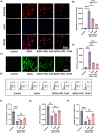
HKL relieved hippocampal neuronal damage and intracellular Aβ aggregation in the hippocampal neuronal model of AD. (A) NeuN/MAP2 staining confirmed the successful isolation of primary mouse hippocampal neurons because they were capable of expressing NeuN and MAP2. (B) Western blot indicated the successful preparation of AβOs. (C) ELISA revealed the inhibitory effect of HKL on intracellular Aβ aggregation in the AβO‐induced hippocampal neuronal model of AD. (D) CCK‐8 assay revealed that HKL treatment improved the viability of the AβO‐induced hippocampal neuronal model of AD. (E) HKL treatment weakened the LDH activity of the AβO‐induced hippocampal neuronal model of AD. (F, G) TUNEL staining suggested the suppressive role of HKL on the apoptosis of the AβO‐induced hippocampal neuronal model of AD. *p < 0.05, **p < 0.01, ***p < 0.001. “ns” represented differences that were not statistically significant.
FIGURE 4
FIGURE 4
HKL up‐regulated SIRT3 and activated mitochondrial autophagy in the hippocampal neuronal model of AD. (A, B) HKL increased SIRT3, LC3II/LC3I, Parkin, and PINK1 proteins, but decreased P62 protein in the AβO‐induced hippocampal neuronal model of AD. (C, D) mRFP–eGFP–LC3 assay suggested the promoting effect of HKL on the formation of autophagosomes in the AβO‐induced hippocampal neuronal model of AD. (E) TEM revealed that HKL treatment facilitated the formation of autophagosomes in the AβO‐induced hippocampal neuronal model of AD. *p < 0.05, **p < 0.01, ***p < 0.001.
FIGURE 5
FIGURE 5
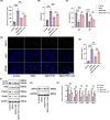
HKL exerted an ameliorative effect on mitochondrial function in the hippocampal neuronal model of AD. (A, B) MitoSOX Red staining revealed the suppressive role of HKL on mitochondrial ROS levels in the AβO‐induced hippocampal neuronal model of AD. (C, D) DCFH‐DA staining revealed that HKL exerted an inhibitory influence on neuronal ROS levels in the AβO‐induced hippocampal neuronal model of AD. (E, F) JC‐1 staining combined with flow cytometry suggested that HKL treatment increased the mitochondrial membrane potential of the AβO‐induced hippocampal neuronal model of AD. (G) HKL treatment enhanced ATP production in the AβO‐induced hippocampal neuronal model of AD. (H) HKL treatment upregulated relative mRNA expression of mtDNA could be upregulated in the AβO‐induced hippocampal neuronal model. *p < 0.05, **p < 0.01, ***p < 0.001.
FIGURE 6
FIGURE 6
HKL possibly ameliorated damage to the hippocampal neuronal model of AD by activating mitochondrial autophagy. (A) ELISA revealed that CsA treatment abrogated the suppressive role of HKL on intracellular Aβ aggregation in the AβO‐induced hippocampal neuronal model of AD. (B) CsA treatment reversed the inhibition of HKL on LDH activity in the AβO‐induced hippocampal neuronal model of AD. (C) CCK‐8 assay indicated that CsA treatment counteracted the promoting effect of HKL on the viability of the AβO‐induced hippocampal neuronal model. (D, E) TUNEL staining revealed that CsA treatment reversed the repression effect of HKL on the apoptosis of the AβO‐induced hippocampal neuronal model of AD. (F‐H) Western blot suggested that CsA treatment attenuated the activation of SIRT3 and mitochondrial autophagy by HKL in the AβO‐induced hippocampal neuronal model of AD. *p < 0.05, **p < 0.01, ***p < 0.001. “ns” represented differences that were not statistically significant.
FIGURE 7
FIGURE 7
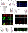
HKL possibly attenuated damage to the hippocampal neuronal model of AD by activating mitochondrial autophagy by upregulating SIRT3. (A) CCK‐8 assay suggested that 3‐TYP treatment reversed the promoting effect of HKL on the viability of the AβO‐induced hippocampal neuronal model of AD. (B) 3‐TYP treatment abrogated the suppression effect of HKL on the LDH activity in the AβO‐induced hippocampal neuronal model of AD. (C, D) TUNEL staining revealed that 3‐TYP treatment counteracted the inhibition of HKL on the apoptosis of the AβO‐induced hippocampal neuronal model of AD. (E, F) Western blot revealed that the activation effect of HKL on SIRT3 expression and mitochondrial autophagy was reversed by 3‐TYP in the AβO‐induced hippocampal neuronal model of AD. (G, H) Immunofluorescence staining revealed that 3‐TYP treatment abrogated the promoting effect of HKL on mitochondrial Parkin expression in the AβO‐induced hippocampal neuronal model of AD. (I, J) MitoSOX Red staining suggested that the suppression effect of HKL on mitochondrial ROS level was abrogated by 3‐TYP in the AβO‐induced hippocampal neuronal model of AD. (K) The enhancement of HKL on ATP production was attenuated by 3‐TYP in the AβO‐induced hippocampal neuronal model of AD. (L, M) DCFH‐DA staining revealed that the repressive role of HKL on neuronal ROS level was reversed by 3‐TYP treatment in the AβO‐induced hippocampal neuronal model of AD. *p < 0.05, **p < 0.01, ***p < 0.001.
FIGURE 8
FIGURE 8
HKL alleviated cognitive impairment and Aβ1–42 plaque deposition in AD mice by activating hippocampal mitochondrial autophagy via increasing SIRT3. (A–D) Morris water maze experiment indicated that the alleviation of HKL on cognitive impairment of AD mice was reversed by 3‐TYP. (E) By Congo‐red staining, HKL reduced plaque deposition in hippocampus and cortex of AD mice, whereas 3‐TYP counteracted this effect of HKL. (F–H) Immunohistochemistry revealed that the suppression of HKL on Aβ1–42 plaque deposition in hippocampus and cortex of AD mice was abrogated by 3‐TYP. (I) Immunofluorescence staining implied the promotion of HKL on hippocampal autophagy in AD mice, but 3‐TYP eliminated this effect of HKL. (J–L) Base on Western blot, HKL activated hippocampal autophagy and autophagy in hippocampal neuronal mitochondria, whereas this effect was abolished by 3‐TYP. *p < 0.05, **p < 0.01, ***p < 0.001. # p < 0.05 vs. the HKL‐H group.

Similar articles

Cited by

References

    1. Jorfi M, Maaser‐Hecker A, Tanzi RE. The neuroimmune axis of Alzheimer's disease. Genome Med. 2023;15(1):6. - PMC - PubMed
    1. Söderberg L, Johannesson M, Nygren P, et al. Lecanemab, aducanumab, and gantenerumab – binding profiles to different forms of amyloid‐beta might explain efficacy and side effects in clinical trials for Alzheimer's disease. Neurotherapeutics. 2023;20(1):195‐206. - PMC - PubMed
    1. Twarowski B, Herbet M. Inflammatory processes in Alzheimer's disease – pathomechanism, diagnosis and treatment: a review. Int J Mol Sci. 2023;24(7):6518. - PMC - PubMed
    1. Iqbal I, Saqib F, Mubarak Z, et al. Alzheimer's disease and drug delivery across the blood‐brain barrier: approaches and challenges. Eur J Med Res. 2024;29(1):313. - PMC - PubMed
    1. Faulin T, Estadella D. Alzheimer's disease and its relationship with the microbiota‐gut‐brain axis. Arq Gastroenterol. 2023;60(1):144‐154. - PubMed

Publication types

MeSH terms

LinkOut - more resources