

Endoscopy-assisted laparoscopic wedge-resection of gastric glomus tumor: A case report
- PMID: 39098173
- PMCID: PMC11345927
- DOI: 10.1016/j.ijscr.2024.110100
Endoscopy-assisted laparoscopic wedge-resection of gastric glomus tumor: A case report
Abstract
Introduction: Glomus tumor is a pericytic mesenchymal neoplasm that most commonly occurs in the extremities. The occurrence in visceral organs is rare and is a differential diagnosis with other gastric submucosal tumors.
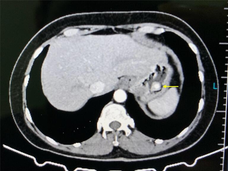
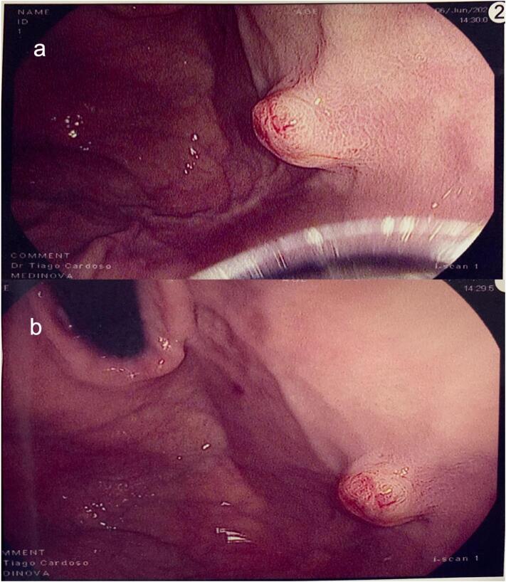
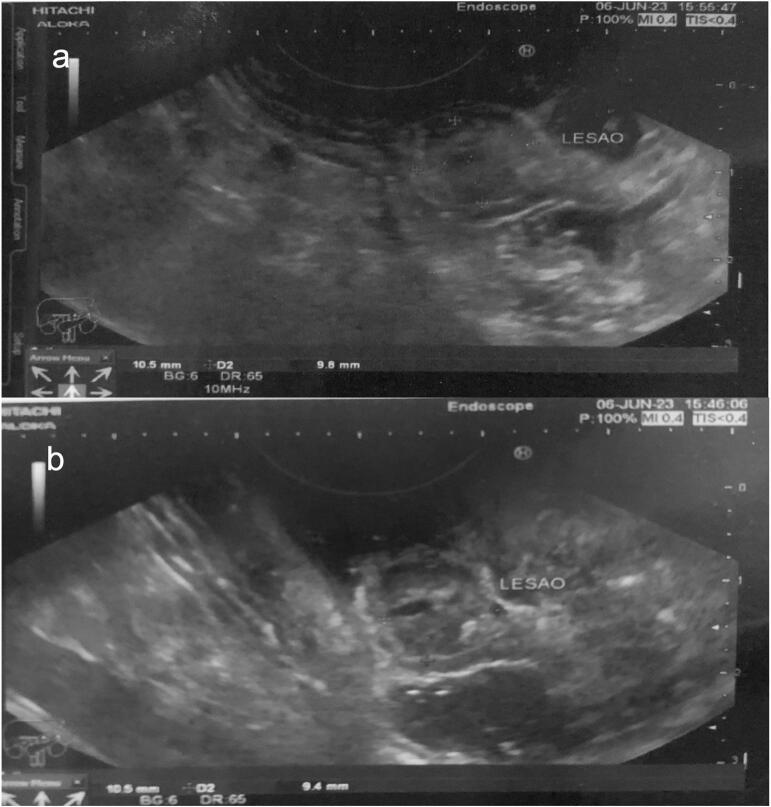
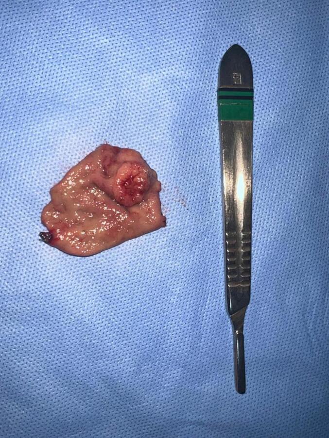
Presentation of case: A woman with epigastric pain underwent esophagogastroduodenoscopy (EGD) which revealed a gastric submucosal tumor. Endoscopic ultrasound with fine-needle aspiration allowed preoperative diagnosis of gastric glomus tumor. Intraoperative EGD-assisted laparoscopic segmental gastrectomy was successfully performed. The patient was discharged in the second postoperative day. There was no evidence of recurrence at 8 months of follow-up.
Discussion: The stomach is a rare location for the glomus tumor, a neoplasm of the glomus body, which is a perivascular structure with thermoregulatory function. Preoperative diagnosis is challenging, and endoscopic ultrasound (EUS) is useful for both assessing malignancy-associated features and biopsy guiding. The treatment is surgical resection with attention to adequate oncological margins while preserving healthy gastric wall.
Conclusion: Immunohistochemical analysis of specimen obtained by EUS fine-needle allows accurate preoperative diagnosis and laparoscopic-endoscopic combined surgery allows good oncological and functional results.
Keywords: Case report; Endoscopy; Glomus tumor; Laparoscopy; Stomach neoplasm.
Copyright © 2024 The Authors. Published by Elsevier Ltd.. All rights reserved.
Conflict of interest statement
Conflict of interest statement All authors declare having no competing interests.
Figures
References
Publication types
LinkOut - more resources
Full Text Sources
Research Materials
