. 2024 Oct;30(4):895-913.
doi: 10.3350/cmh.2024.0364. Epub 2024 Aug 5.

KCTD17-mediated Ras stabilization promotes hepatocellular carcinoma progression

Affiliations

KCTD17-mediated Ras stabilization promotes hepatocellular carcinoma progression

Young Hoon Jung et al. Clin Mol Hepatol. 2024 Oct.

Abstract

Background/aims: Potassium channel tetramerization domain containing 17 (KCTD17) protein, an adaptor for the cullin3 (Cul3) ubiquitin ligase complex, has been implicated in various human diseases; however, its role in hepatocellular carcinoma (HCC) remains elusive. Here, we aimed to elucidate the clinical features of KCTD17, and investigate the mechanisms by which KCTD17 affects HCC progression.

Methods: We analyzed transcriptomic data from patients with HCC. Hepatocyte-specific KCTD17 deficient mice were treated with diethylnitrosamine (DEN) to assess its effect on HCC progression. Additionally, we tested KCTD17-directed antisense oligonucleotides for their therapeutic potential in vivo.

Results: Our investigation revealed the upregulation of KCTD17 expression in both tumors from patients with HCC and mouse models of HCC, in comparison to non-tumor controls. We identified the leucine zipper-like transcriptional regulator 1 (Lztr1) protein, a previously identified Ras destabilizer, as a substrate for KCTD17-Cul3 complex. KCTD17-mediated Lztr1 degradation led to Ras stabilization, resulting in increased proliferation, migration, and wound healing in liver cancer cells. Hepatocyte-specific KCTD17 deficient mice or liver cancer xenograft models were less susceptible to carcinogenesis or tumor growth. Similarly, treatment with KCTD17-directed antisense oligonucleotides (ASO) in a mouse model of HCC markedly lowered tumor volume as well as Ras protein levels, compared to those in control ASO-treated mice.

Conclusion: KCTD17 induces the stabilization of Ras and downstream signaling pathways and HCC progression and may represent a novel therapeutic target for HCC.

Keywords: Antisense oligonucleotides; HCC; KCTD17; Lztr1; Ras.


Conflict of interest statement

Conflicts of Interest

The authors have no conflicts to disclose.

Figures
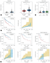
Figure 1.
Figure 1.
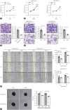
Increased KCTD17 is associated with poor prognosis in hepatocellular carcinoma (HCC). (A) Box and whiskers plots showing expression levels of KCTD17 in non-tumor and tumor from HCC in two independent cohorts (GSE25097, non-tumor=249, tumor=268; GSE36376, non-tumor=193, tumor=240). (B) Box plots showing expression levels of KCTD17 in each stage (stage I=171, II=86, and III+IV=88) in the liver hepatocellular carcinoma (LIHC) from the Cancer Genome Atlas (TCGA) database. Significance is determined using the Wilcoxon rank sum test. (C) Survival probabilities (Kaplan–Meier curves) (left) or cumulative hazard curve (right) based on hepatic KCTD17 expression. The R package, multipleROC, is divided into two groups (i.e., high vs. low KCTD17 expression) according to the optimal gene expression level of KCTD17. (D) Kaplan–Meier curves comparing hepatic KCTD17 expression and overall survival (top) or cumulative hazard curve (bottom) in different stages of HCC. ***P<0.001, ****P<0.0001 as compared to the indicated control by two-way ANOVA. All data are shown as the mean±standard error of the mean.
Figure 2.
Figure 2.
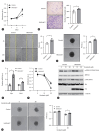
KCTD17 deficiency attenuates hepatocellular carcinoma (HCC) cell growth and migration. (A, B) Cell proliferation of control or Kctd17 KO Hepa1c1c7 cells (A), or doxycycline (Dox)-inducible KCTD17 knockdown Hep3B cells (B). (C, D) Representative pictures and quantitation of transwell migration assay in Kctd17 KO Hepa1c1c7 cells (C), or (D) KCTD17 knockdown Hep3B cells. (E) Representative pictures and quantitation of wound width in KCTD17 knockdown Hep3B cells. (F) Representative pictures and quantitation of three-dimensional spheroid cluster assay in KCTD17 knockdown Hep3B cells. *P<0.05, **P<0.01, ***P<0.001 as compared to the indicated control by two-way ANOVA. All data are shown as the mean±s.e.m.
Figure 3.
Figure 3.
Kctd17 promotes hepatocellular carcinoma (HCC) proliferation, migration and drug resistance. (A–D) Cell proliferation (A), cell migration (B), would healing ability (C), three-dimensional spheroid cluster assay (D) and gene expression of Myc and Bmi1 (E) in control or Kctd17 overexpressing Hepa1c1c7 cells. (F–H) Control or Kctd17-overexpressing cells were treated with different dosages of sorafenib for 48 h, followed by cell proliferation assay (F), Western blot (G) and three-dimensional spheroid cluster assay (H). *P<0.05, **P<0.01 as compared to the indicated control by two-way ANOVA. All data are shown as the mean±s.e.m.
Figure 4.
Figure 4.
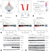
KCTD17 drives Ras signaling. (A) Violin plot based on the expression of KCTD17 (red: 37 top expressed samples, blue: 37 bottom expressed samples). (B, C) Volcano (B) and bubble plots (C) of normalized enrichment score (NES) distribution with RAS signalingrelated gene sets marked. (D) RNA-seq datasets of low-KCTD17 enriched and high-KCTD17 enriched as compared with KRAS and ERK signature in LIHC from TCGA. (E, F) Hepa1c1c7 cells expressing control or HA/Kctd17 (E) or Hep3B cells expressing doxycycline-inducible shKCTD17 (F) were cultured in serum-free medium and then treated with serum for the indicated time, followed by evaluation of Ras/MAPK signaling using Western blot.
Figure 5.
Figure 5.
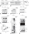
Kctd17 prompts Lztr1 degradation to mediate Ras stabilization. (A) Western blots from 293T cells transfected with H/K-Ras with or without Kctd17. (B) Hepa1c1c7 cells transfected with control or Kctd17 were treated with 50 mg/mL cycloheximide (CHX) and then harvested at the indicated time points after treatment prior to Western blotting and quantitation of endogenous Ras. (C, D) Co-immunoprecipitation of Ras and Lztr1 or Kctd17 from Hepa1c1c7 cells (C) or Western blots in lysate from 293T cells (D). (E) Western blots from Hepa1c1c7 cells transfected with Kctd17 and Lztr1 with or without MG-132. (F) Co-immunoprecipitation of Lztr1 by Kctd17 from Hepa1c1c7 cells. (G) Polyubiquitination of Lztr1 by Kctd17 Myc/Ub-transfected 293T cells with MG-132. (H) Ras ubiquitination in Flag/Ub-transfected 293T cells with or without Kctd17 and Lztr1. (I) Cell proliferation in control or Kctd17 overexpressing cells with or without Lztr1/Flag from Hepa1c1c7 cells. **P<0.01, ***P<0.001 as compared to the indicated control by two-way ANOVA. All data are shown as the mean±s.e.m.
Figure 6.
Figure 6.
Kctd17 is required for hepatocellular carcinoma (HCC) progression in vivo. (A) Experimental schematic for tumor xenograft assay using control or Kctd17 KO Hepa1c1c7 cells. (B–D) Representative pictures (B), tumor size (C) and weight (D) of control or Kctd17 KO xenografts (n=10 per group). (E) Experimental schematic for tumor xenograft assay using doxycycline-inducible KCTD17 knockdown Hep3B cells. (F–H) Representative pictures (F), tumor size (G) and weight (H) of control or doxycycline-inducible shKCTD17 xenografts (n=10 per group). (I) Experimental schematic for hepatocyte-specific Kctd17 KO mice (L-Kctd17). (J–N) Kctd17 expression in tumors and adjacent non-tumors (J), representative images of whole livers (K) and tumor volume (L), Representative images of livers stained with H&E or Ki-67 (M), Western blots from liver tumor lysates (N) of control or L-Kctd17 mice (n=7 to 9 per group), as indicated. Scale bar, 100 or 200 mm. *P<0.05, **P<0.01, ***P<0.001 as compared to the indicated control by two-way ANOVA. All data are shown as the mean±s.e.m.
Figure 7.
Figure 7.
Kctd17-specific ASO decreases hepatocellular carcinoma (HCC) progression. (A) Experimental schematic for oncogene-induced HCC models. After hydrodynamic injection of myr-Akt and N-Ras in WT male mice, control or Kctd17-directed ASO was administered weekly by intraperitoneal injection. (B–E) Kctd17 expression in tumors and adjacent non-tumors (B), representative images of whole liver (C), tumor size (D) and Western blots from liver tumor lysate (E) in control or Kctd17 ASO-treated mice (n=5 or 7 per group). (F) Experimental schematic for DEN/CDAHFD-induced HCC models. Mice were injected with DEN at 2 weeks of life, then started on CDAHFD diet-feeding with concurrent weekly intraperitoneal injections of control or Kctd17 ASO. (G–K) Kctd17 expression in tumors and adjacent non-tumors (G), representative images of whole mouse liver (H), tumor number (I), liver H&E and Ki-67 staining (J), and Western blots in lysate from liver tumor lysate (K), in control or Kctd17 ASO-treated mice (n=6 per group). Scale bar, 100 or 200 mm. (L) Model of Kctd17-mediated HCC progression. *P<0.05, **P<0.01, ***P<0.001 as compared to the indicated control by two-way ANOVA. All data are shown as the mean±s.e.m.
None

References

    1. Niu ZS, Niu XJ, Wang WH. Genetic alterations in hepatocellular carcinoma: an update. World J Gastroenterol. 2016;22:9069–9095. - PMC - PubMed
    1. Zhou J, Sun HC, Wang Z, Cong WM, Wang JH, Zeng MS, et al. Guidelines for diagnosis and treatment of primary liver cancer in China (2017 edition) Liver Cancer. 2018;7:235–260. - PMC - PubMed
    1. Lepage C, Capocaccia R, Hackl M, Lemmens V, Molina E, Pierannunzio D, et al. EUROCARE-5 Working Group Survival in patients with primary liver cancer, gallbladder and extrahepatic biliary tract cancer and pancreatic cancer in Europe 1999-2007: results of EUROCARE-5. Eur J Cancer. 2015;51:2169–2178. - PubMed
    1. Siegel RL, Miller KD, Jemal A. Cancer statistics, 2018. CA Cancer J Clin. 2018;68:7–30. - PubMed
    1. Zhu AX, Kudo M, Assenat E, Cattan S, Kang YK, Lim HY, et al. Effect of everolimus on survival in advanced hepatocellular carcinoma after failure of sorafenib: the EVOLVE-1 randomized clinical trial. JAMA. 2014;312:57–67. - PubMed

MeSH terms

LinkOut - more resources