Case Reports
. 2024 Aug 5;44(8):170.
doi: 10.1007/s10875-024-01774-x.

A Novel Case of IFNAR1 Deficiency Identified a Common Canonical Splice Site Variant in DOCK8 in Western Polynesia: The Importance of Validating Variants of Unknown Significance in Under-Represented Ancestries

Affiliations
Case Reports

A Novel Case of IFNAR1 Deficiency Identified a Common Canonical Splice Site Variant in DOCK8 in Western Polynesia: The Importance of Validating Variants of Unknown Significance in Under-Represented Ancestries

Aimee Huynh et al. J Clin Immunol. .

Erratum in

Abstract

Advanced genomic technologies such as whole exome or whole genome sequencing have improved diagnoses and disease outcomes for individuals with genetic diseases. Yet, variants of unknown significance (VUS) require rigorous validation to establish disease causality or modification, or to exclude them from further analysis. Here, we describe a young individual of Polynesian ancestry who in the first 13 mo of life presented with SARS-CoV-2 pneumonia, severe enterovirus meningitis and adenovirus gastroenteritis, and severe adverse reaction to MMR vaccination. Genomic analysis identified a previously reported pathogenic homozygous variant in IFNAR1 (c.1156G > T, p.Glu386* LOF), which is common in Western Polynesia. Moreover, a new and putatively deleterious canonical splice site variant in DOCK8 was also found in homozygosity (c.3234 + 2T > C). This DOCK8 variant is common in Polynesians and other under-represented ancestries in large genomic databases. Despite in silico bioinformatic predictions, extensive in vitro and ex vivo analysis revealed the DOCK8 variant likely be neutral. Thus, our study reports a novel case of IFNAR1 deficiency, but also highlights the importance of functional validation of VUS, including those predicted to be deleterious, and the pressing need to expand our knowledge of the genomic architecture and landscape of under-represented populations and ancestries.


Conflict of interest statement

The authors declare no competing interests.

Figures
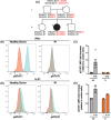
Fig. 1
Fig. 1
IFNAR1 (c.1156G > T, p.(Glu386*) variant is loss-of function in T cells (A) Pedigree of a novel IFNAR1-deficient kindred. Familial segregation of the IFNAR1 c.1156G > T, p.E386X and DOCK8 c.3234 + 2T > C variants are shown. The affected individual P1 is represented by a black symbol (II.2). Individuals of unknown genotype are labelled “E?”. (B-E) T cell blasts were expanded from healthy donors (HD) and P1 by stimulating PBMCs with anti-CD2/CD3/CD28 mAbs. After 14 days, the cells were rested, and then stimulated in the absence or presence of (B, C) IFNb (type I IFN) or (D, E) IL-21. After 15 min, cells were harvested, fixed and permeabilised and then stained with mAb specific for phospho-STAT1. FACS plot (B, C) are representative of pSTAT1 induction in T-cell blasts from HD or P1. The data in (C) and (E) represent the fold-change in pSTAT1 gMFI in T-cell blasts stimulated with IFNβ or IL-21, respectively, relative to unstimulated cells. Each point represents data from an independent experiment
Fig. 2
Fig. 2
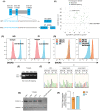
DOCK8 expression is intact in lymphocytes from the IFNAR1-deficient patient harbouring the DOCK8 c.3234 + 2T > C variant (A) schematic of DOCK8 genomic sequence representing the 5’ region of exon 26 (blue), the 3’ region of intron 26 (black) and the variant found in P1 (red). Prediction of the cDNA sequence is reported for both WT DOCK8 and DOCK8 c.3234 + 2T > C. (B) Minor allele frequency (MAF, x-axis) and CADD scores (y-axis) for missense (green circles) and essential splicing (purple circles) variants in DOCK8 reported as homozygous in the public database gnomAD (GRCh37). The DOCK8 c.3234 + 2T > C variant found in P1 (indigo circles) is the only homozygous essential splicing variant previously reported in gnomAD. The mutation significance cut-off (MSC, 95% confidence interval, y axis) is represented by the dotted line. (C, D) PBMCs from healthy donors (HD), P1 and a confirmed DOCK8-deficient patient were fixed and permeabilised and stained with anti-DOCK8 mAb. Intracellular expression of DOCK8 was then determined. (A) DOCK8 expression (blue histogram) in PBMCs from a HD (left) and IFNAR1-deficient patient P1 (right) relative to staining with isotype IgG control mAb. (B) Overlay of DOCK8 expression in PBMCs from HDs (#1-#4), P1 (left) or a DOCK8-deficient patient (right). (E, F) exons 25–27 of DOCK8 were PCR amplified from PBMCs of healthy donors and P1. (E) agarose gel depicting amplified products, including 2 products for P1. (F) Sequencing of amplified PCR products from HD (left) and P1 (right) showing the higher molecular weight band from P1 corresponds to WT DOCK8 cDNA, while the lower band lacks exon 26. (G, H) whole cell lysates were prepared from T blasts expanded from healthy donors (HD), P1 and a confirmed DOCK8-deficient patient. DOCK8 expression was assessed by SDS-PAGE and Western blotting. Detection of GADPH was used as a loading control. (G) representative of western blot from 3 different experiments. (H) summary data depicting expression of DOCK8 protein in lysates from 3 unrelated healthy donors and P1, relative to GAPDH
Fig. 3
Fig. 3
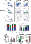
Comparison of the phenotype and function of T cells from patients with IFNAR1-deficiency versus DOCK8-deficiency (A-F) PBMCs from HDs, one DOCK8 deficient patient, 2 IFNAR1-deficient patients or P1 were stained with mAbs against CD3, CD4, CD8, CD45RA, CCR7 and CD57. (A-C) representative FACS plots and (D-E) summary graphs depicting proportions of: (A, D) CD4+ and CD8+ T cells within CD3+ T cells, (B-E) naïve, TCM, TEM and TEMRA subsets within CD4+ (B, D) and CD8+ (C, E) T cells in HDs, and the indicated patients. (F) expression of CD57 on CD8+ naïve, TCM, TEM and TEMRA subsets, and the indicated patients. (G, H) CD45RA memory CD4+ T cells were sorted from healthy donors (HD), a confirmed DOCK8-deficient patient, and P1 and then stimulated in vitro for 5 d with anti-CD2/CD3/CD28 mAb beads. After this time, secretion of (G) Th2 cytokines IL-4, IL-5 and IL-13 and (H) Th17 cytokines IL-17 A, IL-17 F and IL-22 was determined by cytometric bead array

References

    1. Hernandez N, Bucciol G, Moens L, Le Pen J, Shahrooei M, Goudouris E, et al. Inherited IFNAR1 deficiency in otherwise healthy patients with adverse reaction to measles and yellow fever live vaccines. J Exp Med. 2019;216(9):2057–70. - PMC - PubMed
    1. Gothe F, Hatton CF, Truong L, Klimova Z, Kanderova V, Fejtkova M, et al. A novel case of homozygous Interferon Alpha/Beta receptor alpha chain (IFNAR1) Deficiency with Hemophagocytic Lymphohistiocytosis. Clin Infect Dis. 2022;74(1):136–9. - PMC - PubMed
    1. Meyts I, Casanova JL. Viral infections in humans and mice with genetic deficiencies of the type I IFN response pathway. Eur J Immunol. 2021;51(5):1039–61. - PMC - PubMed
    1. Zhang Q, Bastard P, Liu Z, Le Pen J, Moncada-Velez M, Chen J et al. Inborn errors of type I IFN immunity in patients with life-threatening COVID-19. Science. 2020;370(6515). - PMC - PubMed
    1. Zhang Q, Matuozzo D, Le Pen J, Lee D, Moens L, Asano T et al. Recessive inborn errors of type I IFN immunity in children with COVID-19 pneumonia. J Exp Med. 2022;219(8). - PMC - PubMed

Publication types

MeSH terms

Substances

LinkOut - more resources