. 2024 Aug 2:28:0050.
doi: 10.34133/bmr.0050. eCollection 2024.

Targeting Reprogrammed Cancer-Associated Fibroblasts with Engineered Mesenchymal Stem Cell Extracellular Vesicles for Pancreatic Cancer Treatment

Affiliations

Targeting Reprogrammed Cancer-Associated Fibroblasts with Engineered Mesenchymal Stem Cell Extracellular Vesicles for Pancreatic Cancer Treatment

Pengcheng Zhou et al. Biomater Res. .

Abstract

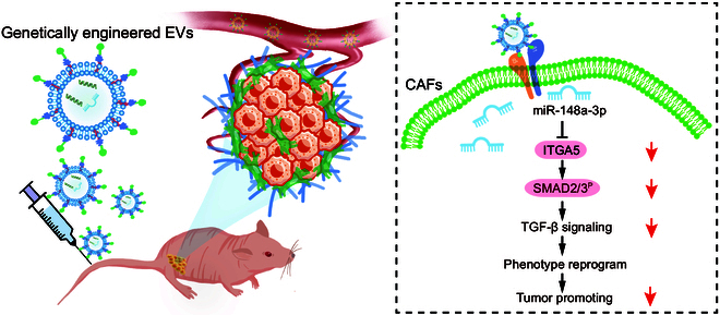
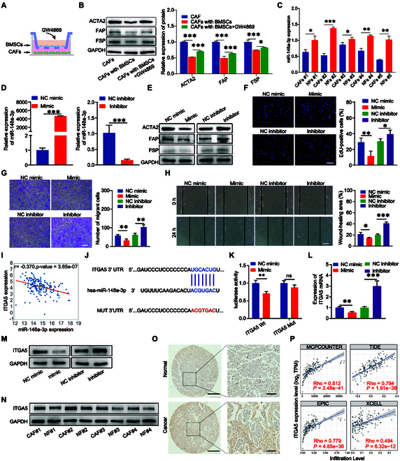
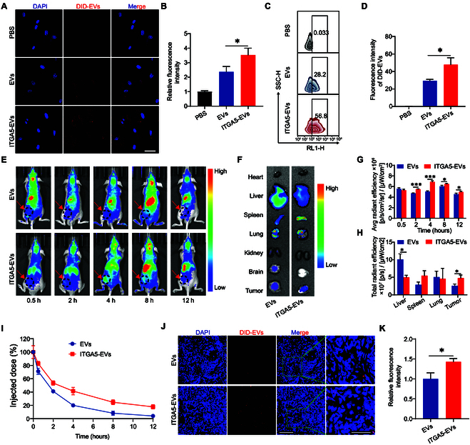
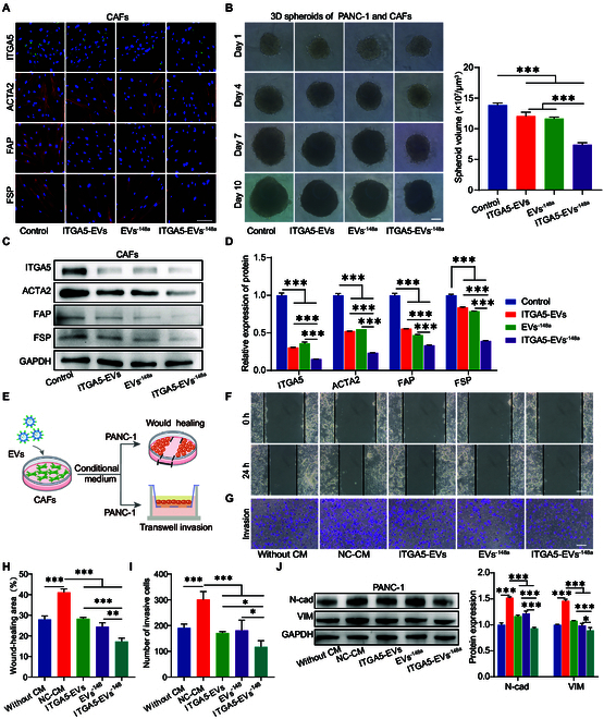
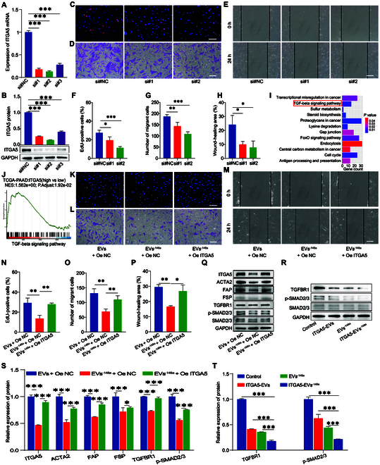
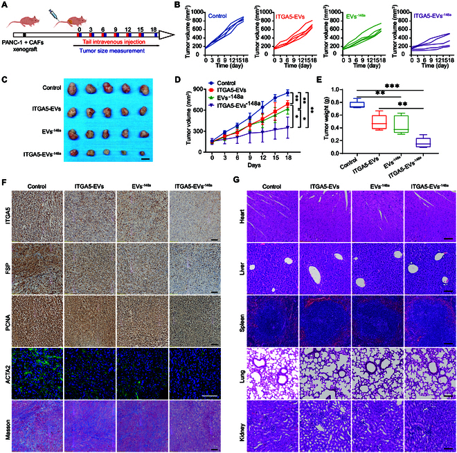
Background: As one of the most aggressive and lethal cancers, pancreatic cancer is highly associated with cancer-associated fibroblasts (CAFs) that influence the development and progression of cancer. Targeted reprogramming of CAFs may be a promising strategy for pancreatic cancer. This study aims to construct engineered extracellular vesicles (EVs) with surface modification of integrin α5 (ITGA5)-targeting peptide and high internal expression of miR-148a-3p by endogenous modification for targeted reprogramming of pancreatic CAFs. Methods: Bone marrow mesenchymal stem cells (BMSCs) and pancreatic CAFs were cocultured to examine the effect of BMSC-derived EVs on the expression levels of CAF markers. miR-148a-3p was identified as a functional molecule. The mechanism of miR-148a-3p was elucidated using the dual-luciferase reporter assay. BMSCs were infected with TERT-encoding and miR-148a-3p-encoding lentiviruses. Subsequently, BMSCs were modified with ITGA5-specific targeting peptide. The supernatant was ultracentrifuged to obtain the engineered EVs (ITGA5-EVs-148a), which were used to reprogram CAFs. Results: BMSCs modulated CAF marker expressions through EVs. miR-148a-3p was up-regulated in BMSCs. The expression of miR-148a-3p in pancreatic CAFs was down-regulated when compared with that in normal fibroblasts (NFs). Mechanistically, ITGA5-EVs-148a effectively suppressed the proliferation and migration of pancreatic CAFs by targeting ITGA5 through the TGF-β/SMAD pathway. ITGA5-EVs-148a was associated with enhanced cellular uptake and exhibited enhanced in vitro and in vivo targeting ability. Moreover, ITGA5-EVs-148a exerted strong reconfiguration effects in inactivating CAFs and reversing tumor-promoting effects in 3D heterospheroid and xenograft pancreatic cancer models. Conclusions: This targeted CAF reprogramming strategy with genetically engineered ITGA5-EVs-148a holds great promise as a precision therapeutics in clinical settings.


Conflict of interest statement

Competing interests: The authors declare that they have no competing interests.

Figures

Fig. 1.
Fig. 1.
The schematic diagram illustrates the process of EVs targeting CAFs and reprogramming CAFs.
Fig. 2.
Fig. 2.
The role of miR-148a-3p in pancreatic CAFs and the expression of downstream target gene ITGA5. (A and B) The activation markers of CAFs were down-regulated upon coculturing CAFs with BMSCs but were up-regulated upon treatment with GW4869 (n = 3). (C) qRT-PCR analysis of miR-148a-3p expression in cultured primary CAFs and normal fibroblasts (NFs) (n = 3). (D) qRT-PCR analysis revealed the transfection efficiency of miR-148a-3p mimic and inhibitor (n = 3). (E) Western blotting analysis of ACTA2, FAP, and FSP protein expression levels in CAFs transfected with miR-148a-3p mimic and inhibitor. (F) EdU assay results of miR-148a-3p-transfected pancreatic CAFs (n = 4). (G) Transwell migration assay results of miR-148a-3p-transfected pancreatic CAFs (n = 4). (H) Wound-healing assay results of miR-148a-3p-transfected pancreatic CAFs (n = 4). (I) Correlation between miR-148a-3p and ITGA5 expression levels in TCGA-pancreatic cancer cohort. (J) Predicted binding site between miR-148a-3p and ITGA5. (K) Dual-luciferase reporter assay results of miR-148a-3p and ITGA5 (n = 3). (L) Effect of miR-148a-3p transfection on ITGA5 mRNA expression (n = 3). (M) Effect of miR-148a-3p transfection on ITGA5 protein expression. (N) ITGA5 protein expression levels in primary cultured CAFs and NFs. (O) TMA of ITGA5 expression in pancreatic cancer and adjacent noncancerous tissues. (P) Correlation between ITGA5 expression and CAF infiltration based on 4 algorithms (MCPOUNTER, TIDE, EPIC, and XCELL). *P < 0.05, **P < 0.01, ***P < 0.001 for all statistical data. Scale bar, 100 μm for all captured pictures.
Fig. 3.
Fig. 3.
Design and establishment of genetically engineered ITGA5-EVs-148a. (A) Schematic diagram illustrating the process of constructing genetically engineered EVs. (B) Proliferation of P6 primary BMSCs and TERT-BMSCs. Scale bar, 100 μm. (C) EdU assay results of P6 primary BMSCs and TERT-BMSCs (n = 3). (D) CCK-8 assay results of P6 primary BMSCs and TERT-BMSCs (n = 3). (E) qRT-PCR analysis of TERT mRNA expression levels in P6 primary BMSCs and TERT-BMSCs (n = 3). (F) TEM analysis of TERT-BMSC-EVs. Scale bar, 100 nm. (G) Western blotting analysis of EV markers in EVs. The BMSC lysate served as a control. (H) Qualification of protein concertation in EVs using the BCA assay (n = 3). (I) NTA of the size distributions of EVs. (J) NTA analysis of the particle numbers of EVs. (K) qRT-PCR analysis of miR-148a-3p expression in BMSCs (n = 3). (L) qRT-PCR analysis of miR-148a-3p expression in EVs (n = 3). (M) Laser scanning confocal microscopy (LSCM) confirmed the incorporation of DSPE-PEG-CRYYRITY into the cell membrane of BMSCs. Scale bar, 100 μm. (N) LSCM confirmed the incorporation of DSPE-PEG-CRYYRITY into EVs. Scale bar, 1 μm. *P < 0.05, **P < 0.01, ***P < 0.001 for all statistical data.
Fig. 4.
Fig. 4.
ITGA5-targeting peptide enhanced the targeting ability of EVs. (A) Internalization of EVs and ITGA5-EVs by CAFs. Scale bar, 100 μm. (B) Qualification of the fluorescence intensity of CAFs incubated with different EVs using LSCM. (C) Flow cytometric analysis of CAFs incubated with different EVs (n = 3). (D) Qualification of the fluorescence intensity of CAFs incubated with different EVs using flow cytometry. (E) In vivo fluorescence images of tumor-bearing mice treated with different EVs or ITGA5-EVs at different time points (n = 3). (F) Ex vivo fluorescence images of major organs and tumor tissues at 12 h after EV administration. (G) Qualification of the fluorescence intensity in tumor sites at different time points (n = 3). (H) Qualification of the fluorescence intensity of major organs at 12 h after EV administration (n = 3). (I) Peripheral blood fluorescence intensity after EV administration. (J) Fluorescence images of DiD-labeled EVs and ITGA5-EVs targeting and penetrating the tumor observed using LSCM. Scale bars, 100 μm (left) and 50 μm (right). (K) Qualification of DiD-labeled EVs and ITGA5-EVs in tumors (n = 3). *P < 0.05, ***P < 0.001 for all statistical data.
Fig. 5.
Fig. 5.
Function of ITGA5-EVs-148a in pancreatic CAFs. (A) Immunofluorescence analysis of ITGA5, ACTA2, FAP, and FSP expression levels in CAFs treated with different EVs using LSCM. (B) Three-dimensional multicellular spheroids treated with different EVs (n = 5). (C and D) The protein expression levels of ITGA5, ACTA2, FAP, FSP, TGFBR1, and p-SMAD2/3 in CAFs treated with different EVs were evaluated using Western blotting analysis (n = 3). (E) Schematic illustration of the process of obtaining conditional medium (CM) from CAFs treated with different EVs after coculture with PANC-1 cells for evaluating the migration and invasion abilities. (F and H) The migration of PANC-1 cells treated with CAF CM was evaluated using the wound-healing assay (n = 3). (G and I) The invasion of PANC-1 cells treated with CAF CM was evaluated using the transwell assay (n = 3). (J) Western blotting analysis of N-cad and VIM levels in PANC-1 cells treated with EVs (n = 3). *P < 0.05, **P < 0.01, ***P < 0.001 for all statistical data. Scale bar, 100 μm for all captured pictures.
Fig. 6.
Fig. 6.
miR-148a-3p/ITGA5 inactivated pancreatic CAFs through the TGF-β/SMAD pathway. (A) The mRNA expression of ITGA5 in CAFs transfected with short-interfering RNA targeting ITGA5 (si-ITGA5) was evaluated using qRT-PCR (n = 3). (B) The protein expression level of ITGA5 in CAFs transfected with si-ITGA5 was evaluated using Western blotting analysis (n = 3). (C to H) The proliferation and migration of CAFs transfected with si-ITGA5 were examined using the EdU (C and F), transwell (D and G), and wound-healing assays (E and H) (n = 4). (I) KEGG pathway enrichment (KEGG) analysis of miR-148a-3p. (J) Gene Set Enrichment Analysis (GSEA) of ITGA5. (K to P) The proliferation and migration of CAFs treated with EVs and transfected with ITGA5 overexpression plasmid were analyzed using the EdU (K and N), transwell (L and O), and wound-healing assays (M and P) (n = 4). (Q and S) The protein expression levels of ITGA5, ACTA2, FAP, FSP, TGFBR1, and p-SMAD2/3 in CAFs treated with EVs and transfected with ITGA5 overexpression plasmid were examined using Western blotting analysis (n = 3). (R and T) Protein expression levels of TGFBR1, p-SMAD2/3, and SMAD2/3 in CAFs treated with different EVs (n = 3). *P < 0.05, **P < 0.01, ***P < 0.001 for all statistical data. Scale bar, 100 μm for all captured pictures.
Fig. 7.
Fig. 7.
ITGA5-EV-148a reprogrammed CAFs and inhibited tumor growth in vivo. (A) Schematic illustration of animal experiments. (B) Individual tumor growth curve in different groups. (C) Tumors of mice at the end of the treatment. Scale bar, 1 cm. (D) Tumor volume growth curve in different groups (n = 5). (E) Tumor weight in different groups at the end of the treatment (n = 5). (F) Analysis of ITGA5, FSP, PCNA, and ACTA2 expression levels and Masson staining of tumors in different groups. Scale bar, 100 μm. (G) HE staining of primary mouse organs harvested from different groups. *P < 0.05, **P < 0.01, ***P < 0.001 for all statistical data. Scale bar, 100 μm for all captured pictures.

Similar articles

Cited by

References

    1. Siegel RL, Miller KD, Jemal A. Cancer statistics, 2020. CA Cancer J Clin. 2020;70(1):7–30. - PubMed
    1. Sung H, Ferlay J, Siegel RL, Laversanne M, Soerjomataram I, Jemal A, Bray F. Global cancer statistics 2020: GLOBOCAN estimates of incidence and mortality worldwide for 36 cancers in 185 countries. CA Cancer J Clin. 2021;71(3):209–249. - PubMed
    1. Balachandran VP, Beatty GL, Dougan SK. Broadening the impact of immunotherapy to pancreatic cancer: Challenges and opportunities. Gastroenterology. 2019;156(7):2056–2072. - PMC - PubMed
    1. Erkan M, Hausmann S, Michalski CW, Fingerle AA, Dobritz M, Kleeff J, Friess H. The role of stroma in pancreatic cancer: Diagnostic and therapeutic implications. Nat Rev Gastroenterol Hepatol. 2012;9(8):454–467. - PubMed
    1. Elahi-Gedwillo KY, Carlson M, Zettervall J, Provenzano PP. Antifibrotic therapy disrupts stromal barriers and modulates the immune landscape in pancreatic ductal adenocarcinoma. Cancer Res. 2019;79(2):372–386. - PMC - PubMed

LinkOut - more resources