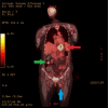
Gastroesophageal Junction Adenocarcinoma With Skeletal Muscle Metastases: A Case Report and Literature Review
- PMID: 39099909
- PMCID: PMC11297803
- DOI: 10.7759/cureus.63855
Gastroesophageal Junction Adenocarcinoma With Skeletal Muscle Metastases: A Case Report and Literature Review
Abstract
Esophageal and gastroesophageal junction (GEJ) malignancies are aggressive, and survival is poor once metastasis occurs. The most common sites of metastatic involvement include the liver, lymph nodes, lung, peritoneum, adrenal glands, bone, and brain, while skeletal muscle (SM) involvement is rare. We report a case of a 68-year-old female who presented with intractable emesis for one month and was found to have a primary GEJ adenocarcinoma measuring up to 6.7 cm. Endoscopic biopsy revealed poorly differentiated GEJ adenocarcinoma with positive AE1/AE3 immunostains. Positron emission tomography/computed tomography and magnetic resonance imaging revealed metastases to the omentum and left lower extremity SMs, including the proximal adductor longus, adductor magnus, and gluteus minimus. This study reviews the literature on SM metastasis in esophageal and GEJ cancer, GEJ cancer classification, incidence, treatment, and prognosis.
Keywords: ac-adenocarcinoma; esophageal cancer (ec); esophagus adenocarcinoma; gastroesophageal adenocarcinoma; gastroesophageal cancer; gastroesophageal junction (gej); muscle metastasis; rare metastasis; skeletal muscle metastasis.
Copyright © 2024, Sabu et al.
Conflict of interest statement
Human subjects: Consent was obtained or waived by all participants in this study. Conflicts of interest: In compliance with the ICMJE uniform disclosure form, all authors declare the following: Payment/services info: All authors have declared that no financial support was received from any organization for the submitted work. Financial relationships: All authors have declared that they have no financial relationships at present or within the previous three years with any organizations that might have an interest in the submitted work. Other relationships: All authors have declared that there are no other relationships or activities that could appear to have influenced the submitted work.
Figures


References
-
- National Cancer Institute: SEER. [ Apr; 2024 ]. 2024. https://seer.cancer.gov/statfacts/html/esoph.html. https://seer.cancer.gov/statfacts/html/esoph.html.
-
- An unusual presentation of metastatic esophageal adenocarcinoma presenting as thigh pain. Norris WE, Perry JL, Moawad FJ, Horwhat JD. https://pubmed.ncbi.nlm.nih.gov/19795036/ J Gastrointestin Liver Dis. 2009;18:371–374. - PubMed
-
- Gastroesophageal junction adenocarcinoma: is there an optimal management? Lin D, Khan U, Goetze TO, et al. Am Soc Clin Oncol Educ Book. 2019;39:0–95. - PubMed
Publication types
LinkOut - more resources
Full Text Sources
Miscellaneous
