. 2024 Jul 11:37:100836.
doi: 10.1016/j.lana.2024.100836. eCollection 2024 Sep.

Human Orthohantavirus disease prevalence and genotype distribution in the U.S., 2008-2020: a retrospective observational study

Affiliations

Human Orthohantavirus disease prevalence and genotype distribution in the U.S., 2008-2020: a retrospective observational study

Shannon L M Whitmer et al. Lancet Reg Health Am. .

Abstract

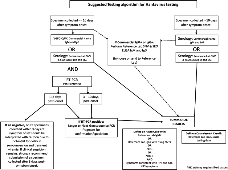
Background: In the United States (U.S.), hantavirus pulmonary syndrome (HPS) and non-HPS hantavirus infection are nationally notifiable diseases. Criteria for identifying human cases are based on clinical symptoms (HPS or non-HPS) and acute diagnostic results (IgM+, rising IgG+ titers, RT-PCR+, or immunohistochemistry (IHC)+). Here we provide an overview of diagnostic testing and summarize human Hantavirus disease occurrence and genotype distribution in the U.S. from 2008 to 2020.

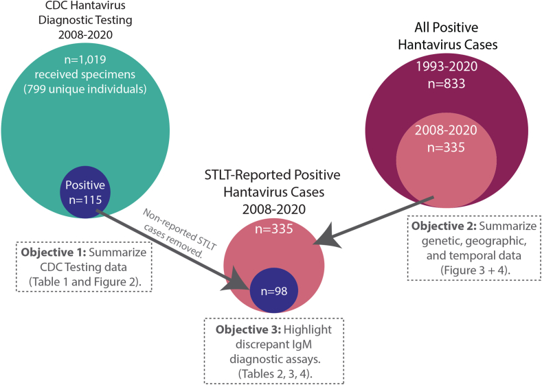
Methods: Epidemiological data from the national hantavirus registry was merged with laboratory diagnostic testing results performed at the CDC. Residual hantavirus-positive specimens were sequenced, and the available epidemiological and genetic data sets were linked to conduct a genomic epidemiological study of hantavirus disease in the U.S.

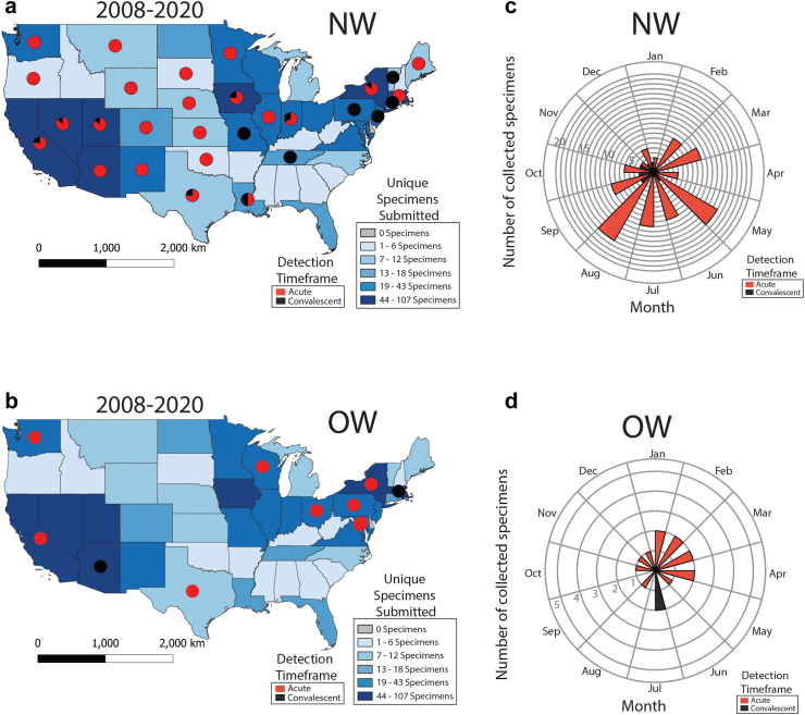
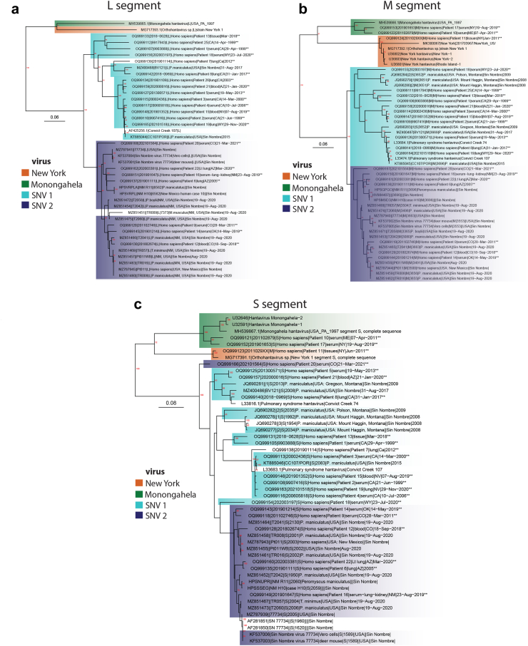
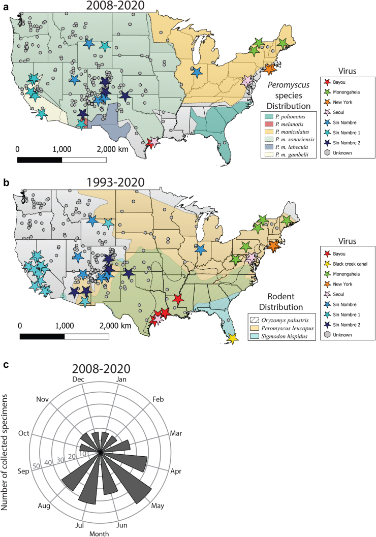
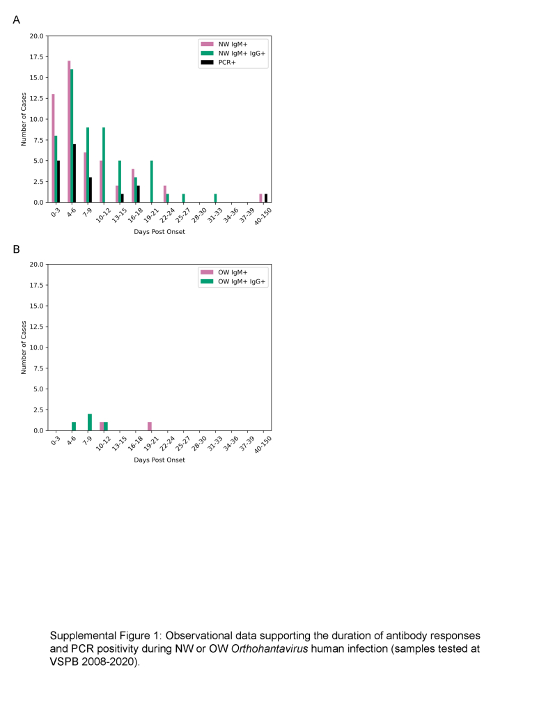
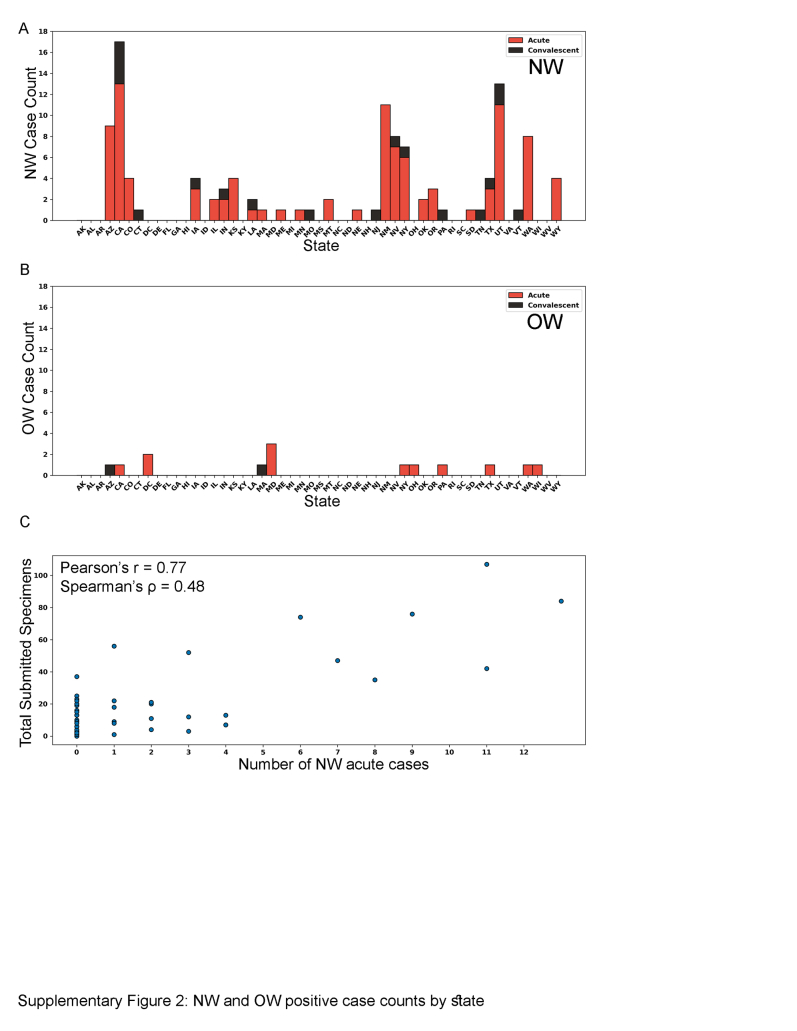
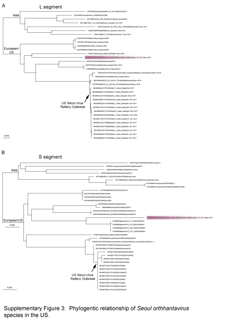
Findings: From 1993 to 2020, 833 human hantavirus cases have been identified, and from 2008 to 2020, 335 human cases have occurred. Among New World (NW) hantavirus cases detected at the CDC diagnostic laboratory (representing 29.2% of total cases), most (85.0%) were detected during acute disease, however, some convalescent cases were detected in states not traditionally associated with hantavirus infections (Connecticut, Missouri, New Jersey, Pennsylvania, Tennessee, and Vermont). From 1993 to 2020, 94.9% (745/785) of U.S. hantaviruses cases were detected west of the Mississippi with 45.7% (359/785) in the Four Corners region of the U.S. From 2008 to 2020, 67.7% of NW hantavirus cases were detected between the months of March and August. Sequencing of RT-PCR-positive cases demonstrates a geographic separation of Orthohantavirus sinnombreense species [Sin Nombre virus (SNV), New York virus, and Monongahela virus]; however, there is a large gap in viral sequence data from the Northwestern and Central U.S. Finally, these data indicate that commercial IgM assays are not concordant with CDC-developed assays, and that "concordant positive" (i.e., commercial IgM+ and CDC IgM+ results) specimens exhibit clinical characteristics of hantavirus disease.

Interpretation: Hantaviral disease is broadly distributed in the contiguous U.S, viral variants are localised to specific geographic regions, and hantaviral disease infrequently detected in most Southeastern states. Discordant results between two diagnostic detection methods highlight the need for an improved standardised testing plan in the U.S. Hantavirus surveillance and detection will continue to improve with clearly defined, systematic reporting methods, as well as explicit guidelines for clinical characterization and diagnostic criteria.

Funding: This work was funded by core funds provided to the Viral Special Pathogens Branch at CDC.

Keywords: Bunyavirus; Disease surveillance; Genetics; HCPS; HFRS; HPS; Hantavirus cardiopulmonary syndrome; Hantavirus haemorrhagic fever with renal syndrome; Hantavirus pulmonary syndrome; Orthohantavirus; Rodent.


Conflict of interest statement

The authors declare no competing interests.

Figures

Fig. 1
Fig. 1
Overview of datasets used to evaluate human Orthohantavirus disease prevalence and genotype distribution in the U.S. Overall 11,019 unique specimens were received at VSPB from 2008 to 2020 for diagnostic testing and 115 unique individuals were identified as hantavirus positive. Seventeen individuals tested by CDC were not reported as hantavirus-positive cases by STLT (n = 98). From 2008 to 2020, 335 unique people were reported as hantavirus-positive cases by STLT and of those VSPB performed diagnostic testing for 98 unique people. Altogether, 833 unique individuals were reported as Hantavirus-positive cases by STLT health departments. The relationships between these datasets, objectives, and figures/tables are highlighted.
Fig. 2
Fig. 2
Initial detection of acute or convalescent human hantavirus cases by VSPB-CDC presented by state and time, 2008–2020. a and b) Map of initial detection of acute or convalescent NW (a) or OW (b) human hantavirus cases. Pie charts indicate ratio of initial acute or convalescent cases detected per state. State color indicates number of unique specimens received per state for hantavirus diagnostic testing. A single specimen was received from Alaska, Puerto Rico, and the U.S. Virgin Islands, and two specimens from Hawaii, but all were hantavirus negative. No other specimens were received from other U.S. territories. c and d) Seasonality of initial detection of acute (red) or convalescent (black) NW (c) or OW (d) human hantavirus cases by VSPB-CDC from 2008 to 2020.
Fig. 3
Fig. 3
Inferred relationships of domestic human Orthohantavirus sinnombreense cases. Viral genotypes, New York virus, Monongahela virus and Sin Nombre virus are highlighted orange, green or blue, respectively (similar to Fig. 4). The two major SNV clades are colored light or dark blue according to the SNV M segment to highlight possible reassortment between segments and sequences without highlight did not match with a specific M segment. New sequences are indicated with “∗∗” at the end of sequence names. Bootstrap support (n = 1000) greater than 70% is labeled in red at internal nodes and scale bars are in units of substitutions/site. a) Phylogenetic tree of all available full length Orthohantavirus sinnombreense species L segments. b) Phylogenetic tree of all available full length Orthohantavirus sinnombreense species M segments. c) Phylogenetic tree of all available full length Orthohantavirus sinnombreense species S segments.
Fig. 4
Fig. 4
Geographic and temporal distribution of STLT-reported human hantaviral cases and viral genotypes from 2008 to 2020 and a comprehensive geographic distribution of all available human hantavirus cases from 1993 to 2020. a) Geographic distribution of human hantaviral cases from 2008 to 2020 (points) and Peromyscus species distributions (shading) from Greenbaum et al. Viral genotypes are indicated by star color and cases without sequence data are labeled as “Unknown.” No human hantavirus cases have been detected in Alaska, Hawaii, or U.S. territories. b) Geographic distribution of human hantaviral cases from 1993 to 2020 (points) and hantavirus rodent vector distributions (shading). Map is colored according to panel A. No human hantavirus cases have been detected in Alaska, Hawaii, or U.S. territories. c) Seasonality of state-reported human hantaviral cases from 2008 to 2020.
Fig. 5
Fig. 5
Suggested testing algorithm for Orthohantavirus diagnostic testing.
Supplemental Figure 1
Observational data supporting the duration of antibody responses and PCR positivity during NW or OW Orthohantavirus human infection (samples tested at VSPB 2008–2020), including duplicate specimens. A. Serological and PCR positivity during NW infection. Bars represent the number of cases for each 3-day binned time point. B. Serological and PCR positivity during OW infection. Bars represent the number of cases for each 3-day binned time point.
Supplemental Figure S2
Summary of human hantavirus-positive cases initially detected by VSPB as acute or convalescent infection by state. A) NW-positive human cases. B) OW-positive human cases. C) Relationship between the total number of submitted specimens per state compared to total number of positive NW acute cases.
Supplemental Figure S3
Inferred relationship of domestic human Orthohantavirus seoulense case (highlighted purple) relative to sequences from the Seoul virus Rattery outbreak (UK, Netherlands, U.S.), historic domestic U.S. sequences (lower clade) and a subset of Seoul sequences from Asia (upper clade).

References

    1. Viruses ICotTo. Bunyaviridae. https://ictv.global/report_9th/RNAneg/Bunyaviridae2023
    1. Lee H.W., French G.R., Lee P.W., Baek L.J., Tsuchiya K., Foulke R.S. Observations on natural and laboratory infection of rodents with the etiologic agent of Korean hemorrhagic fever. Am J Trop Med Hyg. 1981;30(2):477–482. - PubMed
    1. Nunez J.J., Fritz C.L., Knust B., et al. Hantavirus infections among overnight visitors to Yosemite national park, California, USA, 2012. Emerg Infect Dis. 2014;20(3):386–393. - PMC - PubMed
    1. de St Maurice A., Ervin E., Schumacher M., et al. Exposure characteristics of hantavirus pulmonary syndrome patients, United States, 1993-2015. Emerg Infect Dis. 2017;23(5):733–739. - PMC - PubMed
    1. Enria D., Padula P., Segura E.L., et al. Hantavirus pulmonary syndrome in Argentina. Possibility of person to person transmission. Medicina (B Aires) 1996;56(6):709–711. - PubMed

LinkOut - more resources