

Embedded-deep-learning-based sample-to-answer device for on-site malaria diagnosis
- PMID: 39100623
- PMCID: PMC11294195
- DOI: 10.3389/fbioe.2024.1392269
Embedded-deep-learning-based sample-to-answer device for on-site malaria diagnosis
Abstract
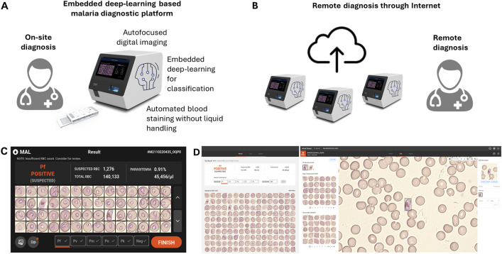
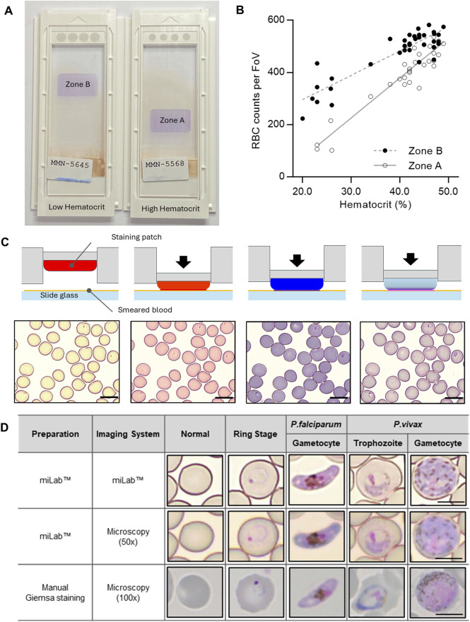
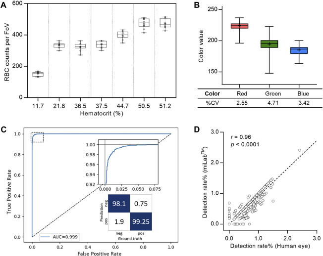
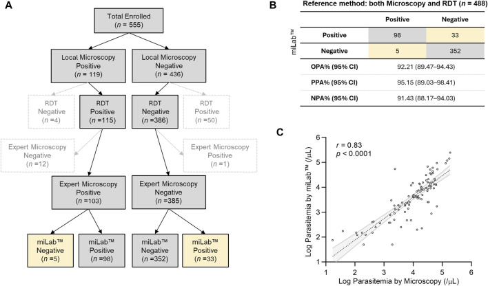
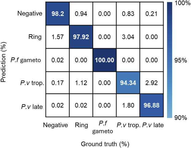
Improvements in digital microscopy are critical for the development of a malaria diagnosis method that is accurate at the cellular level and exhibits satisfactory clinical performance. Digital microscopy can be enhanced by improving deep learning algorithms and achieving consistent staining results. In this study, a novel miLab™ device incorporating the solid hydrogel staining method was proposed for consistent blood film preparation, eliminating the use of complex equipment and liquid reagent maintenance. The miLab™ ensures consistent, high-quality, and reproducible blood films across various hematocrits by leveraging deformable staining patches. Embedded-deep-learning-enabled miLab™ was utilized to detect and classify malarial parasites from autofocused images of stained blood cells using an internal optical system. The results of this method were consistent with manual microscopy images. This method not only minimizes human error but also facilitates remote assistance and review by experts through digital image transmission. This method can set a new paradigm for on-site malaria diagnosis. The miLab™ algorithm for malaria detection achieved a total accuracy of 98.86% for infected red blood cell (RBC) classification. Clinical validation performed in Malawi demonstrated an overall percent agreement of 92.21%. Based on these results, miLab™ can become a reliable and efficient tool for decentralized malaria diagnosis.
Keywords: automated staining process; deep-learning algorithms; digital microscopy; malaria diagnosis; microscopy examination.
Copyright © 2024 Bae, Shin, Kim, Song, Lee, Kim, Lee, Kim, Kanyemba, Lungu, Kang, Han, Beck, Cho, Woo, Lim and Choi.
Conflict of interest statement
Authors CYB, YMS, MK, YS, HJL, KWK, HWL, YJK, B-IK, SH, S-HC, BMW, CYL, K-HC were employed by Noul Co. Ltd. H-PB, DKL were a member of Scientific Advisory Board (Technical Consultant) in Noul Co., Ltd. The remaining author declares that the research was conducted in the absence of any commercial or financial relationships that could be construed as a potential conflict of interest.
Figures
Similar articles
-
Diagnostic accuracy of an automated microscope solution (miLab™) in detecting malaria parasites in symptomatic patients at point-of-care in Sudan: a case-control study.Malar J. 2024 Jun 28;23(1):200. doi: 10.1186/s12936-024-05029-3. Malar J. 2024. PMID: 38943203 Free PMC article.
-
An automatic device for detection and classification of malaria parasite species in thick blood film.BMC Bioinformatics. 2012;13 Suppl 17(Suppl 17):S18. doi: 10.1186/1471-2105-13-S17-S18. Epub 2012 Dec 13. BMC Bioinformatics. 2012. PMID: 23281600 Free PMC article.
-
Tile-based microscopic image processing for malaria screening using a deep learning approach.BMC Med Imaging. 2023 Mar 22;23(1):39. doi: 10.1186/s12880-023-00993-9. BMC Med Imaging. 2023. PMID: 36949382 Free PMC article.
-
Reliable enumeration of malaria parasites in thick blood films using digital image analysis.Malar J. 2009 Sep 23;8:218. doi: 10.1186/1475-2875-8-218. Malar J. 2009. PMID: 19775454 Free PMC article.
-
Advances and challenges in automated malaria diagnosis using digital microscopy imaging with artificial intelligence tools: A review.Front Microbiol. 2022 Nov 15;13:1006659. doi: 10.3389/fmicb.2022.1006659. eCollection 2022. Front Microbiol. 2022. PMID: 36458185 Free PMC article. Review.
Cited by
-
Diagnosis of Plasmodium infections using artificial intelligence techniques versus standard microscopy in a reference laboratory.J Clin Microbiol. 2025 Jan 31;63(1):e0077524. doi: 10.1128/jcm.00775-24. Epub 2024 Dec 10. J Clin Microbiol. 2025. PMID: 39655938 Free PMC article.
-
A large multi-focus dataset for white blood cell classification.Sci Data. 2024 Oct 9;11(1):1106. doi: 10.1038/s41597-024-03938-1. Sci Data. 2024. PMID: 39384810 Free PMC article.
References
LinkOut - more resources
Full Text Sources