

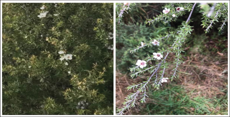
Growth inhibitory effect of Leptospermum scoparium (manuka) chloroform extract on breast and liver cancer cell lines
- PMID: 39101096
- PMCID: PMC11296193
- DOI: 10.5455/javar.2024.k769
Growth inhibitory effect of Leptospermum scoparium (manuka) chloroform extract on breast and liver cancer cell lines
Abstract
Objective: Research has demonstrated that Leptospermum scoparium possesses various therapeutic benefits. This study set out to determine whether or not L. scoparium extracts had any effect on the ability of HepG2 and MCF-7 breast cancer cells to survive.
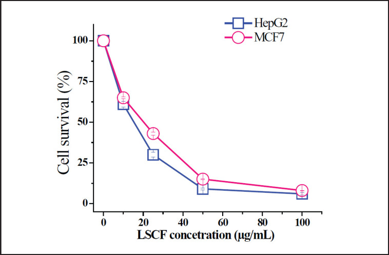
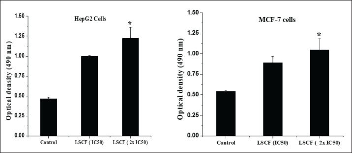
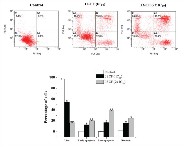
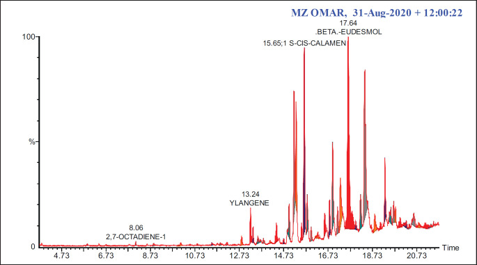
Materials and methods: The antiproliferative activity of L. scoparium extracts was explored using 3-(4,5-dimethylthiazol-2-yl)-2,5-diphenyltetrazolium bromide and lactate dehydrogenase assays. The most active fraction was selected to investigate its effects on apoptosis induction using flow cytometry and quantitative real-time polymerase chain reaction. The constituents of this fraction were characterized using GC-MS analysis.
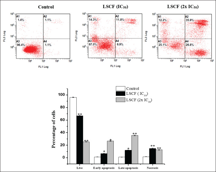
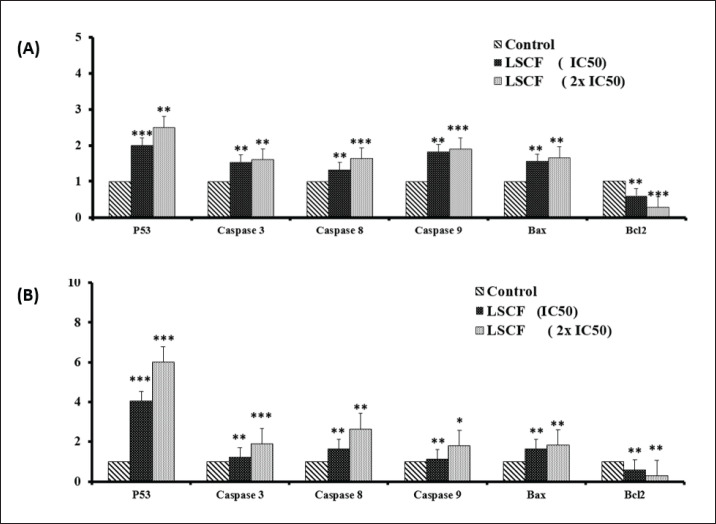
Results: Research demonstrated that the chloroform fraction of L. scoparium (LSCF) significantly impacted the HepG2 and MCF-7 cancer cell lines. Treatment with LSCF led to a notable rise in both early and late apoptotic cells. Furthermore, there was an upregulation in the mRNA levels of P53, Bax, and caspases, while the expression of Bcl-2 mRNA saw a decrease. The analysis of LSCF revealed the primary components to be cis-calamenene, beta-eudesmol, cyclododecane, and alpha-muurolene.
Conclusion: The study showed the promising antiproliferative activity of L. scoparium, suggesting its potential application for cancer treatment.
Keywords: Antiproliferative; GC-MS; LDH; Leptospermum scoparium; MCF-7; MTT; apoptosis.
© The authors.
Conflict of interest statement
The author declares that there is no conflict of interest.
Figures
References
-
- Siegel RL, Miller KD, Jemal A. Cancer statistics, 2020. Cancer J Clin. 2020;70:7–30. https://doi.org/10.3322/caac.21590. - PubMed
-
- Mamdouh AM, Khodeer DM, Tantawy MA, Moustafa YM. In-vitro and in-vivo investigation of amygdalin, metformin, and combination of both against doxorubicin on hepatocellular carcinoma. Life Sci. 2021;285:119961. https://doi.org/10.1016/j.lfs.2021.119961. - PubMed
-
- Zhong L, Li Y, Xiong L, Wang W, Wu M, Yuan T, et al. Small molecules in targeted cancer therapy: advances, challenges, and future perspectives. Signal Transduct Target Ther. 2021;6:1–48. https://doi.org/10.1038/s41392-021-00572-w. - PMC - PubMed
-
- Mathew C, Tesfaye W, Rasmussen P, Peterson GM, Bartholomaeus A, Sharma M, et al. Mānuka oil—a review of antimicrobial and other medicinal properties. Pharmaceuticals. 2020;13:343. https://doi.org/10.3390/ph13110343. - PMC - PubMed
-
- Alsaud N, Shahbaz K, Farid M. Antioxidant and antibacterial evaluation of manuka leaves (Leptospermum scoparium) extracted by hydrophobic deep eutectic solvent. Chem Eng Res Des. 2021;174:96–106. https://doi.org/10.1016/j.cherd.2021.08.004.
LinkOut - more resources
Full Text Sources
Research Materials
Miscellaneous