

Concurrent multiple cerebral cavernous malformations and cauda equina paraganglioma: illustrative case
- PMID: 39102750
- PMCID: PMC11301588
- DOI: 10.3171/CASE24102
Concurrent multiple cerebral cavernous malformations and cauda equina paraganglioma: illustrative case
Abstract
Background: Cauda equina neuroendocrine tumors (CENETs), previously known as cauda equina paragangliomas, and multiple cerebral cavernous malformations (CCMs) are uncommon conditions affecting the central nervous system. To the authors' knowledge, they have not been reported in the same patient.
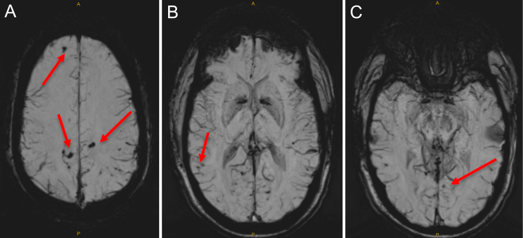
Observations: The authors present the case of a 45-year-old male with CENET and concurrent incidental MRI findings of multiple CCMs. Familial CCMs are associated with mutations in the KRIT1 (CCM1), MGC4607 (CCM2), and PDCD10 (CCM3) genes. Peripheral paragangliomas have been associated with mutations in succinate dehydrogenase (SDHx), RET (multiple endocrine neoplasia 2), VHL (von Hippel-Lindau syndrome), and NF1 (neurofibromatosis type 1) genes. Except for a single case, cauda equina paragangliomas have not been associated with any underlying genetic mutations.
Lessons: It is unclear whether the co-occurrence of these two rare conditions in the same patient is coincidental or suggests a possible shared pathogenesis. https://thejns.org/doi/10.3171/CASE24102.
Keywords: cauda equina; cerebral cavernous malformation; neuroendocrine; paraganglioma; tumor.
Figures


References
-
- Eerola I, Plate KH, Spiegel R, Boon LM, Mulliken JB, Vikkula M. KRIT1 is mutated in hyperkeratotic cutaneous capillary–venous malformation associated with cerebral capillary malformation. Hum Mol Genet. 2000;9(9):1351-1355. - PubMed
-
- Robinson JR, Awad IA, Little JR. Natural history of the cavernous angioma. J Neurosurg. 1991;75(5):709-714. - PubMed
-
- Gross BA, Du R. Hemorrhage from cerebral cavernous malformations: a systematic pooled analysis. J Neurosurg. 2017;126(4):1079-1087. - PubMed
-
- McNicol AM. Adrenal medulla and paraganglia. In: Lloyd RV, ed. Endocrine Pathology: Differential Diagnosis and Molecular Advances. Springer; 2010.
LinkOut - more resources
Full Text Sources
Research Materials
Miscellaneous
