. 2024 Nov;23(11):e14289.
doi: 10.1111/acel.14289. Epub 2024 Aug 5.

Short-term starvation activates AMPK and restores mitochondrial inorganic polyphosphate, but fails to reverse associated neuronal senescence

Affiliations

Short-term starvation activates AMPK and restores mitochondrial inorganic polyphosphate, but fails to reverse associated neuronal senescence

Luca Tagliafico et al. Aging Cell. 2024 Nov.

Abstract

Neuronal senescence is a major risk factor for the development of many neurodegenerative disorders. The mechanisms that drive neurons to senescence remain largely elusive; however, dysregulated mitochondrial physiology seems to play a pivotal role in this process. Consequently, strategies aimed to preserve mitochondrial function may hold promise in mitigating neuronal senescence. For example, dietary restriction has shown to reduce senescence, via a mechanism that still remains far from being totally understood, but that could be at least partially mediated by mitochondria. Here, we address the role of mitochondrial inorganic polyphosphate (polyP) in the intersection between neuronal senescence and dietary restriction. PolyP is highly present in mammalian mitochondria; and its regulatory role in mammalian bioenergetics has already been described by us and others. Our data demonstrate that depletion of mitochondrial polyP exacerbates neuronal senescence, independently of whether dietary restriction is present. However, dietary restriction in polyP-depleted cells activates AMPK, and it restores some components of mitochondrial physiology, even if this is not sufficient to revert increased senescence. The effects of dietary restriction on polyP levels and AMPK activation are conserved in differentiated SH-SY5Y cells and brain tissue of male mice. Our results identify polyP as an important component in mitochondrial physiology at the intersection of dietary restriction and senescence, and they highlight the importance of the organelle in this intersection.

Keywords: dietary restriction; fasting; inorganic polyphosphate; metabolism; mitochondria; polyP; senescence.


Conflict of interest statement

The authors declare no conflict of interest.

Figures

FIGURE 1
FIGURE 1
Depletion of mitochondrial polyP affects cellular morphology and it induces senescence in dSH‐SY5Y cells. 72 h of STS is able to restore mitochondrial polyP levels. (a) Representative images displaying mitochondrial and cellular morphology of dSH‐SY5Y Wt and MitoPPX cells. MitoTracker green fluorescence and dark‐field imaging were utilized to visualize mitochondrial and cellular morphology, respectively, in all experimental conditions. (b) Graph showing the quantification of the mitochondrial area, and the mean length of the mitochondrial branches in single cells. (c) Representative microscope images illustrating β‐galactosidase activity (increased activity of this enzyme has been broadly described as a marker of senescence). (d) Graphs presenting quantification of the intensity of β‐galactosidase staining in different experimental conditions. The mean intensity of the staining adjusted by the area occupied by the cells in each image, and normalized by the levels of the staining in Wt control cells is shown in the graphs. The experiment was conducted under control and 24, 48, and 72 h of STS in Wt and MitoPPX dSH‐SY5Y cells. Based on the findings from this figure, subsequent experiments in this study were conducted at STS 72 h. (e) Graph showing the quantification of the number of cells in all the conditions included in our studies. (f) Graphs showing the ATP/ADP ratio assayed in dSHYSY5Y Wt and MitoPPX cells, under control conditions and after STS. Cells were maintained in STS for 24, 48, and 72 h. (g) PolyP levels were assayed in all experimental conditions using the DAPI‐polyP assay. Please, note that significant differences are observed between Wt and MitoPPX dSH‐SY5Y cells. Moreover, 72 h STS is able to recover the decreased levels of the polymer in MitoPPX cells. Scale bar = 30 μM for images in panel A and 75 μM for images in panel c. Data are presented as mean ± SEM of at least three independent experiments. *p ≤ 0.05, **p ≤ 0.01, and ***p ≤ 0.001.
FIGURE 2
FIGURE 2
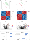
The proteome of Wt and MitoPPX dSH‐SY5Y cells is significantly different both under control conditions and after STS. (a) PCAs and heat maps (displaying the top 25 differently expressed proteins) showing the differences in the proteome between Wt and MitoPPX dSH‐SY5Y cells, under control conditions and after STS. (b) Volcano plot representations of the analysis of the same data. (c) Graphs presenting the z‐scores of the main diseases and functions relevant to this study and predicted to be differently affected in Wt and MitoPPX dSH‐SY5Y cells, under control and STS conditions. Predictions were conducted using IPA. Only data with p ≤ 0.05 were included. Mass spectrometry was conducted in biological quadruplicates.
FIGURE 3
FIGURE 3
Effects of STS on the mitochondrial physiology of dSH‐SY5Y Wt and MitoPPX cells. (a) Representative immunoblots and quantification of the signal of some main proteins involved in the regulation of mitochondrial physiology. Note the increased levels of Bax in dSH‐SY5Y MitoPPX cells under both control conditions and after STS, and the significant rise in the pAMPK/AMPK ratio in dSH‐SY5Y MitoPPX cells after STS. (b) Representative immunoblots and quantification of the signal of the complexes of the mitochondrial ETC. Complexes I and III exhibit significant differences in MitoPPX cells under STS. Please, note that some membranes were re‐blotted for multiple antibodies and therefore, the same immunoblot showing the signal for β‐actin is used for more than one protein. All uncropped membranes that were quantified are included in a Supplementary File. Data are presented as mean ± SEM of at least three independent experiments. *p ≤ 0.05, **p ≤ 0.01, and ****p ≤ 0.0001.
FIGURE 4
FIGURE 4
Intermittent fasting increases polyP levels in male C57BJ/6 mice. (a) Graph displaying the variation in mice weight during the different cycles of intermittent fasting. Measurements include both female and male C57BJ/6 mice. (b) Quantification of glucose and lactate levels, as well as of the presence of ketone bodies at the end of the final cycle of intermittent fasting. Also in this case, measurements include both female and male C57BJ/6 mice. (c) Graphs showing the results from the glucose tolerance test in each of the sexes. This test was conducted at the end of the third cycle of fasting. (d) Graphs showing the results from the assay of non‐fasting glucose levels at baseline and immediately before the last cycle of intermittent fasting. This parameter was evaluated independently for males and females. (e) Assay of polyP levels in the brains of male and female C57BJ/6 mice. Measurements were conducted immediately after euthanasia using the DAPI‐polyP method. (f) Representative immunoblots and quantification of the signal of the pAMPK/AMPK ratio, as well as of the pCREB/CREB ratio in male and female C57BJ/6 mice. Data are presented as mean ± SEM of at least three mice. *p ≤ 0.0.05, **p ≤ 0.01, and ****p ≤ 0.0001.

References

    1. Abramov, A. Y. , Fraley, C. , Diao, C. T. , Winkfein, R. , Colicos, M. A. , Duchen, M. R. , French, R. J. , & Pavlov, E. (2007). Targeted polyphosphatase expression alters mitochondrial metabolism and inhibits calcium‐dependent cell death. Proceedings of the National Academy of Sciences of the United States of America, 104, 18091–18096. 10.1073/pnas.0708959104 - DOI - PMC - PubMed
    1. Alirezaei, M. , Kemball, C. C. , Flynn, C. T. , Wood, M. R. , Whitton, J. L. , & Kiosses, W. B. (2010). Short‐term fasting induces profound neuronal autophagy. Autophagy, 6, 702–710. 10.4161/auto.6.6.12376 - DOI - PMC - PubMed
    1. Angelova, P. R. , Agrawalla, B. K. , Elustondo, P. A. , Gordon, J. , Shiba, T. , Abramov, A. Y. , Chang, Y. T. , & Pavlov, E. V. (2014). In situ investigation of mammalian inorganic polyphosphate localization using novel selective fluorescent probes JC‐D7 and JC‐D8. ACS Chemical Biology, 9, 2101–2110. 10.1021/cb5000696 - DOI - PubMed
    1. Angelova, P. R. , Baev, A. Y. , Berezhnov, A. V. , & Abramov, A. Y. (2016). Role of inorganic polyphosphate in mammalian cells: From signal transduction and mitochondrial metabolism to cell death. Biochemical Society Transactions, 44, 40–45. 10.1042/BST20150223 - DOI - PubMed
    1. Baev, A. Y. , Angelova, P. R. , & Abramov, A. Y. (2020). Inorganic polyphosphate is produced and hydrolyzed in F0F1‐ATP synthase of mammalian mitochondria. Biochemical Journal, 477, 1515–1524. 10.1042/bcj20200042 - DOI - PMC - PubMed

Substances

LinkOut - more resources