. 2025 Jul:73:501-515.
doi: 10.1016/j.jare.2024.08.001. Epub 2024 Aug 3.

GDF11 protects against mitochondrial-dysfunction-dependent NLRP3 inflammasome activation to attenuate osteoarthritis

Affiliations

GDF11 protects against mitochondrial-dysfunction-dependent NLRP3 inflammasome activation to attenuate osteoarthritis

Pengfei Zhang et al. J Adv Res. 2025 Jul.

Abstract

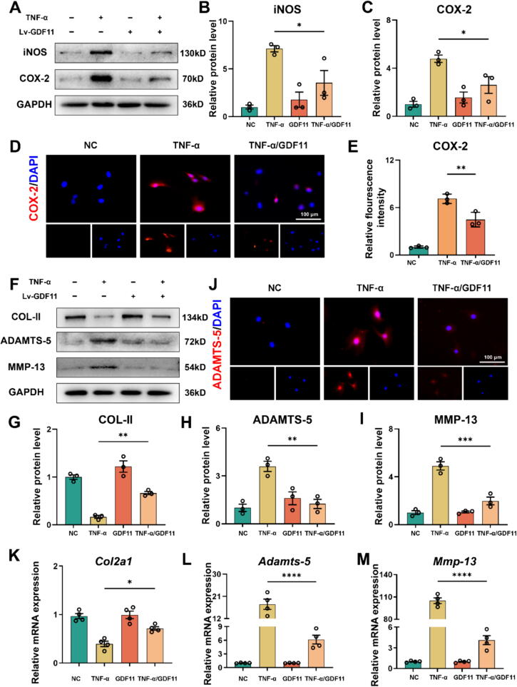
Introduction: Osteoarthritis (OA) is a highly prevalent degenerative disease worldwide, and tumor necrosis factor (TNF-α) is closely associated with its development. Growth differentiation factor 11 (GDF11) has demonstrated anti-injury and anti-aging abilities in certain tissues; however, its regulatory role in OA remains unclear and requires further investigation.

Objectives: To identify whether GDF11 can attenuate osteoarthritis. To exploring the the potential mechanism of GDF11 in alleviating osteoarthritis.

Methods: In this study, we cultured and stimulated mouse primary chondrocytes with or without TNF-α, analyzing the resulting damage phenotype through microarray analysis. Additionally, we employed GDF11 conditional knockout mice OA model to examine the relationship between GDF11 and OA. To investigate the target of GDF11's function, we utilized NLRP3 knockout mice and its inhibitor to verify the potential involvement of the NLRP3 inflammasome.

Results: Our in vitro experiments demonstrated that endogenous overexpression of GDF11 significantly inhibited TNF-α-induced cartilage matrix degradation and inflammatory expression in chondrocytes. Furthermore, loss of GDF11 led to NLRP3 inflammasome activation, inflammation, and metabolic dysfunction. In an in vivo surgically induced mouse model, intraarticular administration of recombinant human GDF11 alleviated OA pathogenesis, whereas GDF11 conditional knockout reversed this effect. Additionally, findings from the NLRP3-knockout DMM mouse model revealed that GDF11 exerted its protective effect by inhibiting NLRP3.

Conclusion: These findings demonstrate the ability of GDF11 to suppress TNF-α-induced inflammation and cartilage degeneration by preventing mitochondrial dysfunction and inhibiting NLRP3 inflammasome activation, suggesting its potential as a promising therapeutic drug for osteoarthritis.

Keywords: GDF11; NLRP3 inflammasome; Osteoarthritis; TNF-α.


Conflict of interest statement

Declaration of competing interest The authors declare that they have no known competing financial interests or personal relationships that could have appeared to influence the work reported in this paper.

Figures

None
Schematic model of GDF11 Protects Against Mitochondrial-Dysfunction-Dependent NLRP3 Inflammasome Activation to Attenuate Osteoarthritis.
Fig. 1
Fig. 1
GDF11 expression level is diminished in osteoarthritis. (A) Representative X-ray images of the knee joint were obtained from both normal patients and individuals diagnosed with severe osteoarthritis (OA), classified as David Grade IV. (B) The expression of synovial fluid GDF11 level of patients with anterior cruciate ligament injury (Normal patients) and OA patients (David Grade Ⅰ-IV) (C) The expression of synovial fluid GDF8 level of patients with Normal patients and OA patients (David Grade Ⅰ-IV). (D-E) The GDF11 protein levels in cartilage samples of weight-bearing area and non-weight-bearing area. (F) Real-time PCR of GDF11. (G) Microarray heatmap of the Control group and TNF-α group (n = 3 for each group). (I) GDF11 expression with TNF-α stimulation for 24 h, 48 h, 72 h. (J-K) Immunohistochemistry images displaying the expression of GDF11 in knee joint tissue were captured from the sham group and the DMM rat model group. Scale bars, 200 μm (low field), 100 μm (high field). Error bar represents mean ± SD; ***p < 0.001, **p < 0.01, *p < 0.05.
Fig. 2
Fig. 2
GDF11 promotes against inflammatory stress, retained cell metabolism and alleviated degeneration in chondrocytes. (A-C) Protein levels and quantification of iNOS and COX-2 in GDF11-lentiviruses transduced primary chondrocytes after treatment of TNF-α, n = 3 for each group. (D-E) Immunofluorescence staining and quantification of COX-2 in GDF11-lentiviruses transduced primary chondrocytes after treatment of TNF-α, n = 3 for each group. Scale bar, 100 μm. (F) Protein levels of COL-II, ADAMTS-5 and MMP-13 in GDF11-lentiviruses transfected primary chondrocytes after treatment of TNF-α. (G-I) Quantification of Western Blot. (J) Immunofluorescence staining of ADAMTS-5 in GDF11-lentiviruses transfected primary chondrocytes after treatment of TNF-α, n = 3 for each group. Scale bar, 100 μm. (K-M) Relative mRNA expression of Col2a1, Adamts-5, and Mmp-13 expression by RT-PCR, n = 4 for each group. Error bar represents mean ± SD; ***p < 0.001, **p < 0.01, *p < 0.05.
Fig. 3
Fig. 3
GDF11 Mitochondrial Dysfunction and Reduced ATP Production induced by TNF-α. (A) Representative transmission electron microscopy (TEM) images of chondrocytes obtained from various groups. Scale bar, 1 μm (low field). (B-D) Representative mitotracker and JC-1 images of chondrocyte mitochondria using fluorescence microscopy. Scale bar, mitotracker: 100 μm and JC-1: 200 μm. (E) ATP levels in chondrocytes after treatment of TNF-α and recombinant GDF11. (F-G) The protein levels of NLRP3, OPA-1, DRP-1 and MFN-2 in GDF11-lentiviruses transfected chondrocytes using Western blot, n = 3 for each group. (H) The Western Blot (J) quantification ration of p-P65/P65 level in GDF11-lentiviruses transfected chondrocytes, n = 4 for each group. (I) The NLRP3 expression levels of chondrocytes after treatment of TNF-α and recombinant GDF11 examined by immunofluorescence staining. Scale bar, 10 μm. (J) The protein levels of p-P65, P65, p-IκBα and IκBα using Western blot. (K-L) ROS level and quantification of primary chondrocytes treatment with TNF-α and recombinant GDF11, n = 3 for each group. Scale bar, 200 μm. Error bar represents mean ± SD; ***p < 0.001, **p < 0.01, *p < 0.05.
Fig. 4
Fig. 4
GDF11 conditional knockout aggravate OA progression in vivo. (A) Flowchart of animal experiment. (B) The micro-CT representative images of the Sham, WT, GDF11-CKO and WT mice injected with rh GDF11 after DMM model. And arrows show the osteophytes formation. (C) Micro-CT was employed to conduct a joint space height assay. (n = 6 for each group). (D) Micro-CT was employed to conduct an osteophyte number assay (n = 6 for each group). (E) The HE staining representative images of the Sham. WT, GDF11-CKO and WT mice injected with rh GDF11 after DMM model. Scale bars, 200 μm (low field), 100 μm (high field). (F) The safranin O fast green staining representative images of different indicate groups after DMM model. Scale bars, 200 μm (low field), 100 μm (high field). (G) Osteoarthritis Research Society International (OARSI) score of OA based on the safranin O staining results (n = 6 for each group). (H) Cartilage thickness in different indicate groups after DMM model based on the results of safranin O staining (n = 6 for each group). Error bar represents mean ± SD; ***p < 0.001, **p < 0.01, *p < 0.05. (For interpretation of the references to colour in this figure legend, the reader is referred to the web version of this article.)
Fig. 5
Fig. 5
Lack of GDF11 led to NLRP3 inflammasome activation, inflammation and metabolic dysfunction. (A-C) Relative protein levels and quantification of iNOS and COX-2 in silence-GDF11 lentiviruses transfected chondrocytes with treatment of TNF-α and Mcc950, n = 3 for each group. (D-E) Immunofluorescence staining and quantification of COX-2 in silence-GDF11 lentiviruses transfected primary chondrocytes after treatment of TNF-α and Mcc950, n = 3 for each group. Scale bar, 100 μm. (F) Relative protein levels of COL-II, ADAMTS-5, MMP-13, NLRP3, CASP-1 and IL-1β in silence-GDF11 lentiviruses transfected primary chondrocytes after treatment of TNF-α and Mcc950. (G-K) Protein level quantification of COL-II, ADAMTS-5, MMP-13, CASP-1 and IL-1β in silence-GDF11 lentiviruses transfected primary chondrocytes after treatment of TNF-α and Mcc950, n = 3 for each group. (L) Immunofluorescence staining and quantification of ADAMTS-5 in silence-GDF11 lentiviruses transfected primary chondrocytes after treatment of TNF-α and Mcc950, n = 3 for each group. Scale bar, 100 μm. Error bar represents mean ± SD; ***p < 0.001, **p < 0.01, *p < 0.05.
Fig. 6
Fig. 6
GDF11 exerts protective effects on knee joint cartilage through the NLRP3 activation. (A) Micro-CT representative images of the DMM model mice in Sham, WT, NLRP3 KO group and NLRP3 KO+Si-GDF11 group. Arrows show the formation of osteophytes. (B) Joint space height assay based on Micro-CT (n = 6 for each group). (C) Osteophyte number assay based on Micro-CT (n = 6 for each group). (D) The HE staining representative images in Sham, WT, NLRP3 KO group and NLRP3 KO+Si-GDF11 group. Scale bars, 200 μm (low field), 100 μm (high field). (E) Osteoarthritis Research Society International (OARSI) score of OA based on the safranin O staining results (F). (G) Cartilage thickness in different indicate groups after DMM model based on the results of safranin O staining (n = 6 for each group). Error bar represents mean ± SD; ***p < 0.001, **p < 0.01, *p < 0.05.

References

    1. Katz J.N., Arant K.R., Loeser R.F. Diagnosis and treatment of hip and knee osteoarthritis: a review. JAMA. 2021;325(6):568–578. doi: 10.1001/jama.2020.22171. - DOI - PMC - PubMed
    1. Tevlin R, desJardins-Park H, Huber J, DiIorio SE, Longaker MT, Wan DC. Musculoskeletal tissue engineering: Adipose derived stromal cell implementation for the treatment of osteoarthritis. Biomaterials. 2022;286:121544. doi: 10.1016/j.biomaterials.2022.121544. - PMC - PubMed
    1. Lee Y.T., Yunus M.H.M., Ugusman A., Yazid M.D. Natural Compounds Affecting Inflammatory Pathways of Osteoarthritis. Antioxidants (Basel) 2022;11(9) doi: 10.3390/antiox11091722. - DOI - PMC - PubMed
    1. Ashford S., Williard J. Osteoarthritis: A review. Nurse Pract. 2014;39(5):1–8. doi: 10.1097/01.NPR.0000445886.71205.c4. - DOI - PubMed
    1. Wolfstadt J.I., Cole B.J., Ogilvie-Harris D.J., Viswanathan S., Chahal J. Current concepts: the role of mesenchymal stem cells in the management of knee osteoarthritis. Sports Health. 2015;7(1):38–44. doi: 10.1177/1941738114529727. - DOI - PMC - PubMed

MeSH terms

Substances

LinkOut - more resources