. 2024 Sep;37(5):1593-1601.
doi: 10.1007/s13577-024-01113-7. Epub 2024 Aug 5.

Development of 3D-iNET ORION: a novel, pre-clinical, three-dimensional in vitro cell model for modeling human metastatic neuroendocrine tumor of the pancreas

Affiliations

Development of 3D-iNET ORION: a novel, pre-clinical, three-dimensional in vitro cell model for modeling human metastatic neuroendocrine tumor of the pancreas

Jan Strnadel et al. Hum Cell. 2024 Sep.

Abstract

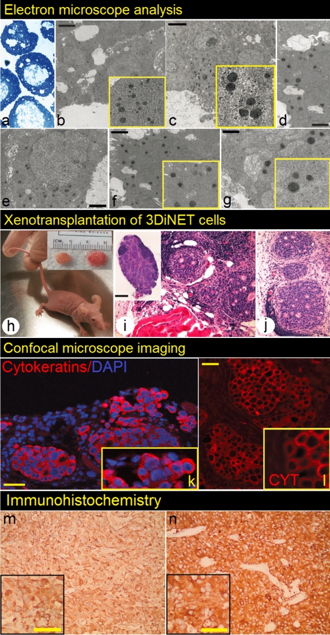
Neuroendocrine tumors (NETs) of the pancreas are rare neoplasms that present complex challenges to diagnosis and treatment due to their indolent course. The incidence of pancreatic neuroendocrine tumors has increased significantly over the past two decades. A limited number of pancreatic neuroendocrine cell lines are currently available for the research. Here, we present 3D-iNET ORION, a novel 3-dimensional (spheroid) cell line, isolated from human pancreatic neuroendocrine tumor liver metastasis. Three-dimensionally grown (3D) cancer cell lines have gained interest over the past years as 3D cancer cell lines better recapitulate the in vivo structure of tumors, and are more suitable for in vitro and in vivo experiments. 3D-iNET ORION cancer cell line showed high potential to form tumorspheres when embedded in Matrigel matrix and expresses synaptophysin and EpCAM. Electron microscopy analysis of cancer cell line proved the presence of dense neurosecretory granules. When xenografted into athymic mice, 3D-iNET ORION cells produce slow-growing tumors, positive for chromogranin and synaptophysin. Human Core Exome Panel Analysis has shown that 3DiNET ORION cell line retains the genetic aberration profile detected in the original tumor. In conclusion, our newly developed neuroendocrine cancer cell line can be considered as a new research tool for in vitro and in vivo experiments.

Keywords: 3D cancer cell lines; Neuroendocrine tumor; Pancreas.


Conflict of interest statement

All authors declare no conflict of interest.

Figures

Fig. 1
Fig. 1
3DiNET ORION cell line was derived from a 83-year-old male patient with pancreatic neuroendocrine tumor liver metastasis (a). Histological evaluation of tumor shows the infiltration of liver parenchyma by sheets of small, uniform tumor cells focally arranged in rosettes (bd). The tumor cells showed strong positivity for chromogranin (eg) and synaptophysin (mo). NET tumor was also positive for CEA (h), CD99 (p), CK7 (q), and CK19 (r). Ki-67 marker was found positive in approximately 70% of the tumor cells, consistent with high proliferation rate (ik). Patchy positivity was detected for CD56 (l). Ki-67 marker was found positive in approximately 70% of the tumor cells, consistent with a high proliferation rate (ik), although mitotic figures were not prominent. Images were captured at 4 × , 10 × and 20 × magnification. Scale bars represent 50 µm (b, c, e, f and i), 20 µm (d, g, k and o) and 100 µm (h, l and p)
Fig. 2
Fig. 2
Primary 3D spheroid cell line was derived with two different strategies—metastatic tumor tissue (a) was processed as described previously [23] and also with modified protocol that utilizes cancer-associated fibroblasts or stromal cells, isolated from the same tumor, as a “feeder layer” (see Material and Methods section for detailed protocol description). A continuously growing cell line (c and d, scale bar represents 500 µm) was finally isolated with the use of the first approach (b left part of the scheme) utilizing Matrigel-embedded spheroids [22]. For growth curve construction and doubling time analysis, cells growing in the form of spheroids (at passage No.22) were used (e). Confocal microscope imaging of cryosectioned spheres showed strong positivity of 3DiNET ORION cells for cytokeratins (f, g) and synaptophysin (h, i). Image j shows hematoxylin–eosin staining of cryosectioned sphere (bar represents 500 µm). Expression of several transcription factors expressed by 3DiNET ORION cells was detected by antibody array protein chip (k). Expression of selected CD markers (CD10, CD56 and EpCAM) was evaluated by flow cytometry (l), while expression of synaptophysin was verified in lysates from three independent experiments (cells from passage No.7 (1st line), No.13 (2nd line) and No.20 (3rd line) was used) by Western blot analysis (GAPDH served as loading control), m)
Fig.3
Fig.3
Characterization of 3DiNET ORION cell line at in vitro and in vivo level. Ultrathin (50–60 nm), resin-embedded sections (a) prepared from 3D spheroids and stained with uranyl acetate and Sato´s lead stain, were analyzed using transmission electron microscope. The presence of dense neurosecretory granules (evidence for neuroendocrine origin of the tumor) was detected (bg).Scale bars 1000 nm. represent Subcutaneous bilateral xenotransplantation of 3DiNET ORION cell line in the form of spheroids resulted into the formation of tumors, approximately 3 months after xenografting into athymic (nude) mice (h, i and j shows H/E staining of tumors). Confocal imaging of cryosections (k, l) prepared from xenografts showed strong positivity for cytokeratins. m shows positivity for chromogranin and n for synaptophysin Scale bars represent 50 µm

References

    1. Rindi G, Mete O, Uccella S, Basturk O, La Rosa S, Brosens LAA, et al. Overview of the 2022 WHO classification of neuroendocrine neoplasms. Endocr Pathol. 2022;33(1):115–54. 10.1007/s12022-022-09708-2. 10.1007/s12022-022-09708-2 - DOI - PubMed
    1. Oronsky B, Ma PC, Morgensztern D, Carter CA. Nothing but NET: a review of neuroendocrine tumors and carcinomas. Neoplasia (New York, NY). 2017;19(12):991–1002. 10.1016/j.neo.2017.09.002. 10.1016/j.neo.2017.09.002 - DOI - PMC - PubMed
    1. Clift AK, Kidd M, Bodei L, Toumpanakis C, Baum RP, Oberg K, et al. Neuroendocrine neoplasms of the small bowel and pancreas. Neuroendocrinology. 2020;110(6):444–76. 10.1159/000503721. 10.1159/000503721 - DOI - PMC - PubMed
    1. Chen M, Van Ness M, Guo Y, Gregg J. Molecular pathology of pancreatic neuroendocrine tumors. J Gastrointest Oncol. 2012;3(3):182–8. 10.3978/j.issn.2078-6891.2012.018. 10.3978/j.issn.2078-6891.2012.018 - DOI - PMC - PubMed
    1. Chung C. Management of neuroendocrine tumors. Am J Health-Syst Pharm : AJHP : Off J Am Soc Health-Syst Pharm. 2016;73(21):1729–44. 10.2146/ajhp150373.10.2146/ajhp150373 - DOI - PubMed

LinkOut - more resources