. 2024 Aug 5;23(1):158.
doi: 10.1186/s12943-024-02048-1.

BRCA1 secondary splice-site mutations drive exon-skipping and PARP inhibitor resistance

Collaborators, Affiliations

BRCA1 secondary splice-site mutations drive exon-skipping and PARP inhibitor resistance

Ksenija Nesic et al. Mol Cancer. .

Abstract

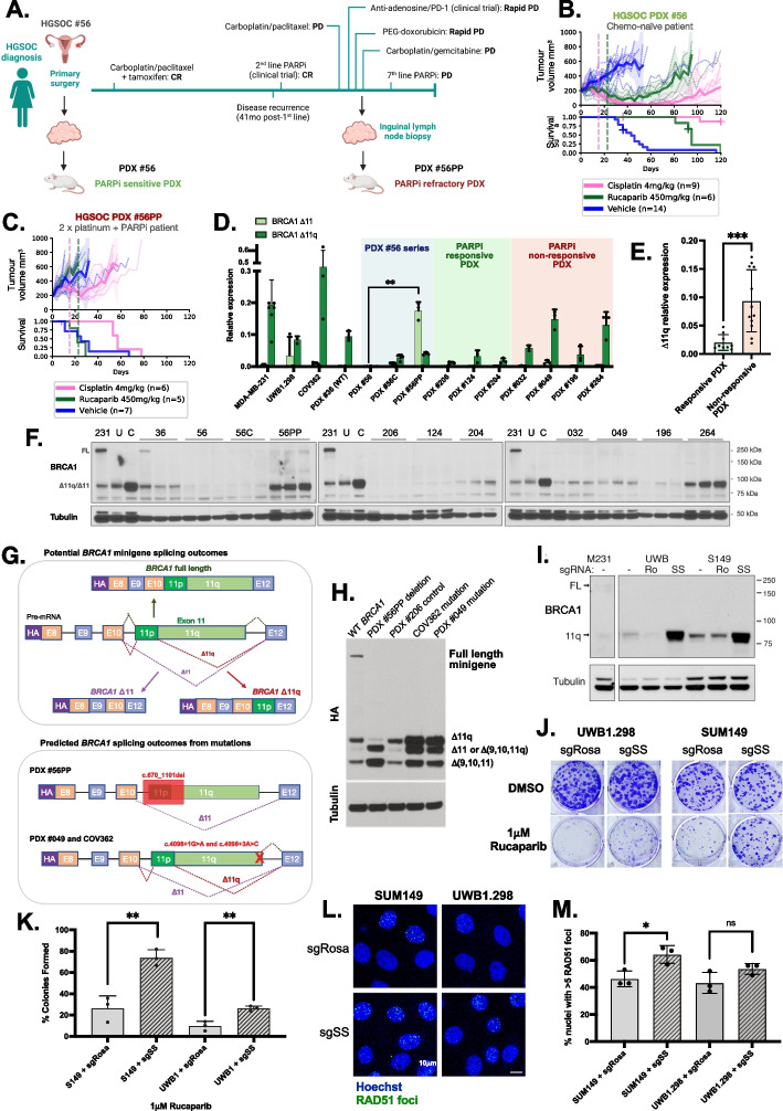
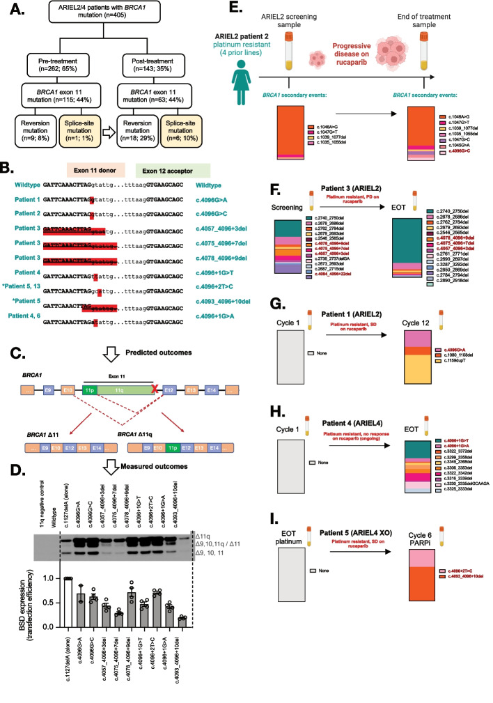
PARP inhibitor (PARPi) therapy has transformed outcomes for patients with homologous recombination DNA repair (HRR) deficient ovarian cancers, for example those with BRCA1 or BRCA2 gene defects. Unfortunately, PARPi resistance is common. Multiple resistance mechanisms have been described, including secondary mutations that restore the HR gene reading frame. BRCA1 splice isoforms △11 and △11q can contribute to PARPi resistance by splicing out the mutation-containing exon, producing truncated, partially functional proteins. However, the clinical impacts and underlying drivers of BRCA1 exon skipping are not fully understood.We analyzed nine ovarian and breast cancer patient derived xenografts (PDX) with BRCA1 exon 11 frameshift mutations for exon skipping and therapy response, including a matched PDX pair derived from a patient pre- and post-chemotherapy/PARPi. BRCA1 exon 11 skipping was elevated in PARPi resistant PDX tumors. Two independent PDX models acquired secondary BRCA1 splice site mutations (SSMs) that drive exon skipping, confirmed using qRT-PCR, RNA sequencing, immunoblotting and minigene modelling. CRISPR/Cas9-mediated disruption of splicing functionally validated exon skipping as a mechanism of PARPi resistance. SSMs were also enriched in post-PARPi ovarian cancer patient cohorts from the ARIEL2 and ARIEL4 clinical trials.Few PARPi resistance mechanisms have been confirmed in the clinical setting. While secondary/reversion mutations typically restore a gene's reading frame, we have identified secondary mutations in patient cohorts that hijack splice sites to enhance mutation-containing exon skipping, resulting in the overexpression of BRCA1 hypomorphs, which in turn promote PARPi resistance. Thus, BRCA1 SSMs can and should be clinically monitored, along with frame-restoring secondary mutations.


Conflict of interest statement

K. Nesic reports nonfinancial support from Clovis Oncology during the conduct of the study. O. Kondrashova reports personal fees from XING Technologies outside the submitted work. K. Nesic, C. Vandenberg, M.J. Wakefield, C.L. Scott, K. Shield-Artin, A. Farrell, E. Kyran and H.E. Barker all receive research support outside of this study from Eisai Inc, AstraZeneca, Boehringer Ingelheim and Ideaya Biosciences. N. Traficante reports grants from AstraZeneca Pty Ltd. during the conduct of the study and grants from AstraZeneca Pty Ltd. outside the submitted work. D. Bowtell reports grants from AstraZeneca Pty Ltd. during the conduct of the study and research support grants from AstraZeneca, Roche-Genentech and BeiGene (paid to institution) outside the submitted work; and personal consulting fees from Exo Therapeutics, that are outside the submitted work. Australian Ovarian Cancer Study reports grants from AstraZeneca Pty Ltd. during the conduct of the study and grants from AstraZeneca Pty Ltd. outside the submitted work. There are no other conflicts of interest in relation to the work under consideration for publication, nor other relationships / conditions / circumstances that present a potential conflict of interest. A. DeFazio reports grants from AstraZeneca outside the submitted work. A. Dobrovic reports grants from National Breast Cancer Foundation of Australia during the conduct of the study. T.C. Harding, K. Lin and T. Kwan were employees of Clovis Oncology. R. Kristeleit reports funding from Clovis Oncology, AstraZeneca, GSK, Pharma&, and was a Co-ordinating Investigator for the ARIEL4 and ATHENA trials. I. McNeish reports advisory boards for Clovis Oncology, AstraZeneca, GSK, OncoC4, Theolytics, Epsila Bio, Duke St Bio, Scancell, Roche, Takeda, and also institutional grant income from AstraZeneca. E.M. Swisher is on the Scientific Advisory Board for Ideaya Biosciences. C.L. Scott reports research support from AstraZeneca Pty Ltd, Boehringer Ingelheim and Eisai Inc, and other support from Clovis Oncology and Sierra Oncology outside the submitted work; and unpaid advisory boards: AstraZeneca, Clovis Oncology, Roche, Eisai, Sierra Oncology, Takeda, MSD. No disclosures were reported by the other authors.

Figures

Fig. 1
Fig. 1
Splice-site mutations (SSMs) drive alternative splicing and PARPi resistance in PDX and cell line models of ovarian and breast cancer. A Patient #56 timeline, showing generation of the matched HGSOC PDX #56 (chemo-naïve) and #56PP (post-chemotherapy/PARPi patient). Created with BioRender.com. CR = Complete response; PD = Progressive disease; SD = Stable disease; C6 = cycle 6. B In vivo treatment data for HGSOC PDX #56 (previously published [3]), classified as PARPi responsive (P = 0.005). C HGSOC PDX #56PP was derived from patient #56 following multiple lines of therapy, including PARPi inhibitor, and was refractory to rucaparib (P = 0.375). Mean PDX tumor volume (mm3) ± 95% CI (hashed lines are individual mice) and corresponding Kaplan–Meier survival analysis. Censored events are represented by crosses on Kaplan–Meier plot; n = individual mice. Detailed patient clinical data can be found in Supplementary Table 1. Details of time to harvest, time to progression and Log-Rank test P values for each PDX can be found in Supplementary Table 2. D Relative △11 and △11q expression for each PDX (mean ± SD). PDX #56PP had the highest △11 expression relative to all other PDX (P = 0.0079 compared to matched PDX #56), while PDX #049 and #264 had the highest △11q levels relative to other PDX (classifications in Supplementary Tables 3 and 4). E △11q levels in PARPi responsive PDX models (< 2 prior lines of platinum in patient) were lower than levels in non-responsive PDX models (≥ 2 prior lines of platinum + PARPi) (P = 0.0007). Statistical comparisons of gene expression were made using an unpaired t-test with Welch’s correction. F Lysates from nuclear extracts from 3 independent tumors were probed for BRCA1 expression by immunoblotting. Bands at the anticipated sizes for full length (FL) BRCA1 and the △11/△11q isoforms are marked. Tubulin immunoblotting is included as a loading control. Gels were run simultaneously with cell line lysates included as controls for each gel. MDA-MB-231 [231] cells are a BRCA1 wild-type control with full-length (FL) and △11q expression, UWB1.289 (U) and COV362 (C) cells with exon 11 variants and △11/△11q expression. G Schematic of BRCA1 mini-gene design, and splicing outcomes predicted for each secondary splice site mutation found in PDX and cell line models. H Splicing predictions for each secondary splice-site mutation modelled by the mini-gene were confirmed by immunoblotting for the HA tag. The PDX #56PP deletion was confirmed to drive high ∆11 and potentially also isoform ∆(9,10,11q) expression. COV362 and PDX #049 secondary splice site mutations were confirmed to drive high ∆11q relative to the wild-type (WT) BRCA1 control and the primary deleterious BRCA1 mutation found in PDX #206. I Cells exposed to sgSS (SS) were found to have elevated BRCA1 D11q protein compared to untreated (-) and sgRosa (Ro) cells. J Example image of colony forming assays of UWB1.289 or SUM149 cells exposed to sgSS or sgRosa treated with DMSO control or 1µM Rucaparib. K Quantification of n = 3 colony forming experiments described in part (J). Mean ± SEM plotted; Ns = Not statistically significant; **p < 0.01; using unpaired, two-tailed t-test. L Representative image of RAD51 foci in UWB1.289 or SUM149 cells exposed to sgSS or sgRosa treated with 10 Gy dose of irradiation. M Quantification of nuclei with > 5 RAD51 foci in UWB1.289 (P = 0.11 compared to control) or SUM149 (P = 0.02 compared to control) cells exposed to sgSS or sgRosa and treated with 10 Gy dose of irradiation. Mean ± SEM plotted; Ns = Not statistically significant; *p < 0.05; using unpaired, two-tailed t-test
Fig. 2
Fig. 2
BRCA1 secondary splice-site mutations are enriched in ARIEL2/4 clinical trial patient samples following PARPi treatment. BRCA1 secondary splice-site mutations increased form 1% (pre-PARPi) to 10% (post-PARPi) in patient tumor/plasma samples from the ARIEL2 and ARIEL4 clinical trials. B The BRCA1 (NM_007294.4) exon 11 donor splice-site mutations identified in these patients and the DNA sequence context are presented. *Patient 5 is an ARIEL4 chemotherapy to PARPi cross-over (XO) arm patient, not included in part A (n = 5). C-D The predicted outcomes on BRCA1 gene splicing based on these disruptions of the exon 11 donor splice site (detailed in Supplementary Table 9) were confirmed for most mutations (D) using the previously described BRCA1 minigene system, with the c.1127delA used as a primary mutation control and included in all SSM minigenes [6]. Mutations driving lower levels of ∆11/∆11q also had a reduced minigene transfection efficiencies relative to other samples (measured as BSD (blasticidin resistance gene) expression). E Summary of BRCA1 secondary events detected in Patient 2 (ARIEL2) before and after PARPi therapy, and their relative proportions in each sample (by colour). F A number of BRCA1 secondary events were also detected in the screening biopsy for platinum and rucaparib resistant Patient 3 (ARIEL2) prior to PARPi, and the number increased at end of rucaparib treatment (EOT). G In contrast, platinum resistant (4 prior lines of platinum) Patient 1 (ARIEL2) had no secondary BRCA1 events detected at first cycle of rucaparib, and had stable disease (SD) on treatment. Three secondary events were detected at cycle 12 of treatment, including a splice-site mutation c.4096G > A. H Patient 4 (ARIEL4) was partially platinum sensitive (2 prior lines) with no secondary BRCA1 events detected at cycle 1 of rucaparib. The EOT plasma sample was positive for multiple reversion events and two splice site mutations (4096 + 1G > T and 4096 + 1G > A) confirmed by minigene to alter splicing (D). I Patient 5 (ARIEL4) was platinum resistant and was enrolled in the chemotherapy arm of ARIEL4. They then crossed over (XO arm) to receive rucaparib treatment where they had stable disease. There were no secondary events detected prior to starting rucaparib, but at cycle 6 there were two BRCA1 splice-site mutations detected (c.4096 + 2 T > C and c.4093_4096 + 10del), without other reversion events. c.4096 + 2 T > C was found to drive alternative BRCA1 splicing (D). Red bold text indicates SSMs detected in patient samples (E-I), while other events presented are exonic secondary/reversion variants predicted to restore the BRCA1 reading frame

Update of

References

    1. Veneziani AC, Scott C, Wakefield MJ, Tinker AV, Lheureux S. Fighting resistance: post-PARP inhibitor treatment strategies in ovarian cancer. Ther Adv Med Oncol. 2023;15:17588359231157644. 10.1177/17588359231157644. 10.1177/17588359231157644 - DOI - PMC - PubMed
    1. Pettitt SJ, Krastev DB, Brandsma I, Drean A, Song F, Aleksandrov R, et al. Genome-wide and high-density CRISPR-Cas9 screens identify point mutations in PARP1 causing PARP inhibitor resistance. Nat Commun. 2018;9(1):1849. 10.1038/s41467-018-03917-2. 10.1038/s41467-018-03917-2 - DOI - PMC - PubMed
    1. Kondrashova O, Topp M, Nesic K, Lieschke E, Ho GY, Harrell MI, et al. Methylation of all BRCA1 copies predicts response to the PARP inhibitor rucaparib in ovarian carcinoma. Nat Commun. 2018;9(1):3970. 10.1038/s41467-018-05564-z. 10.1038/s41467-018-05564-z - DOI - PMC - PubMed
    1. Cong K, Peng M, Kousholt AN, Lee WTC, Lee S, Nayak S, et al. Replication gaps are a key determinant of PARP inhibitor synthetic lethality with BRCA deficiency. Mol Cell. 2021;81(15):3128–44.e7. 10.1016/j.molcel.2021.06.011. 10.1016/j.molcel.2021.06.011 - DOI - PMC - PubMed
    1. Bolton KL, Chenevix- Trench G, Goh C, Sadetzki S, Ramus SJ, Karlan BY, et al. Association between BRCA1 and BRCA2 mutations and survival in women with invasive epithelial ovarian cancer. JAMA. 2012;307(4):382–9. 10.1001/jama.2012.20. 10.1001/jama.2012.20 - DOI - PMC - PubMed

MeSH terms

LinkOut - more resources