. 2024 Aug 5;22(1):724.
doi: 10.1186/s12967-024-05492-1.

Transplantation of miR-145a-5p modified M2 type microglia promotes the tissue repair of spinal cord injury in mice

Affiliations

Transplantation of miR-145a-5p modified M2 type microglia promotes the tissue repair of spinal cord injury in mice

Penghui Li et al. J Transl Med. .

Abstract

Background: The traumatic spinal cord injury (SCI) can cause immediate multi-faceted function loss or paralysis. Microglia, as one of tissue resident macrophages, has been reported to play a critical role in regulating inflammation response during SCI processes. And transplantation with M2 microglia into SCI mice promotes recovery of motor function. However, the M2 microglia can be easily re-educated and changed their phenotype due to the stimuli of tissue microenvironment. This study aimed to find a way to maintain the function of M2 microglia, which could exert an anti-inflammatory and pro-repair role, and further promote the repair of spinal cord injury.

Methods: To establish a standard murine spinal cord clip compression model using Dumont tying forceps. Using FACS, to sort microglia from C57BL/6 mice or CX3CR1GFP mice, and further culture them in vitro with different macrophage polarized medium. Also, to isolate primary microglia using density gradient centrifugation with the neonatal mice. To transfect miR-145a-5p into M2 microglia by Lipofectamine2000, and inject miR-145a-5p modified M2 microglia into the lesion sites of spinal cord for cell transplanted therapy. To evaluate the recovery of motor function in SCI mice through behavior analysis, immunofluorescence or histochemistry staining, Western blot and qRT-PCR detection. Application of reporter assay and molecular biology experiments to reveal the mechanism of miR-145a-5p modified M2 microglia therapy on SCI mice.

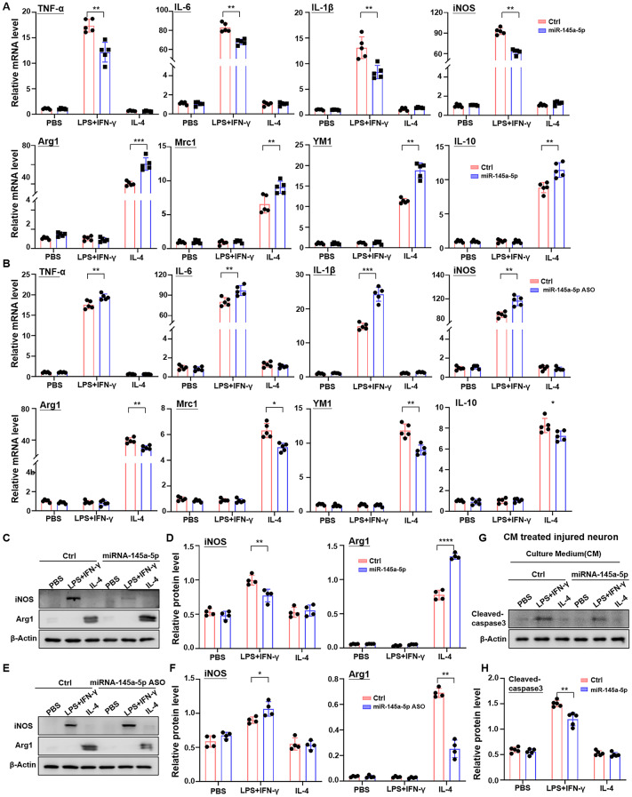
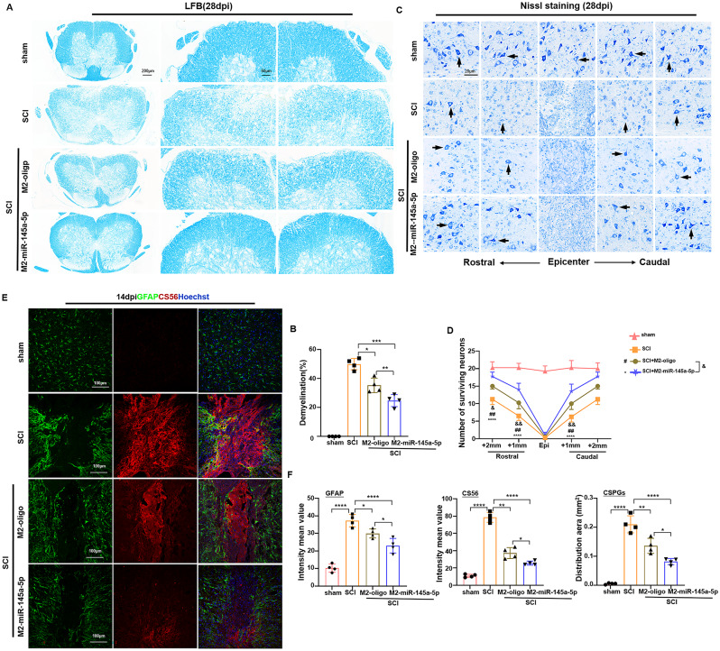
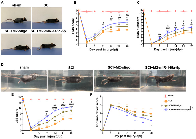
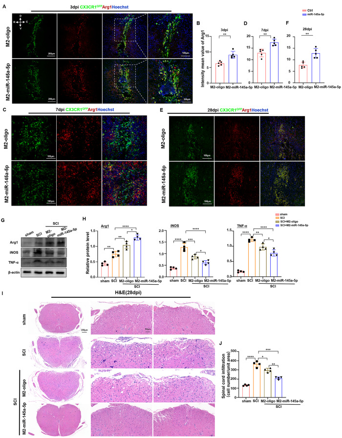
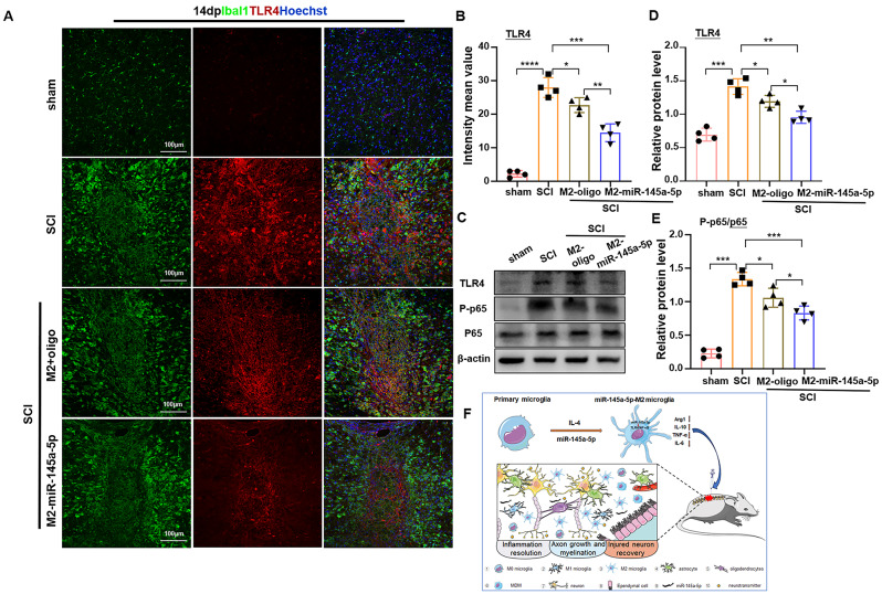
Results: With in vitro experiments, we found that miR-145a-5p was highly expressed in M2 microglia, and miR-145a-5p overexpression could suppress M1 while promote M2 microglia polarization. And then delivery of miR-145a-5p overexpressed M2 microglia into the injured spinal cord area significantly accelerated locomotive recovery as well as prevented glia scar formation and neuron damage in mice, which was even better than M2 microglia transplantation. Further mechanisms showed that overexpressed miR-145a-5p in microglia inhibited the inflammatory response and maintained M2 macrophage phenotype by targeting TLR4/NF-κB signaling.

Conclusions: These findings indicate that transplantation of miR-145a-5p modified M2 microglia has more therapeutic potential for SCI than M2 microglia transplantation from epigenetic perspective.

Keywords: M2 microglia; Neuroinflammation; Spinal cord injury; miR-145a-5p.


Conflict of interest statement

The authors declare that they have no competing interests.

Figures

Fig. 1
Fig. 1
miR-145a-5p promoted M2 while inhibited M1 microglia polarization leading to the reduced oxidative stress-induced neuron damage. (A) BV2 cells were transfected with miR-145a-5p mimics or Ctrl and stimulated with PBS, LPS + IFN-γ or IL-4 for 24 h, respectively. The mRNA level of M1 markers including TNF-α, IL-6, IL-1β and iNOS, as well as M2 markers containing Arg1, Mrc1, YM1 and IL-10 were determined by qRT-PCR, respectively (n = 5). (B) BV2 cells were transfected with miR-145a-5p ASO or Ctrl and treated as (A). The mRNA level of M1 and M2 markers were determined by qRT-PCR, respectively (n = 5). (C, D) BV2 cells were treated as (A), and then collected and lysed. The protein level of iNOS and Arg1 were detected by Western blot. Representative image (C) and quantitative data (D) were shown (n = 4). (E, F) BV2 were treated as (B), and then collected and lysed. The protein level of iNOS and Arg1 were detected by Western blot. Representative image (E) and quantitative data (F) were shown (n = 4). (G, H) The supernatant of BV2 cells treated as (A) was collected, and then co-cultured with H2O2-treated hippocampal neurons. The activated cleaved-caspase 3 of neuron was determined by Western blot. Representative image (G) and quantitative data (H) were shown (n = 4). Data shown as mean ± SEM. *, p < 0.05; **, p < 0.01; ***, p < 0.001; ****, p < 0.0001 by unpaired student’s t-test. ASO, antisense oligonucleotides
Fig. 2
Fig. 2
Injured spinal cord was obviously repaired after transplantation miR-145a-5p overexpressed M2 microglia. (A) Representative images of Luxol Fast Blue (LFB) staining with coronal spinal cord sections after cell transplantation 28 days. Scale bars = 200 μm. (B) Quantitative analysis of the demyelination in SCI tissue sections with different treatment shown in (A) (n = 4). (C) Representative images of Nissl’s staining in SCI tissue sections with different treatment 28 days. Scale bars = 20 μm. (D) Quantitative analysis of the number of survival neurons at different distance from the epicenter of injury in each group shown in (C) (n = 4). (E) Representative images of immunofluorescent analysis of CS56 (red), GFAP (green) and Hoechst (blue) on injured spinal cord tissue sections with different treatment 2 weeks. Scale bars = 100 μm. (F) Quantitative analysis of mean intensity value of CS56 and GFAP as well as the CSPGs+ area in different group shown in (E) (n = 4). Data shown as mean ± SEM. *, p < 0.05; **, p < 0.01; ***, p < 0.001; ****p < 0.0001 by one-way ANOVA with Turkey’s multiple comparison test
Fig. 3
Fig. 3
miR-145a-5p overexpressed M2 microglia transplantation improved the locomotor function of SCI mice. (A) Mouse movement behaviors on a plank were photographed in different group including sham, SCI, SCI with M2 + oligo microglia delivery, and SCI with miR-145a-5p overexpressed M2 microglia delivery after 28 days. (B, C) The Basso mouse scale (BMS) score (B) and subscore (C) in (A) were analyzed and quantitatively compared (n = 6). (D) Mouse swimming in water in different group was recorded. (E) LSS score in (D) was statistically analyzed (n = 6). (F) hindlimb reflex scoring was statistically analyzed (n = 6). Data shown as mean ± SEM, *, p < 0.05; **, p < 0.01; ***, p < 0.001; ****, p < 0.0001, SCI mice with M2-miR-145a-5p microglia treatment versus SCI mice without treatment. #, p < 0.05; ##, p < 0.01; ###, p < 0.001; ####, p < 0.0001, SCI mice with M2 microglia treatment versus SCI mice without treatment. &, p < 0.05; &&, p < 0.01; &&&, p < 0.001; &&&&, p < 0.0001, SCI mice with M2-miR-145a-5p microglia treatment versus SCI mice with M2 microglia treatment. Statistical analysis was performed using a two-way repeated ANOVA with Bonferroni’s post hoc test
Fig. 4
Fig. 4
Delivery of M2 microglia transfected with miR-145a-5p alleviated inflammation in SCI mice. (A-F) CX3CR1GFP/+ microglia were transfected with miR-145a-5p mimics or Ctrl and stimulated with IL-4 to induce M2 microglia, and then to delivery these microglia into SCI mice, respectively. After transplantation 3, 7 and 28 days, Arg1, as an M2 marker, was stained (A, C, E). Nucleic was costained with Hoechst. The mean inflorescence intensity of Arg1+ microglia was measured and quantitatively compared (B, D, F) (n = 5). (G, H) The protein level of the Arg1, iNOS, and TNF-α in injured spinal cord was detected by Western blot after cell transplantation 7 days (G). The quantitative data were calculated and shown in (H) (n = 4). (I) Representative images of HE staining with SCI sections after cell treatment 28 days. Scale bars = 200 μm. (J) The infiltrated inflammatory cells were counted and quantitatively compared (n = 4). Data shown as mean ± SEM. *, p < 0.05; **, p < 0.01; ***, p < 0.001; ****, p < 0.0001 by one-way ANOVA with Turkey’s multiple comparison test
Fig. 5
Fig. 5
miR-145a-5p suppressed inflammation by targeting TLR4/ NF-κB signaling in vitro. (A) Schematic diagrams showing the predicted binding sequence between miR-145a-5p and wide-type (WT) or mutant 3’-UTRs of TLR4. (B) Dual luciferase assay was performed in HEK293T cells after co-transfected with reporter plasmids containing WT or mutant 3’-UTRs of TLR4 as well as miR-145a-5p mimics or Ctrl (n = 4). (C) Representative Western blot showing the expression of TLR4 and NF-kB signal-related molecules such as p65 in BV2 that were transfected with miR-145a-5p mimics or Ctrl followed by PBS, LPS + IFN-γ or IL-4 stimulation for 24 h. (D) Quantitative analysis of the protein level of TLR4 and p65 as well as P-p65 in (D) (n = 4). (E) BV2 was transfected with miR-145a-5p inhibitor or Ctrl and siTLR4 followed by LPS + IFN-γ stimulation for 24 h. The expression of TLR4, p65 and P-p65, as well as TNF-α, IL-1β and iNOS was detected by Western blot (n = 4). (F) Quantitative analysis of the protein level in (E) (n = 4). Data shown as mean ± SEM, *, p < 0.05; **, p < 0.01; ***, p < 0.001; ****, p < 0.0001 by one-way ANOVA with Turkey’s multiple comparison test
Fig. 6
Fig. 6
Transplantation of miR-145a-5p overexpressed M2 microglia attenuated SCI by inhibiting TLR4/NF-κB pathway in vivo. (A) Tissue section was stained with Ibal1(Green), TLR4 (Red) and DNA(Hoechst) after different treatment for SCI on day 7. (B) Quantitative analysis and comparison of the intensity mean value of TLR4 in (A) (n = 4). (C) The expression of TLR4 and p65 as well as P-p65 were detected by Western blot with the spinal cord lysis from SCI mice with different treatment as (A). (D, E) The protein level of TLR4 (D), p65 and P-p65 (E) in (C) was quantitatively compared (n = 4). (F) The Scheme of miR-145a-5p overexpressed M2 microglia transplantation promoted the tissue repair of SCI by targeting TLR4/NF-κB signaling and abolishing inflammation response. Data shown as mean ± SEM. *, p < 0.05; **, p < 0.01; ***, p < 0.001; ****, p < 0.0001 by one-way ANOVA with Turkey’s multiple comparison test

References

    1. Khorasanizadeh M, Yousefifard M, Eskian M, Lu Y, Chalangari M, Harrop JS, Jazayeri SB, Seyedpour S, Khodaei B, Hosseini M, et al. Neurological recovery following traumatic spinal cord injury: a systematic review and meta-analysis. J Neurosurg Spine. 2019;15:1–17. - PubMed
    1. Wichmann TO, Kasch H, Dyrskog S, Høy K, Møller BK, Krog J, Hviid CVB, Hoffmann HJ, Rasmussen MM. The inflammatory response and blood-spinal cord barrier integrity in traumatic spinal cord injury: a prospective pilot study. Acta Neurochir. 2022;164:3143–53. 10.1007/s00701-022-05369-6 - DOI - PubMed
    1. Hemley SJ, Tu J, Stoodley MA. Role of the blood-spinal cord barrier in posttraumatic syringomyelia. J Neurosurg Spine. 2009;11:696–704. 10.3171/2009.6.SPINE08564 - DOI - PubMed
    1. Anjum A, Yazid MD, Fauzi Daud M, Idris J, Ng AMH, Selvi Naicker A, Ismail OHR, Athi Kumar RK, Lokanathan Y. Spinal cord Injury: pathophysiology, Multimolecular interactions, and underlying recovery mechanisms. Int J Mol Sci. 2020;21:7533. 10.3390/ijms21207533 - DOI - PMC - PubMed
    1. Hellenbrand DJ, Quinn CM, Piper ZJ, Morehouse CN, Fixel JA, Hanna AS. Inflammation after spinal cord injury: a review of the critical timeline of signaling cues and cellular infiltration. J Neuroinflamm. 2021;18:284.10.1186/s12974-021-02337-2 - DOI - PMC - PubMed

LinkOut - more resources