. 2026 Jan 1;21(1):406-416.
doi: 10.4103/NRR.NRR-D-23-01741. Epub 2024 Jul 29.

Contribution of ferroptosis and SLC7A11 to light-induced photoreceptor degeneration

Affiliations

Contribution of ferroptosis and SLC7A11 to light-induced photoreceptor degeneration

Xiaoxu Huang et al. Neural Regen Res. .

Abstract

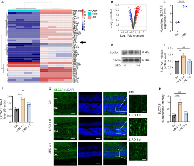
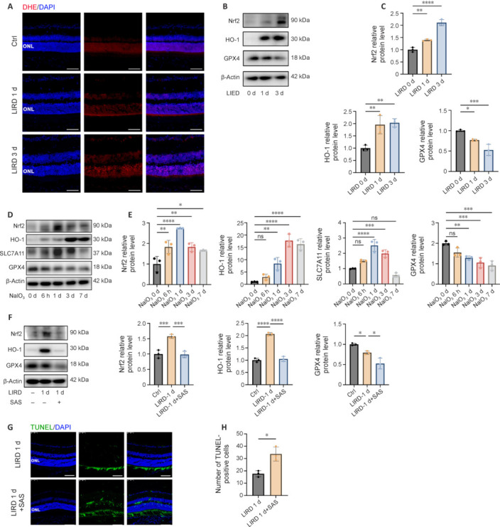
JOURNAL/nrgr/04.03/01300535-202601000-00043/figure1/v/2025-06-09T151831Z/r/image-tiff Progressive photoreceptor cell death is one of the main pathological features of age-related macular degeneration and eventually leads to vision loss. Ferroptosis has been demonstrated to be associated with retinal degenerative diseases. However, the molecular mechanisms underlying ferroptosis and photoreceptor cell death in age-related macular degeneration remain largely unexplored. Bioinformatics and biochemical analyses in this study revealed xC - , solute carrier family 7 member 11-regulated ferroptosis as the predominant pathological process of photoreceptor cell degeneration in a light-induced dry age-related macular degeneration mouse model. This process involves the nuclear factor-erythroid factor 2-related factor 2-solute carrier family 7 member 11-glutathione peroxidase 4 signaling pathway, through which cystine depletion, iron ion accumulation, and enhanced lipid peroxidation ultimately lead to photoreceptor cell death and subsequent visual function impairment. We demonstrated that solute carrier family 7 member 11 overexpression blocked this process by inhibiting oxidative stress in vitro and in vivo . Conversely, solute carrier family 7 member 11 knockdown or the solute carrier family 7 member 11 inhibitor sulfasalazine and ferroptosis-inducing agent erastin aggravated H 2 O 2 -induced ferroptosis of 661W cells. These findings indicate solute carrier family 7 member 11 may be a potential therapeutic target for patients with retinal degenerative diseases including age-related macular degeneration.

Keywords: age-related macular degeneration; ferroptosis; light exposure damage; oxidative stress; pathway; photoreceptor; programmed cell death; solute carrier family 7 member 11 (SLC7A11).


Conflict of interest statement

Conflicts of interest: There are no conflicts of interest to disclose.

Figures

Figure 1
Figure 1
LIRD induced an increase in photoreceptor death in mice. (A–C) Cell death tests revealed an increase in PI-positive cells (red) and TUNEL-positive cells (green) in retinal cryosections after LIRD compared with the control group. Nuclei were stained with DAPI (blue) (n = 3). Scale bars: 50 µm. (D) The concentration of Fe2+ and Fe2+/total Fe in the retina increased after 3 days of LIRD (n = 3). (E, F) Both the a-wave and b-wave of ERG recordings decreased after 3 days of LIRD compared with 0 day (n = 5). Data in (F) are shown as mean ± SD. *P < 0.05, **P < 0.01, ***P < 0.001, ****P < 0.0001. All experiments were conducted in triplicate. Ctrl: Control; DAPI: 4′,6-diamidino-2-phenylindole; INL: inner nuclear layer; LIRD: light-induced retinal damage; ONL: outer nuclear layer; PI: propidium iodide; TUNEL: terminal deoxynucleotidyl transferase-mediated dUTP nick-end labeling.
Figure 2
Figure 2
The expression of SLC7A11 increases in the retina of LIRD mice. (A) Hierarchical clustering analysis of the DEGs between the Ctrl group and LIRD group. (B) Volcano plot of the DEGs (|LogFC| > 1 and P < 0.05). (C) SLC7A11 expression in the LIRD group was significantly higher than that in the Ctrl group in the GEO database. (D, E) Western blot showing the protein levels of SLC7A11 in the retina increased after LIRD (n = 3). (F) SLC7A11 mRNA levels in the retina increased at day 1 after LIRD (n = 3). (G, H) Retinal sections from the LIRD group and control group stained with anti-SLC7A11 (green). Data in H are shown as mean ± SD. **P < 0.01, ***P < 0.001, ****P < 0.0001. Scale bars: 25 µm for original images, 5 µm for enlarged images. All experiments were conducted in triplicate. Ctrl: Control; DAPI: 4′,6-diamidino-2-phenylindole; DEGs: differentially expressed genes; INL: inner nuclear layer; LIRD: light-induced retinal damage; ns: not significant; ONL: outer nuclear layer; SLC7A11: solute carrier family 7 member 11.
Figure 3
Figure 3
Ferroptosis and oxidative stress is induced in response to light damage in vivo. (A) DHE staining (red) showing a time-dependent increase in ROS levels from 0 to 3 days after LIRD. (B, C) Western blot analysis and data quantification showed that the expression levels of Nrf2, HO-1, and GPX4 in mouse retina were significantly elevated after LIRD (n = 3). (D, E) Western blot showing the expression levels of protein Nrf2, HO-1, SLC7A11, and GPX4 in mouse retina after NaIO3 treatment (n = 3). (F) Western blot showed that pre-treatment of sulfasalazine (SAS, a SLC7A11 inhibitor; 8 mg/kg) before LIRD reduced the expression of Nrf2, HO-1, and GPX4 proteins compared with levels in the LIRD group. (G, H) Cell death in ONL after SAS treatment and LIRD. TUNEL immunoreactivity (green) was increased in retinal sections of SAS-treated LIRD mice compared with the LIRD group (n = 3). Data are shown as mean ± SD. *P < 0.05, **P < 0.01, ***P < 0.001, ****P < 0.0001. Scale bars: 50 µm. All experiments were conducted in triplicate. Ctrl: Control; DAPI: 4′,6-diamidino-2-phenylindole; DHE: dihydroethidium; GPX4: glutathione peroxidase 4; HO-1: heme oxygenase-1; LIRD: light-induced retinal damage; NRF2: nuclear factor-erythroid factor 2-related factor 2; ONL: outer nuclear layer; SAS: sulfasalazine; TUNEL: terminal deoxynucleotidyl transferase-mediated dUTP nick-end labeling.
Figure 4
Figure 4
SLC7A11 expression was upregulated in 661W cells with H2O2-treatment and the SLC7A11 inhibitor SAS promoted 661W ferroptosis. (A–D) Western blot showing SLC7A11 protein levels in 661w cells with H2O2 treatment at different concentration and time (n = 3). (E, F) The BODIPY TM581/591 staining showed the increased lipid peroxidation level after H2O2 treatment (n = 3). Scale bars: 25 μm. (G) The GSH level of 661W cells significantly decreased after SAS treatment in a concentration-dependent form (n = 4). (H, I) The CCK8 assay showed no significant change in cell viability of 661W cells at different SAS concentrations ranging from 0 to 400 μM while SAS combined with H2O2 treatment led to decreased cell viability in a dose-dependent manner (n = 5). (J, K) Western blot showing the expression levels of protein Nrf2, GPX4 and HO-1 in 661W cells after treatment of H2O2 and SAS (n = 3). Data are shown as mean ± SD. *P < 0.05, **P < 0.01, ****P < 0.0001. All experiments were conducted in triplicate independently. Ctrl: Control; DAPI: 4′,6-diamidino-2-phenylindole; GPX4: glutathione peroxidase 4; HO-1: heme oxygenase-1; NRF2: nuclear factor-erythroid factor 2-related factor 2; ns: not significant; SAS: sulfasalazine; SLC7A11: solute carrier family 7 member 11.
Figure 5
Figure 5
SLC7A11 knockdown promotes ferroptosis of 661W cells in vitro. (A) Western blot analysis of SLC7A11 in 661W cells transfected with various SLC7A11-siRNAs. (B) SLC7A11 mRNA levels in 661W cells transfected with various SLC7A11-siRNAs (n = 3). (C, D) Western blot and data quantification of the expression levels of Nrf2, HO-1, and GPX4 in siSLC7A11#3-treated 661W cells exposed to H2O2 (n = 3). (E) CCK-8 assay revealed no significant change in cell viability in 661W cells after treatment with Fer-1 at different concentrations (n = 5). (F) siSLC7A11 transfection significantly decreased the viability of cells treated with 40 µM Fer-1 and H2O2 (n = 5). Data are shown as mean ± SD. ns, non-significant. *P < 0.05, **P < 0.01, ***P < 0.001, ****P < 0.0001. All experiments were conducted in triplicate. Ctrl: Control; Fer-1: ferrostatin-1 (ferroptosis inhibitor); GPX4: glutathione peroxidase 4; HO-1: heme oxygenase-1; Nrf2: nuclear factor-erythroid factor 2-related factor 2; ns: not significant; SLC7A11: solute carrier family 7 member 11.
Figure 6
Figure 6
SLC7A11 overexpression enhanced ferroptosis resistance in vitro. (A–D) Western blot of Nrf2, HO-1, SLC7A11, and GPX4 in 661W cells transfected with Slc7a11 plasmid and treated with or without 400 µM H2O2 (n = 3). (E, F) Cell Counting Kit-8 assay revealed a significant decrease in cell viability in 661W cells after treatment of Erastin (ferroptosis inducer) or RSL3 (ferroptosis inducer) in a concentration-dependent manner (n = 5). (G, H) SLC7A11 overexpression rescued the cell viability of 10 µM Erastin– or 10 µM RSL3–treated 661W cells after exposure to H2O2 (n = 5). Data are shown as mean ± SD. *P < 0.05, **P < 0.01, ***P < 0.001, ****P < 0.0001. All experiments were conducted in triplicate. Ctrl: Control; GPX4: glutathione peroxidase 4; HO-1: heme oxygenase-1; NRF2: nuclear factor-erythroid factor 2-related factor 2; SLC7A11: solute carrier family 7 member 11.
Figure 7
Figure 7
SLC7A11 overexpression in photoreceptors suppresses retinal degeneration of LIRD mice. (A) Immunofluorescence of the retinal sections after subretinal injection of AAV2/8-Slc7a11-GFP or AAV2/8-vehicle-GFP for 2 weeks showed AAV transfection reached the photoreceptors. Scale bars: 50 µm. (B, C) Protein expression levels of Nrf2, HO-1, SLC7A11, and GPX4 in retina after LIRD pretreatment with the transfection of AAV2/8-Slc7a11 or AAV2/8-vehicle (n = 3). (D, E) Both the a-wave and b-wave of ERG recordings from mice with 3-day LIRD were increased after AAV2/8-Slc7a11 transfection (n = 3). (F, G) TUNEL (red) and PI (red) staining of retinal sections showed transfection of AAV2/8-Slc7a11 reduced the cell death of photoreceptors caused by light exposure. Scale bars: 50 µm. Data in C and E are shown as mean ± SD. *P < 0.05, **P < 0.01. All experiments were conducted in triplicate. AAV: Adeno-associated virus; DAPI: 4′,6-diamidino-2-phenylindole; GFP: green fluorescent protein; GPX4: glutathione peroxidase 4; HO-1: heme oxygenase-1; LIRD: light-induced retinal damage; NRF2: nuclear factor-erythroid factor 2-related factor 2; SLC7A11: solute carrier family 7 member 11.

References

    1. Alborzinia H, Ignashkova TI, Dejure FR, Gendarme M, Theobald J, Wölfl S, Lindemann RK, Reiling JH. Golgi stress mediates redox imbalance and ferroptosis in human cells. Commun Biol. 2018;1:210. - PMC - PubMed
    1. Ashok A, Singh N, Chaudhary S, Bellamkonda V, Kritikos AE, Wise AS, Rana N, McDonald D, Ayyagari R. Retinal degeneration and Alzheimer’s disease: an evolving link. Int J Mol Sci. 2020;21:7290. - PMC - PubMed
    1. Baksheeva VE, Tiulina VV, Tikhomirova NK, Gancharova OS, Komarov SV, Philippov PP, Zamyatnin AA, Jr, Senin II, Zernii EY. Suppression of light-induced oxidative stress in the retina by mitochondria-targeted antioxidant. Antioxidants (Basel) 2018;8:3. - PMC - PubMed
    1. Blasiak J. Senescence in the pathogenesis of age-related macular degeneration. Cell Mol Life Sci. 2020;77:789–805. - PMC - PubMed
    1. Boyd P, Hyde DR. Iron contributes to photoreceptor degeneration and Müller glia proliferation in the zebrafish light-treated retina. Exp Eye Res. 2022;216:108947. - PMC - PubMed

LinkOut - more resources