

Incidental diagnosis of a large left ventricular pseudoaneurysm
- PMID: 39104447
- PMCID: PMC11299581
- DOI: 10.1016/j.radcr.2024.06.039
Incidental diagnosis of a large left ventricular pseudoaneurysm
Abstract
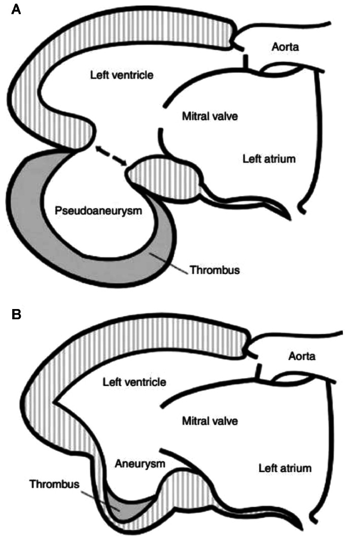
Left ventricular pseudoaneurysm is a rare complication of myocardial infarction and represent a myocardial rupture contained within a pericardial space limited by adhesions. Differentiating it from a left ventricular aneurysm can be a real diagnostic challenge. We report a case of a 50-year-old man admitted for symptoms of left heart failure. Transthoracic echocardiography and cardiac computed tomography scan incidentally showed a large lateral left ventricular pseudoaneurysm measuring 75/50 mm in diameter. Patch closure was carried out under cardiopulmonary bypass. Postoperative follow up was uneventful. This case demonstrates the increasing detection of «incidental» left ventricular pseudoaneurysm with more frequent use of multimodality imaging techniques including cardiac CT scan.
Keywords: Imaging; Left ventricle; Myocardial infarction; Pseudoaneurysm.
© 2024 The Authors. Published by Elsevier Inc. on behalf of University of Washington.
Figures


References
-
- Antman EM. In: Braunwalds heart disease: a textbook of cardiovascular medicine. 7th ed. Zipes, et al., editors. Elsevier Saunders; Philadelphia: 2005. ST-elevation myocardial infarction: management; pp. 1167–1226. editors.
-
- Stewart S, Huddle R, Stuard I, Schreiner BF, DeWeese JA. False aneurysm and pseudo-false aneurysm of the left ventricle (etiology, pathology, diagnosis, and operative management) Ann Thorac Surg. 1981;31:259–265. - PubMed
-
- Davidson KH, Parisi AG, Harrington JJ, Barsamian EM, Fishbein MC. Pseudo aneurysm of the left ventricle (an unusual echocardiographic presentation) Ann Intern Med. 1977;86:430–433. - PubMed
-
- March KL, Sawada SG, Tarver RD, Kesler KA, Armstrong WF. Current concepts of left ventricular pseudoaneurysm: pathophysiology, therapy, and diagnostic imaging methods. Clin Cardiol. 1989;12:531–540. - PubMed
-
- Frances C, Romero A, Grady D. Left ventricular pseudoaneurysm. J Am Coll Cardiol. 1998;32:557–561. - PubMed
Publication types
LinkOut - more resources
Full Text Sources
